A Meningioma With Extensive Peritumoral Edema Mimicking Metastatic Brain Tumor: A Case Report
- Affiliations
-
- 1Department of Neurosciences, East Avenue Medical Center, Quezon City, Philippines
- 2Department of Pathology, East Avenue Medical Center, Quezon City, Philippines
- 3Department of Neurosurgery, East Avenue Medical Center, Quezon City, Philippines
- KMID: 2542086
- DOI: http://doi.org/10.14791/btrt.2022.0029
Abstract
- Meningioma is the most common brain tumor among all histologically reported malignant and non-malignant tumors of the central nervous system. Angiomatous meningioma is one of the subtypes of meningioma that is rarely reported. In this paper, we present a case of a 67-year-old female patient who sought consultation due to seizure, cognitive decline, and parkinsonism. Contrast-enhanced MRI showed a well-defined tumor in the left frontal lobe convexity with extensive perilesional edema. A tumor excision was done and histopathology studies revealed an angiomatous meningioma subtype. This case is reportable because angiomatous meningioma is a recognized rare entity. It is important to share this entity with other medical professionals and start to consider this condition in differential diagnosis when diagnosing a patient with an intracranial mass with an extensive peritumoral edema. Furthermore, the patient’s unusual presentation of parkinsonian features and its occurrence with colorectal cancer history suggest a possible association between these conditions.
Keyword
Figure
-
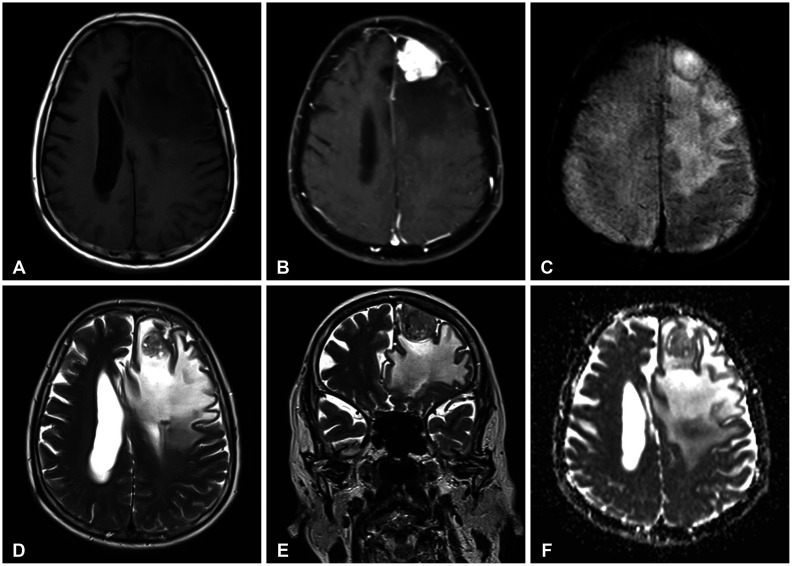
Fig. 1 Neuroimaging MRI with contrast enhancement. A: The tumor lesion is isointense on the T1-weighted (T1W) sequence located at the left frontal lobe, measuring 3×2.8×3.4 cm. B: There are marked intense enhancements of the lesion after contrast as depicted in the T1W sequence. This abuts the dura and compresses the left frontal horn. C: Susceptibility-weighted image sequence does not show magnetic susceptibility. D and E: There are some signal voids of blood vessels seen in the T2-weighted sequences and it demonstrated marked surrounding edema. F: The region does not show abnormally low apparent diffusion coefficient values.
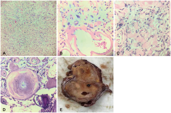
Fig. 2 Histopathologic and gross examination. A-D: Hematoxylin and eosin stain. A: Tumor composed of numerous vascular spaces and meningothelial cells (×100 magnification). B: Vascular spaces lined by endothelial cells with intervening areas of round to ovoid meningothelial cells. Some meningothelial cells are wrapped around the blood vessels, displaying degenerative nuclear atypia with intranuclear pseudo-inclusions (×400 magnification). C: Meningothelial cells with eosinophilic cytoplasm and several hyaline globules within spaces (×400 magnification). D: Psammoma bodies demonstrating concentric calcifications (×400 magnification). E: Gross examination shows an encapsulated tumor (0.2-cm thick capsule) with a dark brown, smooth cut surface with punctate hemorrhages.
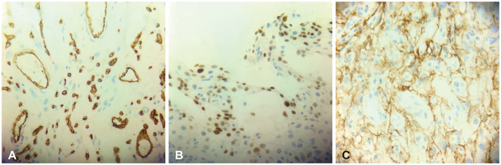
Fig. 3 Immunohistochemical study. A: Tumor cells show negative immunoreactivity for CD34 but positive for endothelial cells lining the blood vessels (CD34, ×400 magnification). B: Tumor cells show focal, strong nuclear expression on progesterone receptors (progesterone, ×400 magnification). C: Tumor cells exhibiting diffuse, strong, cytoplasmic staining on epithelial membrane antigen (EMA, ×400 magnification).
Reference
-
1. Ostrom QT, Patil N, Cioffi G, Waite K, Kruchko C, Barnholtz-Sloan JS. CBTRUS statistical report: primary brain and other central nervous system tumors diagnosed in the United States in 2013-2017. Neuro Oncol. 2020; 22(12 Suppl 2):iv1–iv96. PMID: 33123732.2. Hasselblatt M, Nolte KW, Paulus W. Angiomatous meningioma: a clinicopathologic study of 38 cases. Am J Surg Pathol. 2004; 28:390–393. PMID: 15104303.3. Bansal D, Diwaker P, Gogoi P, Nazir W, Tandon A. Intraparenchymal angiomatous meningioma: a diagnostic dilemma. J Clin Diagn Res. 2015; 9:ED07–ED08.4. Hua L, Luan S, Li H, Zhu H, Tang H, Liu H, et al. Angiomatous meningiomas have a very benign outcome despite frequent peritumoral edema at onset. World Neurosurg. 2017; 108:465–473. PMID: 28844928.5. Louis DN, Perry A, Wesseling P, Brat DJ, Cree IA, Figarella-Branger D, et al. The 2021 WHO classification of tumors of the central nervous system: a summary. Neuro Oncol. 2021; 23:1231–1251. PMID: 34185076.6. Yang L, Ren G, Tang J. Intracranial angiomatous meningioma: a clinicopathological study of 23 cases. Int J Gen Med. 2020; 13:1653–1659. PMID: 33408502.7. Ben Nsir A, Chabaane M, Krifa H, Jeme H, Hattab N. Intracranial angiomatous meningiomas: a 15-year, multicenter study. Clin Neurol Neurosurg. 2016; 149:111–117. PMID: 27513979.8. Liu Z, Wang C, Wang H, Wang Y, Li JY, Liu Y. Clinical characteristics and treatment of angiomatous meningiomas: a report of 27 cases. Int J Clin Exp Pathol. 2013; 6:695–702. PMID: 23573316.9. Hwang J, Kong DS, Seol HJ, Nam DH, Lee JI, Choi JW. Clinical and radiological characteristics of angiomatous meningiomas. Brain Tumor Res Treat. 2016; 4:94–99. PMID: 27867918.10. WHO Classification of Tumours Editorial Board. Central nervous system tumours. 5th ed. Lyon: International Agency for Research on Cancer;2021.11. Bodla AA, Mehta P, Mushtaq F, Durrani OM. Angiomatous meningioma of orbit mimicking as malignant neoplasm: a case report and literature review. Orbit. 2011; 30:183–185. PMID: 21780930.12. Malmer B, Tavelin B, Henriksson R, Grönberg H. Primary brain tumours as second primary: a novel association between meningioma and colorectal cancer. Int J Cancer. 2000; 85:78–81. PMID: 10585587.
- Full Text Links
-
- Actions
-
Cited
- CITED
-
- Close
- Share
-
- Similar articles
-
- Failed First Craniotomy and Tumor Removal of Parasagittal Meningioma with Severe Peritumoral Brain Edema
- Meningioma With Partial and Spontaneous Regression of Peritumoral Edema on Long-Term Follow Up
- Radiological Characteristics of Peritumoral Edema in Meningiomas
- Factors Influencing the Development of Peritumoral Brain Edema in Menigiomas
- Crush Cytology of Secretory Meningioma: A Case Report
