Crush Cytology of Secretory Meningioma: A Case Report
- Affiliations
-
- 1Department of Pathology, Gachon University Gil Medical Center, Incheon, Korea. hicho@gilhospital.com
- 2Department of Neurosurgery, Gachon University Gil Medical Center, Incheon, Korea.
- KMID: 2114664
- DOI: http://doi.org/10.14791/btrt.2015.3.2.147
Abstract
- Secretory meningioma, a histologic subtype of meningioma of World Health Organization grade 1, is clinically significant because it is frequently accompanied by peritumoral brain edema. The patient was a 53-year-old woman suffering from dysarthria and motor weakness of the right arm. Enhanced magnetic resonance images showed an enhancing mass measuring 2.5 cm in size located in the right parietal convexity. Intraoperative squash cytology showed moderately cellular smears composed mainly of clusters of ovoid cells with scattered whorl formations. The cells had round nuclei and a moderate amount of eosinophilic cytoplasm with ill-defined cell borders. Neither atypia nor mitosis was observed. Some scattered round shaped eosinophilic refractile hyaline globules, measuring from 5 to 25 microm, were observed, and a periglobular halo was occasionally observed. The diagnosis of secretory meningioma should be made as early as possible so that neurosurgeons can prevent postoperative aggravation of peritumoral edema. We emphasize that cytologic findings including eosinophilic, non-fibrillary cytoplasm with eosinophilic refractile hyaline globules are helpful in differentiating secretory meningioma from other subtypes of meningioma, primary and metastatic brain tumors.
Keyword
MeSH Terms
Figure
-
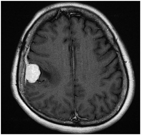
Fig. 1 T1-weighted MR image shows an enhancing mass located in the right parietal convexity with peritumoral edema.

Fig. 2 Intraoperative crush and squash cytology of the resected mass. A: Cellular smear shows sheets of cohesive polygonal tumor cells forming occasional whorls (hematoxylin and eosin stain, ×200). Arrow and inset indicate eosinophilic globules (hematoxylin and eosin stain, ×1,000). B: Intracytoplasmic inclusions of variable size (white arrows) have a centrally located eosinophilic homogeneous core and a well-defined, thin, clear rim. Note frequent nuclear pseudoinclusions (black arrows, hematoxylin and eosin stain, ×1,000). C: Left: a resected mass is composed of large lobules of syncytially arranged ovoid shaped cells with periodic acid-Schiff-positive eosinophilic granular bodies in aggregated or singly scattered form. Note a periglobular thin halo-like rim (periodic acid-Schiff stain, ×400). Right: cells containing cytoplasmic secretory inclusions are occasionally positive for pancytokeratin (pancytokeratin immunostain, ×400).
Cited by 1 articles
-
Intracranial Metaplastic Meningioma : Clinical and Radiological Characteristics of 11 Cases
Taehoon Kim, Jin Wook Kim, So Young Ji, Ho Kang, Kyung-Min Kim, Yong Hwy Kim, Chul-Kee Park, Seung Hong Choi, Sung-Hye Park
J Korean Neurosurg Soc. 2020;63(5):657-663. doi: 10.3340/jkns.2020.0151.
Reference
-
1. Perry A, Louis DN, Scheithauer BW, Budka H, von Deimling A. Meningiomas. In : Louis DN, Ohgaki H, Wiestler OD, Cavenee WK, editors. Pathology and Genetics, World Health Organization Classification of Tumours of the Central Nervous System. 4th ed. Lyon: IARC press;2007. p. 164–172.2. Osawa T, Tosaka M, Nagaishi M, Yoshimoto Y. Factors affecting peritumoral brain edema in meningioma: special histological subtypes with prominently extensive edema. J Neurooncol. 2013; 111:49–57.
Article3. Regelsberger J, Hagel C, Emami P, Ries T, Heese O, Westphal M. Secretory meningiomas: a benign subgroup causing life-threatening complications. Neuro Oncol. 2009; 11:819–824.
Article4. Bitzer M, Nägele T, Geist-Barth B, et al. Role of hydrodynamic processes in the pathogenesis of peritumoral brain edema in meningiomas. J Neurosurg. 2000; 93:594–604.
Article5. Seok JY, Kim NR, Cho HY, Chung DH, Yee GT, Kim EY. Crush cytology of microcystic meningioma with extensive sclerosis. Korean J Pathol. 2014; 48:77–80.
Article6. Hinton DR, Kovacs K, Chandrasoma PT. Cytologic features of secretory meningioma. Acta Cytol. 1999; 43:121–125.
Article7. Kim SH, Lee KG, Kim TS. Cytologic features of secretory meningioma in squash preparation: a case report. Korean J Cytopathol. 2004; 15:52–55.8. Siddiqui MT, Mahon BM, Cochran E, Gattuso P. Cytologic features of meningiomas on crush preparations: a review. Diagn Cytopathol. 2008; 36:202–206.
Article9. Taraszewska A, Matyja E. Secretory meningiomas: immunohistochemical pattern of lectin and ultrastructure of pseudopsammoma bodies. Folia Neuropathol. 2014; 52:141–150.10. Takei H, Bhattacharjee MB, Adesina AM. Chordoid glioma of the third ventricle: report of a case with cytologic features and utility during intraoperative consultation. Acta Cytol. 2006; 50:691–696.11. Ribourtout B, Zandecki M. Plasma cell morphology in multiple myeloma and related disorders. Morphologie. 2015; 99:38–62.
Article12. Jaskólski D, Papierz T, Liberski PP, Sikorska B. Ultrastructure of meningiomas: autophagy is involved in the pathogenesis of "intranuclear vacuoles". Folia Neuropathol. 2012; 50:187–193.13. Browne TJ, Goumnerova LC, De Girolami U, Cibas ES. Cytologic features of pilocytic astrocytoma in cerebrospinal fluid specimens. Acta Cytol. 2004; 48:3–8.
Article14. Kim SH, Kim TS. Squash smear findings of eosinophilic granular bodies in pilocytic astrocytoma. Acta Cytol. 2005; 49:112–114.15. Monabati A, Kumar PV, Kamkarpour A. Intraoperative cytodiagnosis of metastatic brain tumors confused clinically with brain abscess. A report of three cases. Acta Cytol. 2000; 44:437–441.
Article16. Schmitt HP. Metastases of malignant neoplasms to intracranial tumours: the "tumour-in-a-tumour" phenomenon. Virchows Arch A Pathol Anat Histopathol. 1984; 405:155–160.
Article17. Hockley AD. Metastatic carcinoma in a spinal meningioma. J Neurol Neurosurg Psychiatry. 1975; 38:695–697.
Article
- Full Text Links
-
- Actions
-
Cited
- CITED
-
- Close
- Share
-
- Similar articles
-
- Secretory meningioma: a case report with histopathological, immunohistochemical and ultrastructural analysis
- Secretory Meningioma: A case report
- A Case of Crush Syndrome after Physical Assaults
- Crush Cytology Features and Differential Diagnosis of Meningiomas and Schwannomas in Central Nervous System
- Secretory Meningioma with Elevated Serum Carcinoembryonic Antigen
