Investig Magn Reson Imaging.
2019 Dec;23(4):395-400. 10.13104/imri.2019.23.4.395.
Dilated Cardiomyopathy in Acromegaly: a Case Report with Cardiac MR Findings
- Affiliations
-
- 1Department of Radiology, Inha University Hospital, Incheon, Korea.
- 2Department of Radiology, Seoul National University Hospital, Seoul, Korea. hyewon101@hotmail.com
- KMID: 2468059
- DOI: http://doi.org/10.13104/imri.2019.23.4.395
Abstract
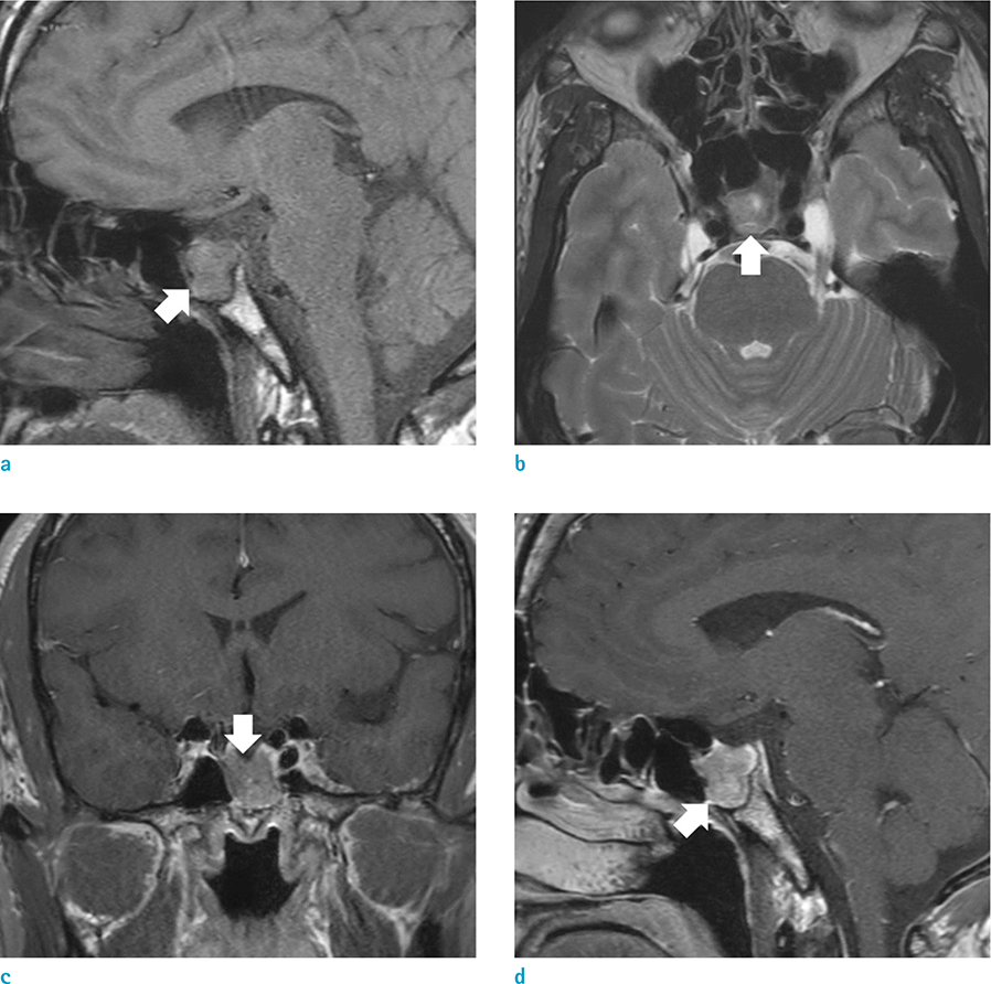
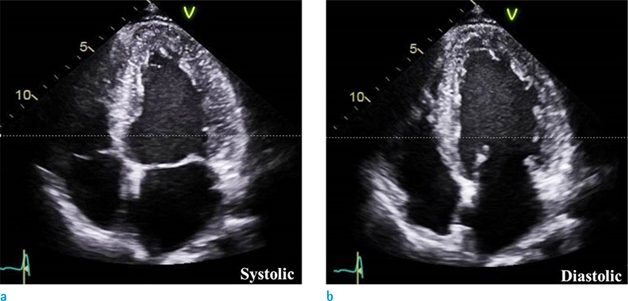
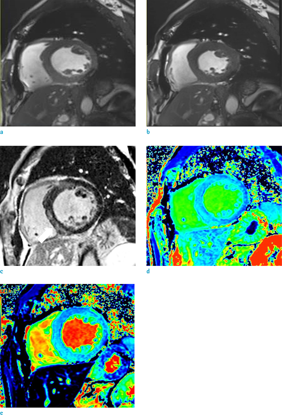
- Acromegaly is a rare endocrine disorder caused by excessive secretion of the growth hormone. There is a wide range of clinical manifestations from somatic symptoms to respiratory or cardiac failure. Among them, cardiovascular involvement is a leading cause of morbidity and mortality. There are relatively few cases reporting cardiac magnetic resonance imaging (CMR) findings of cardiomyopathy in patients with acromegaly. Thus, we report a case of acromegaly showing dilated cardiomyopathy focusing on the findings of CMR.
MeSH Terms
Figure
Reference
-
1. Lavrentaki A, Paluzzi A, Wass JA, Karavitaki N. Epidemiology of acromegaly: review of population studies. Pituitary. 2017; 20:4–9.2. Lugo G, Pena L, Cordido F. Clinical manifestations and diagnosis of acromegaly. Int J Endocrinol. 2012; 2012:540398.3. Bihan H, Espinosa C, Valdes-Socin H, et al. Long-term outcome of patients with acromegaly and congestive heart failure. J Clin Endocrinol Metab. 2004; 89:5308–5313.4. dos Santos Silva CM, Gottlieb I, Volschan I, et al. Low frequency of cardiomyopathy using cardiac magnetic resonance imaging in an acromegaly contemporary cohort. J Clin Endocrinol Metab. 2015; 100:4447–4455.5. Sharma AN, Tan M, Amsterdam EA, Singh GD. Acromegalic cardiomyopathy: epidemiology, diagnosis, and management. Clin Cardiol. 2018; 41:419–425.6. Dabir D, Child N, Kalra A, et al. Reference values for healthy human myocardium using a T1 mapping methodology: results from the International T1 multicenter cardiovascular magnetic resonance study. J Cardiovasc Magn Reson. 2014; 16:69.7. Haaf P, Garg P, Messroghli DR, Broadbent DA, Greenwood JP, Plein S. Cardiac T1 mapping and extracellular volume (ECV) in clinical practice: a comprehensive review. J Cardiovasc Magn Reson. 2016; 18:89.8. Dargad RR, Parekh JD, Dargad RR. Acromegaly with dilated cardiomyopathy. J Assoc Physicians India. 2016; 64:96–97.9. Giesbrandt KJ, Bolan CW, Shapiro BP, Edwards WD, Mergo PJ. Diffuse diseases of the myocardium: MRI-pathologic review of nondilated cardiomyopathies. AJR Am J Roentgenol. 2013; 200:W266–W273.
- Full Text Links
- Actions
-
Cited
- CITED
-
- Close
- Share
- Similar articles
-
- Acromegaly improved with sandostatin LAR treatment
- Continuous Positive Airway Pressure Therapy in a Obstructive SleepApnea Syndrome associated with Active Acromegaly
- Maternally Inherited Familial Hypertrophic Cardiomyopathy Manifested by Pregnancy Related Early Progression and Sudden Cardiac Death
- A case of acromegaly associated with moyamoya disease, coronary arterial stenosis, and dilated cardiomyopathy
- Dilated Cardiomyopathy Associated with Clozapine


