Combination Effect of Titrated Extract of Centella asiatica and Astaxanthin in a Mouse Model of Phthalic Anhydride-Induced Atopic Dermatitis
- Affiliations
-
- 1College of Pharmacy and Medical Research Center, Chungbuk National University, Cheongju, Korea. jinthong@chungbuk.ac.kr
- KMID: 2448757
- DOI: http://doi.org/10.4168/aair.2019.11.4.548
Abstract
- PURPOSE
In our previous study, we demonstrated that both titrated extract of Centella asiatica (TECA) and astaxanthin (AST) have anti-inflammatory effects in a 5% phthalic anhydride (PA) mouse model of atopic dermatitis (AD). The increasing prevalence of AD demands new therapeutic approaches for treating the disease. We investigated the therapeutic efficacy of the ointment form of TECA, AST and a TECA + AST combination in a mouse model of AD to see whether a combination of the reduced doses of 2 compounds could have a synergistic effect.
METHODS
An AD-like lesion was induced by the topical application of 5% PA to the dorsal ear and back skin of an Hos:HR-1 mouse. After AD induction, TECA (0.5%), AST (0.5%) and the TECA (0.25%) + AST (0.25%) combination ointment (20 μg/cm2) were spread on the dorsum of the ear or back skin 3 times a week for 4 weeks. We evaluated dermatitis severity, histopathological changes and changes in protein expression by Western blotting for inducible nitric oxide synthase (iNOS), cyclocxygenase (COX)-2, and nuclear factor (NF)-κB activity. We also measured the concentrations of tumor necrosis factor (TNF)-α, interleukin (IL)-6 and immunoglobulin E (IgE) in the blood of AD mice by enzyme-linked immunosorbent assay (ELISA).
RESULTS
PA-induced skin morphological changes and ear thickness were significantly reduced by TECA, AST and TECA + AST treatments, but these inhibiting effects were more pronounced in the TECA + AST treatment. TECA, AST and the TECA+AST reatments inhibited the expression of iNOS and COX-2; NF-κB activity; and the release of TNF-α, IL-6 and IgE. However, the TECA+AST treatment showed additive or synergistic effects on AD.
CONCLUSIONS
Our results demonstrate that the combination of TECA and AST could be a promising therapeutic agent for AD by inhibiting NF-κB signaling.
MeSH Terms
-
Animals
Blotting, Western
Centella*
Dermatitis
Dermatitis, Atopic*
Ear
Enzyme-Linked Immunosorbent Assay
Immunoglobulin E
Immunoglobulins
Inflammation
Interleukin-6
Interleukins
Mice*
Nitric Oxide Synthase Type II
Prevalence
Skin
Tumor Necrosis Factor-alpha
Immunoglobulin E
Immunoglobulins
Interleukin-6
Interleukins
Nitric Oxide Synthase Type II
Tumor Necrosis Factor-alpha
Figure
-
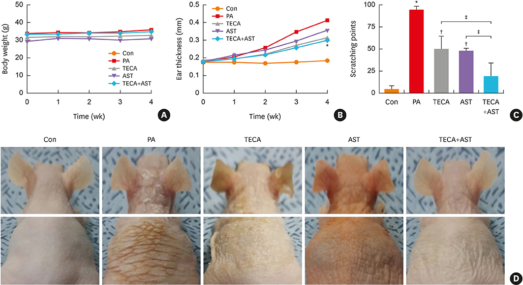
Fig. 1 Differences in body weight, dorsal ear skin thickness and phenotypes, as well as back skin phenotypes. Body weights of mice in the 5 groups were measured using a chemical balance (A). PA solution was repeatedly applied to the dorsal ear and back skin during the topical application of Centella asiatica phytosome. After 4 weeks, dorsal ear skin thickness (B), the number of times scratching at the end of the application experiment (C) and phenotypes (D) were observed following the procedure described in Materials and Methods. Data shown are the mean ± standard deviation (n = 10). Con, control; PA, phthalic anhydride; TECA, titrated extract of Centella asiatica; AST, astaxanthin. *P < 0.05 is the significance level compared to the control group. †P < 0.05 is the significance level compared to the PA treatment group. ‡P < 0.05 is the significance level compared to the TECA or AST treatment alone group.
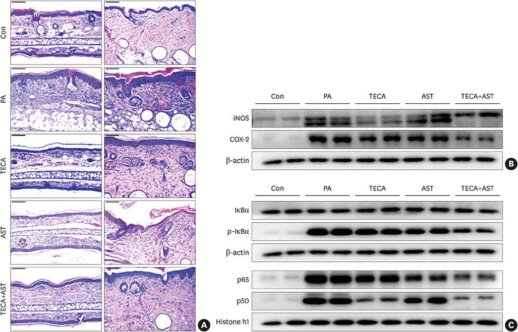
Fig. 2 Histopathological analysis of dorsal ear and back skin tissues and the anti-inflammatory effects of the TECA + AST treatment through inhibiting NF-κB in the back skin. Histopathology of dorsal ear and back skin tissues (A). PA solution was repeatedly applied to the dorsal ear and back skin during the topical application of TECA, AST and the TECA+AST combination. Histopathological changes were examined in the slide sections of dorsal ear and back skin tissues by staining with H&E followed by observation at 200× magnification (scale bars, 100 μm). Alterations in the expression of iNOS and COX-2 proteins of the back skin were measured by Western blotting (B). The effect of TECA, AST and the TECA + AST combination on NF-κB (p50 and p65) subunit translocation into the nucleus and IκBα phosphorylation in back skin cytosol (C). Equal amounts of nuclear proteins (20 μg/lane) or total proteins (20 μg/lane) were subjected to 10% SDS-PAGE, and the expressions of p50, p65, IκBα and p-IκBα proteins were detected by Western blotting using specific antibodies. Histone h1 protein and β-actin protein were used here as internal controls. Data shown are the mean ± standard deviation (n = 10). Con, control; TECA, titrated extract of Centella asiatica; AST, astaxanthin; NF-κB, nuclear factor-κB; PA, phthalic anhydride; H&E, hematoxylin and eosin; iNOS, inducible nitric oxide synthase; COX, cyclooxygenase; SDS-PAGE, sodium dodecyl sulfate-polyacrylamide gel electrophoresis.
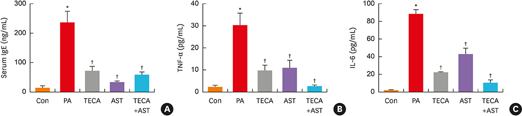
Fig. 3 Changes in serum cytokine concentration. After the final treatment, mice from each group were euthanized under anesthesia. The serum used to measure the cytokine concentration was prepared from blood samples collected from the abdominal veins of mice. Serum IgE (A), TNF-α and IL-6 (B) concentrations were quantified by ELISA. Data shown are the mean ± standard deviation (n = 10). Con, control; PA, phthalic anhydride; TECA, titrated extract of Centella asiatica; AST, astaxanthin; IgE, immunoglobulin E; TNF, tumor necrosis factor; IL, interleukin; ELISA, enzyme-linked immunosorbent assay. *P < 0.05 is the significance level compared to the control group. †P < 0.05 is the significance level compared to the PA treatment group.
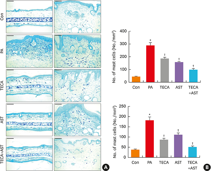
Fig. 4 The inhibition of mast cell infiltration by topical application of the TECA + AST combination in dorsal ear and back skin. Mast cell infiltrations in dorsal ear and back skin (A). Slide sections of dorsal ear and back tissues were examined by staining with 0.25% toluidine blue followed by observation at 200× magnification (scale bars, 100 μm). The number of infiltrated mast cells per specific area was measured as described in Materials and Methods (B). Data shown are obtained from the same mice treated as shown in Fig. 1. Data shown are the mean ± standard deviation (n=10). *P < 0.05 is the significance level compared to the control group. Con, control; PA, phthalic anhydride; TECA, titrated extract of Centella asiatica; AST, astaxanthin. *P < 0.05 is the significance level compared to the control group. †P < 0.05 is the significance level compared to the PA treatment group.
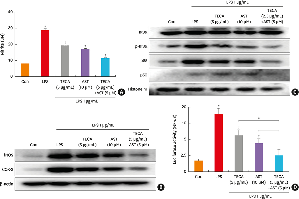
Fig. 5 Effects of the TECA + AST treatment on NO production and the expression of iNOS and COX-2 in LPS-treated RAW 264.7 macrophages. The cells were treated with 1 μg/mL LPS alone or LPS with TECA (5 μg/mL), AST (10 μM) and the combination of TECA (2.5 μg/mL) + AST (5 μM) for 24 hours. At the end of incubation, 50 μL of the medium was removed to measure NO production (A). Control values were obtained in the absence of LPS. In the culture medium, NO production was measured by the Griess reaction as described in Materials and Methods. Equal amounts of total proteins (20 μg/lane) were subjected to 10% SDS-PAGE, and alterations in the expression of iNOS and COX-2 proteins were detected by Western blotting using specific antibodies (B). The β-actin protein was used here as an internal control. The effect of the TECA + AST treatment on the LPS-induced translocation of the NF-κB subunits (p50 and p65) into the nucleus and the phosphorylation of IκBα in cytosol (C). Equal amounts of nuclear proteins (20 μg/lane) or total proteins (20 μg/lane) were subjected to 10% SDS-PAGE, and the expressions of p50, p65, IκBα and p-IκBα proteins were detected by Western blotting using specific antibodies. Histone h1 protein and β-actin protein were used here as internal controls. The effect of the TECA + AST treatment on the LPS-induced NF-κB transcriptional activity (D). Data shown are the mean ± standard deviation from 3 experiments in duplicate. Con, control; LPS, lipopolysaccharides; TECA, titrated extract of Centella asiatica; AST, astaxanthin; NF-κB, nuclear factor-κB; iNOS, inducible nitric oxide synthase; COX, cyclooxygenase; SDS-PAGE, sodium dodecyl sulfate-polyacrylamide gel electrophoresis. *P < 0.05 is the significance level compared to the control group. †P < 0.05 is the significance level compared to the PA treatment group. ‡P < 0.05 is the significance level compared to the TECA or AST treatment alone group.
Reference
-
1. Leung DY, Boguniewicz M, Howell MD, Nomura I, Hamid QA. New insights into atopic dermatitis. J Clin Invest. 2004; 113:651–657.
Article2. Goindi S, Kumar G, Kumar N, Kaur A. Development of novel elastic vesicle-based topical formulation of cetirizine dihydrochloride for treatment of atopic dermatitis. AAPS PharmSciTech. 2013; 14:1284–1293.
Article3. Kim BE, Leung DYM. Significance of skin barrier dysfunction in atopic dermatitis. Allergy Asthma Immunol Res. 2018; 10:207–215.
Article4. El Hachem M, Gesualdo F, Ricci G, Diociaiuti A, Giraldi L, Ametrano O, et al. Topical corticosteroid phobia in parents of pediatric patients with atopic dermatitis: a multicentre survey. Ital J Pediatr. 2017; 43:22.
Article5. Lee CS, Lee SA, Kim YJ, Seo SJ, Lee MW. 3,4,5-tricaffeoylquinic acid inhibits tumor necrosis factor-α-stimulated production of inflammatory mediators in keratinocytes via suppression of Akt- and NF-κB-pathways. Int Immunopharmacol. 2011; 11:1715–1723.
Article6. Umezawa K, Chaicharoenpong C. Molecular design and biological activities of NF-kappaB inhibitors. Mol Cells. 2002; 14:163–167.7. Park CS, Kim TB, Moon KA, Bae YJ, Lee HR, Jang MK, et al. Chlamydophila pneumoniae enhances secretion of VEGF, TGF-beta and TIMP-1 from human bronchial epithelial cells under Th2 dominant microenvironment. Allergy Asthma Immunol Res. 2010; 2:41–47.8. Karuppagounder V, Arumugam S, Thandavarayan RA, Pitchaimani V, Sreedhar R, Afrin R, et al. Modulation of HMGB1 translocation and RAGE/NFκB cascade by quercetin treatment mitigates atopic dermatitis in NC/Nga transgenic mice. Exp Dermatol. 2015; 24:418–423.
Article9. Park JH, Yeo IJ, Han JH, Suh JW, Lee HP, Hong JT. Anti-inflammatory effect of Astaxanthin in phthalic anhydride-induced atopic dermatitis animal model. Exp Dermatol. 2018; 27:378–385.
Article10. Hamasaka A, Yoshioka N, Abe R, Kishino S, Umezawa K, Ozaki M, et al. Topical application of dehydroxymethylepoxyquinomicin improves allergic inflammation via NF-kappaB inhibition. J Allergy Clin Immunol. 2010; 126:400–403.11. Tanaka A, Muto S, Jung K, Itai A, Matsuda H. Topical application with a new NF-kappaB inhibitor improves atopic dermatitis in NC/NgaTnd mice. J Invest Dermatol. 2007; 127:855–863.12. Park JH, Choi JY, Son DJ, Park EK, Song MJ, Hellström M, et al. Anti-inflammatory effect of titrated extract of Centella asiatica in phthalic anhydride-induced allergic dermatitis animal model. Int J Mol Sci. 2017; 18:E738.13. Devi VK, Jain N, Valli KS. Importance of novel drug delivery systems in herbal medicines. Pharmacogn Rev. 2010; 4:27–31.14. Hong SW, Kim MR, Lee EY, Kim JH, Kim YS, Jeon SG, et al. Extracellular vesicles derived from Staphylococcus aureus induce atopic dermatitis-like skin inflammation. Allergy. 2011; 66:351–359.15. Tinnell SB, Jacobs-Helber SM, Sterneck E, Sawyer ST, Conrad DH. STAT6, NF-kappaB and C/EBP in CD23 expression and IgE production. Int Immunol. 1998; 10:1529–1538.
Article16. Marquardt DL, Walker LL. Dependence of mast cell IgE-mediated cytokine production on nuclear factor-kappaB activity. J Allergy Clin Immunol. 2000; 105:500–505.17. Yoshihisa Y, Andoh T, Matsunaga K, Rehman MU, Maoka T, Shimizu T. Efficacy of astaxanthin for the treatment of atopic dermatitis in a murine model. PLoS One. 2016; 11:e0152288.
Article18. Leung DY. Atopic dermatitis: new insights and opportunities for therapeutic intervention. J Allergy Clin Immunol. 2000; 105:860–876.
Article19. Bieber T. Atopic dermatitis. N Engl J Med. 2008; 358:1483–1494.
Article20. Kawakami T, Ando T, Kimura M, Wilson BS, Kawakami Y. Mast cells in atopic dermatitis. Curr Opin Immunol. 2009; 21:666–678.
Article21. Jeong HJ, Koo HN, Na HJ, Kim MS, Hong SH, Eom JW, et al. Inhibition of TNF-alpha and IL-6 production by aucubin through blockade of NF-kappaB activation RBL-2H3 mast cells. Cytokine. 2002; 18:252–259.22. Matsumoto M, Yamada T, Yoshinaga SK, Boone T, Horan T, Fujita S, et al. Essential role of NF-kappa B-inducing kinase in T cell activation through the TCR/CD3 pathway. J Immunol. 2002; 169:1151–1158.23. Wullaert A, Bonnet MC, Pasparakis M. NF-κB in the regulation of epithelial homeostasis and inflammation. Cell Res. 2011; 21:146–158.
Article24. Park JH, Kim MS, Jeong GS, Yoon J. Xanthii fructus extract inhibits TNF-α/IFN-γ-induced Th2-chemokines production via blockade of NF-κB, STAT1 and p38-MAPK activation in human epidermal keratinocytes. J Ethnopharmacol. 2015; 171:85–93.25. Tanaka A, Konno M, Muto S, Kambe N, Morii E, Nakahata T, et al. A novel NF-kappaB inhibitor, IMD-0354, suppresses neoplastic proliferation of human mast cells with constitutively activated c-kit receptors. Blood. 2005; 105:2324–2331.
- Full Text Links
-
- Actions
-
Cited
- CITED
-
- Close
- Share
-
- Similar articles
-
- Antifungal Activity of Methanolic of Centella asiatica and Andrographis panicuiata
- Role of IL-10 in the Trimellitic Anhydride-induced Contact Dermatitis
- Centella asiatica enhances neurogenesis and protects neuronal cells against H2O2-induced oxidative injury
- First Report of Septoria centellae Associated with Leaf Spot of Centella asiatica in Korea
- Inhibitory Effect of Carnosol on Phthalic Anhydride-Induced Atopic Dermatitis via Inhibition of STAT3
