A large thymic mass with persistent active tissue in an elderly cadaver
- Affiliations
-
- 1Department of Anatomy, AIIMS Bhubaneswar, Odisha, India.
- 2Department of Anatomy, AIIMS Patna, Bihar, India. binitachaudhary18@gmail.com
- KMID: 2442337
- DOI: http://doi.org/10.5115/acb.2019.52.1.93
Abstract
- Thymus is an encapsulated organ having its bilateral origin from the third pharyngeal pouch. It appears to be a single organ but actually it is bilobed. It attains its maximum development at puberty and then it begins to involute. The parenchyma is replaced by adipocytes and lymphocyte production declines. Here we present a large thymus with a small area of persistent active tissue in it which was obtained during routine undergraduate dissection class. Tissues taken from different quadrants of the large thymic mass were processed, embedded in paraffin and sections were taken for hematoxylin and eosin staining which showed presence of thymic tissue in only one quadrant. Further sections from that quadrant was treated with cytokeratin to confirm its epithelial origin. Therefore knowledge of a large persistent thymus will be helpful to the radiologists and surgeons for making differential diagnosis and in avoiding unnecessary surgical intervention.
MeSH Terms
Figure
-
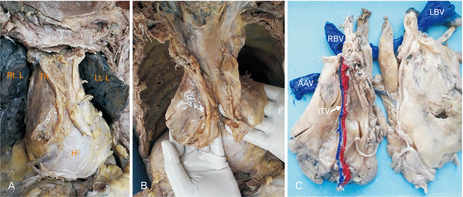
Fig. 1 (A) Presence of a large thymic mass in the anterior mediastinum lying in front of pericardium and in between right (Rt. L) and left (Lt. L) lung. (B) Right and left lobes of the thymus were separated only in its lower part. (C) Thymus removed along with its blood supply. AAV, arch of azygous vein; ITV, internal thoracic vessel; LBV, left brachiocephalic vein; RBV, right brachiocephalic vein; Th, thymus; H, heart.
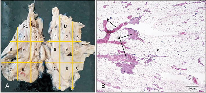
Fig. 2 (A) The right (Rt.) and left (Lt.) lobes of the large thymic mass were divided into nine quadrants. (B) Hematoxylin and eosin stained section of eighth quadrant of Rt. lobe of thymus showing persistence of thymic tissue (a), blood vessel (b), and fatty infiltration (c). Scale bar=10 µm.

Fig. 3 (A–C) Epithelial cells stained intensely with cytokeratin antibody in section obtained from the eighth quadrant of right lobe of thymus suggesting persistence of active tissue in it.
Reference
-
1. Dutta AK. Principles of general anatomy. 2nd ed. Kolkata: K P Basu Publishing Co.;2015.2. Kumar V, Abbas AK, Fausto N, Aster JC. Robbins and Cotran pathologic basis of disease. 8th ed. Gurgaon: Elsevier;2013.3. Sinnatamby CS. Last's anatomy: regional and applied. 12th ed. Edinburg: Churchill Livingstone;2011.4. Susimitha SV, Anitha V. Presence of large bilobed thymus in adult male cadaver. Int J Anat Res. 2014; 2:541–544.5. Nayak SB, Kumar N, Aithal PA, Shetty SD, Rao SS, Guru A. Possibly active persistent thymus found in a human adult cadaver: a morpho-histological study. Online J Health Allied Sci. 2015; 14:15.6. Araki T, Nishino M, Gao W, Dupuis J, Hunninghake GM, Murakami T, Washko GR, O'Connor GT, Hatabu H. Normal thymus in adults: appearance on CT and associations with age, sex, BMI and smoking. Eur Radiol. 2016; 26:15–24.
Article7. Nasseri F, Eftekhari F. Clinical and radiologic review of the normal and abnormal thymus: pearls and pitfalls. Radiographics. 2010; 30:413–428.
Article
- Full Text Links
-
- Actions
-
Cited
- CITED
-
- Close
- Share
-
- Similar articles
-
- Thymic Cysts: Two cases report
- Cervical thymic cyst in the elderly: a case report
- Thymic Carcinoma Presenting Two Independent Nodules: Case Report
- A Case of Ectopic Lateral Cervical Thymic Cyst Mimicking as a Second Branchial Cleft Cyst
- Spontaneous Thymic Cyst Hemorrhage Manifesting as a Mediastinal Mass
