Effects of different surface finishing protocols for zirconia on surface roughness and bacterial biofilm formation
- Affiliations
-
- 1Department of Prosthodontics, School of Dentistry, ITRD, Kyungpook National University, Daegu, Republic of Korea.
- 2Department of Microbiology and Immunology, School of Dentistry, Kyungpook National University, Daegu, Republic of Korea.
- 3Department of Periodontics and Endodontics, State University of New York at Buffalo, Buffalo, New York, United states of America.
- 4Department of Dentistry, Yonsei University Wonju College of Medicine, Wonju, Republic of Korea.
- 5Department of Prosthodontics, College of Dentistry, Yonsei University, Seoul, Republic of Korea. kwlee@yuhs.ac
- KMID: 2438930
- DOI: http://doi.org/10.4047/jap.2019.11.1.41
Abstract
- PURPOSE
Surface finishing of a zirconia restoration is essential after clinical adjustment. Herein, we investigated the effects of a surface finishing protocol for monolithic zirconia on final roughness and bacterial adherence.
MATERIALS AND METHODS
Forty-eight disk-shaped monolithic zirconia specimens were fabricated and divided into four groups (n = 12) based on initial surface treatment, finishing, and polishing protocols: diamond bur+polishing bur (DP group), diamond bur+stone grinding bur+polishing bur (DSP group), no diamond bur+polishing bur (NP group), and no diamond bur+stone grinding bur+polishing bur (NSP group). Initial and final surface roughness was measured with a profilometer, and shown using scanning electron microscope. Bacterial adhesion was evaluated by quantifying Streptococcus mutans in the biofilm. Kruskal-Wallis and Mann-Whitney U tests were used to compare results among groups, and two-way analysis of variance was used to evaluate the effects of grinding burs on final roughness (α=.05).
RESULTS
The DP group had the highest final Ra value, followed by the DSP, NP, and NSP groups. Use of the stone grinding bur as a coarse-finishing step significantly decreased final Ra values when a diamond bur was used (P < .001). Omission of the stone grinding bur increased biofilm formation on specimen surfaces. Combining a stone grinding bur with silicone polishing burs produced the smallest final biofilm values, regardless of the use of a diamond bur in initial surface treatment.
CONCLUSION
Coarse finishing of monolithic zirconia with a stone grinding bur significantly decreased final Ra values and bacterial biofilm formation when surfaces had been roughened by a diamond bur.
MeSH Terms
Figure
-
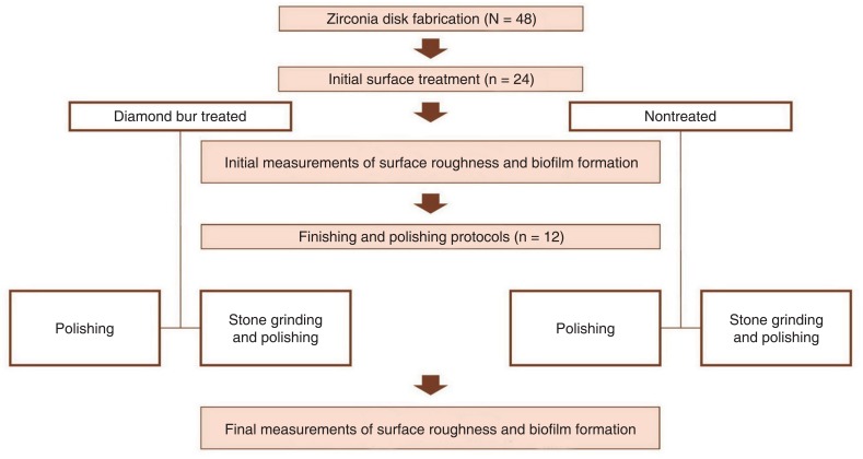
Fig. 1 Workflow of study.
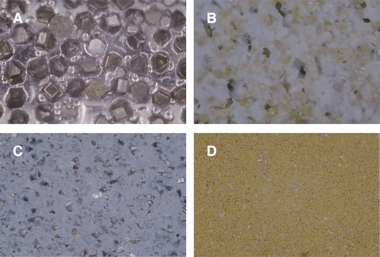
Fig. 2 Microscopic views of burs at ×300 magnification. (A) Diamond bur, (B) Stone grinding bur, (C) Silicone polishing bur, (D) Fine silicone polishing bur.
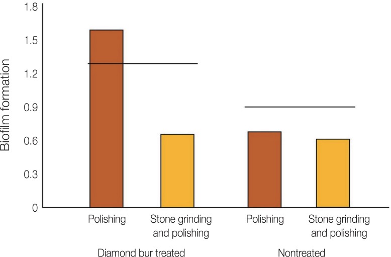
Fig. 3 Biofilm formation on zirconia specimen from each group after finishing and polishing. Black lines indicate initial biofilm formation for diamond bur-treated and nontreated specimens.
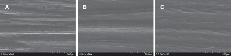
Fig. 4 Scanning electron microscope images of diamond bur-treated specimens. (A) Surface after use of diamond bur alone, (B) Surface after use of diamond and polishing burs, (C) Surface after use of diamond bur, stone grinding bur, and polishing bur.
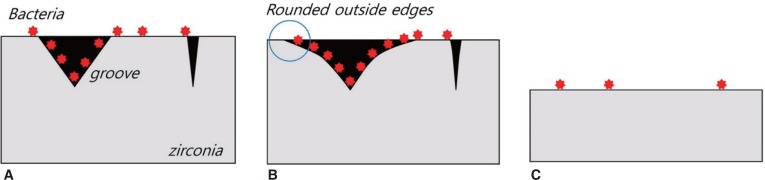
Fig. 5 Schematic image of bacterial adhesion to zirconia; heavy bacterial inhabitation is observed in the partially altered surface treated by diamond and polishing burs. (A) Surface after use of diamond bur alone, (B) Surface after use of diamond and polishing burs, (C) Surface after use of diamond bur, stone grinding bur, and polishing bur.
Reference
-
1. Caglar I, Ates SM, Yesil Duymus Z. The effect of various polishing systems on surface roughness and phase transformation of monolithic zirconia. J Adv Prosthodont. 2018; 10:132–137. PMID: 29713434.
Article2. Chavali R, Lin CP, Lawson NC. Evaluation of different polishing systems and speeds for dental zirconia. J Prosthodont. 2017; 26:410–418. PMID: 26618785.
Article3. Kim HK, Kim SH, Lee JB, Ha SR. Effects of surface treatments on the translucency, opalescence, and surface texture of dental monolithic zirconia ceramics. J Prosthet Dent. 2016; 115:773–779. PMID: 26809221.
Article4. Kim MJ, Oh SH, Kim JH, Ju SW, Seo DG, Jun SH, Ahn JS, Ryu JJ. Wear evaluation of the human enamel opposing different Y-TZP dental ceramics and other porcelains. J Dent. 2012; 40:979–988. PMID: 22892464.
Article5. Teughels W, Van Assche N, Sliepen I, Quirynen M. Effect of material characteristics and/or surface topography on biofilm development. Clin Oral Implants Res. 2006; 17(Suppl 2):68–81. PMID: 16968383.
Article6. Jones CS, Billington RW, Pearson GJ. The in vivo perception of roughness of restorations. Br Dent J. 2004; 196:42–45. PMID: 14966503.
Article7. Yuzugullu B, Celik C, Burak Ozcelik T, Erkut S, Yurdakul P, Ocal Y, Sener B. The effect of different polishing sequences on the adhesion of Streptococcus mutans to feldspathic Porcelain. J Adhes. 2016; 92:939–949.8. Bollen CM, Lambrechts P, Quirynen M. Comparison of surface roughness of oral hard materials to the threshold surface roughness for bacterial plaque retention: a review of the literature. Dent Mater. 1997; 13:258–269. PMID: 11696906.9. Lee BC, Jung GY, Kim DJ, Han JS. Initial bacterial adhesion on resin, titanium and zirconia in vitro. J Adv Prosthodont. 2011; 3:81–84. PMID: 21814616.10. Miyazaki T, Nakamura T, Matsumura H, Ban S, Kobayashi T. Current status of zirconia restoration. J Prosthodont Res. 2013; 57:236–261. PMID: 24140561.
Article11. Huh YH, Park CJ, Cho LR. Evaluation of various polishing systems and the phase transformation of monolithic zirconia. J Prosthet Dent. 2016; 116:440–449. PMID: 27061631.
Article12. Preis V, Grumser K, Schneider-Feyrer S, Behr M, Rosentritt M. The effectiveness of polishing kits: influence on surface roughness of zirconia. Int J Prosthodont. 2015; 28:149–151. PMID: 25822299.
Article13. Ho CM, Ding H, Chen X, Tsoi JK, Botelho MG. The effects of dry and wet grinding on the strength of dental zirconia. Ceram Int. 2018; 44:10451–10462.
Article14. Kosmac T, Oblak C, Jevnikar P, Funduk N, Marion L. Strength and reliability of surface treated Y-TZP dental ceramics. J Biomed Mater Res. 2000; 53:304–313. PMID: 10898871.15. Kosmac T, Oblak C, Jevnikar P, Funduk N, Marion L. The effect of surface grinding and sandblasting on flexural strength and reliability of Y-TZP zirconia ceramic. Dent Mater. 1999; 15:426–433. PMID: 10863444.16. Lee KR, Choe HC, Heo YR, Lee JJ, Son MK. Effect of different grinding burs on the physical properties of zirconia. J Adv Prosthodont. 2016; 8:137–143. PMID: 27141258.
Article17. Gilan I, Sivan A. Extracellular DNA plays an important structural role in the biofilm of the plastic degrading actinomycete Rhodo-coccus ruber. Adv Microbiol. 2013; 3:543–551.18. O'Toole GA. Microtiter dish biofilm formation assay. J Vis Exp. 2011; 47:2437.19. Han A, Tsoi JKH, Matinlinna JP, Chen Z. Influence of grit-Blasting and hydrofluoric acid etching treatment on surface characteristics and biofilm formation on zirconia. Coatings. 2017; 7:130.
Article
- Full Text Links
-
- Actions
-
Cited
- CITED
-
- Close
- Share
-
- Similar articles
-
- Effects of different finishing/polishing protocols and systems for monolithic zirconia on surface topography, phase transformation, and biofilm formation
- Surface Roughness and Microbial Adhesion After Finishing of Alkasite Restorative Material
- Comparison of biofilm on titanium and zirconia surfaces: in vivo study
- Effect of sintering programs and surface treatments on monolithic zirconia
- Investigation of effect of zirconia on osseointegration by surface treatments
