Korean J Physiol Pharmacol.
2018 Nov;22(6):721-729. 10.4196/kjpp.2018.22.6.721.
Regional difference in spontaneous firing inhibition by GABA(A) and GABA(B) receptors in nigral dopamine neurons
- Affiliations
-
- 1Department of Physiology, Sungkyunkwan University School of Medicine, Suwon 16419, Korea. mkpark@skku.edu
- 2Samsung Biomedical Research Institute, Samsung Medical Center, Seoul 06351, Korea.
- KMID: 2430095
- DOI: http://doi.org/10.4196/kjpp.2018.22.6.721
Abstract
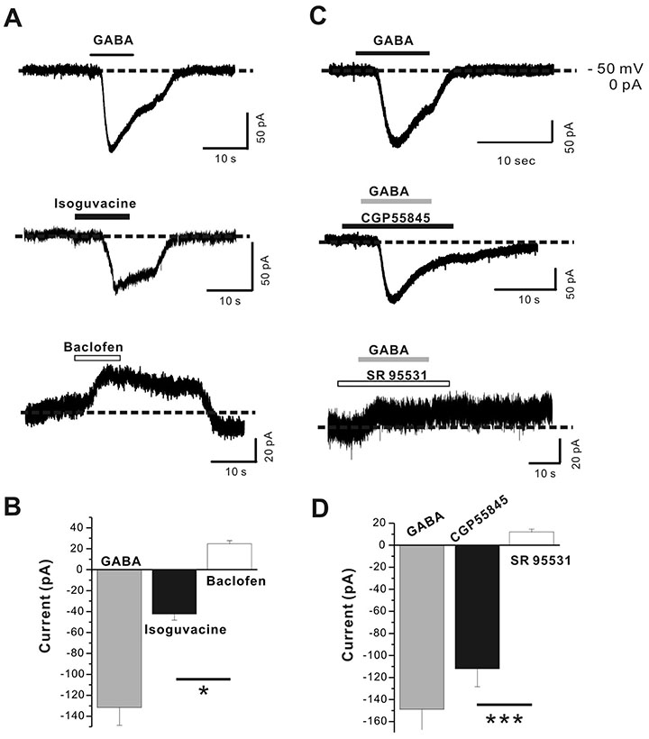
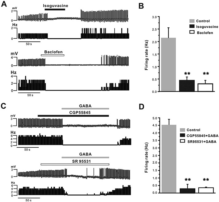
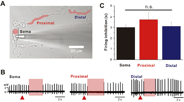
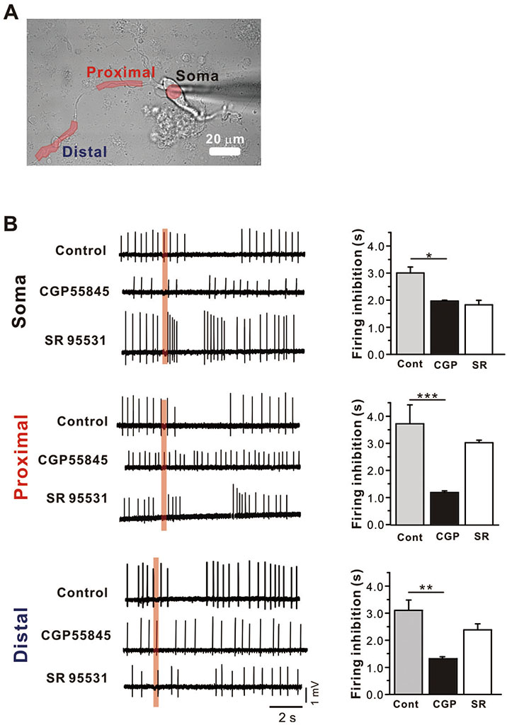
- GABAergic control over dopamine (DA) neurons in the substantia nigra is crucial for determining firing rates and patterns. Although GABA activates both GABA(A) and GABA(B) receptors distributed throughout the somatodendritic tree, it is currently unclear how regional GABA receptors in the soma and dendritic compartments regulate spontaneous firing. Therefore, the objective of this study was to determine actions of regional GABA receptors on spontaneous firing in acutely dissociated DA neurons from the rat using patch-clamp and local GABA-uncaging techniques. Agonists and antagonists experiments showed that activation of either GABA(A) receptors or GABA(B) receptors in DA neurons is enough to completely abolish spontaneous firing. Local GABA-uncaging along the somatodendritic tree revealed that activation of regional GABA receptors limited within the soma, proximal, or distal dendritic region, can completely suppress spontaneous firing. However, activation of either GABA(A) or GABA(B) receptor equally suppressed spontaneous firing in the soma, whereas GABA(B) receptor inhibited spontaneous firing more strongly than GABA(A) receptor in the proximal and distal dendrites. These regional differences of GABA signals between the soma and dendritic compartments could contribute to our understanding of many diverse and complex actions of GABA in midbrain DA neurons.
Keyword
MeSH Terms
Figure
Reference
-
1. Grace AA, Onn SP. Morphology and electrophysiological properties of immunocytochemically identified rat dopamine neurons recorded in vitro. J Neurosci. 1989; 9:3463–3481.
Article2. Jang J, Um KB, Jang M, Kim SH, Cho H, Chung S, Kim HJ, Park MK. Balance between the proximal dendritic compartment and the soma determines spontaneous firing rate in midbrain dopamine neurons. J Physiol. 2014; 592:2829–2844.
Article3. Schultz W. Behavioral dopamine signals. Trends Neurosci. 2007; 30:203–210.
Article4. Missale C, Nash SR, Robinson SW, Jaber M, Caron MG. Dopamine receptors: from structure to function. Physiol Rev. 1998; 78:189–225.
Article5. Sivilotti L, Nistri A. GABA receptor mechanisms in the central nervous system. Prog Neurobiol. 1991; 36:35–92.
Article6. Eder M, Rammes G, Zieglgänsberger W, Dodt HU. GABAA and GABAB receptors on neocortical neurons are differentially distributed. Eur J Neurosci. 2001; 13:1065–1069.7. Tepper JM, Martin LP, Anderson DR. GABAA receptor-mediated inhibition of rat substantia nigra dopaminergic neurons by pars reticulata projection neurons. J Neurosci. 1995; 15:3092–3103.8. Bolam JP, Smith Y. The GABA and substance P input to dopaminergic neurones in the substantia nigra of the rat. Brain Res. 1990; 529:57–78.
Article9. Henny P, Brown MT, Northrop A, Faunes M, Ungless MA, Magill PJ, Bolam JP. Structural correlates of heterogeneous in vivo activity of midbrain dopaminergic neurons. Nat Neurosci. 2012; 15:613–619.
Article10. Grace AA, Floresco SB, Goto Y, Lodge DJ. Regulation of firing of dopaminergic neurons and control of goal-directed behaviors. Trends Neurosci. 2007; 30:220–227.
Article11. Lobb CJ, Troyer TW, Wilson CJ, Paladini CA. Disinhibition bursting of dopaminergic neurons. Front Syst Neurosci. 2011; 5:25.
Article12. Erhardt S, Mathé JM, Chergui K, Engberg G, Svensson TH. GABAB receptor-mediated modulation of the firing pattern of ventral tegmental area dopamine neurons in vivo. Naunyn Schmiedebergs Arch Pharmacol. 2002; 365:173–180.13. McKernan RM, Whiting PJ. Which GABAA-receptor subtypes really occur in the brain? Trends Neurosci. 1996; 19:139–143.14. Sieghart W. Structure, pharmacology, and function of GABAA receptor subtypes. Adv Pharmacol. 2006; 54:231–263.15. Lüscher C, Jan LY, Stoffel M, Malenka RC, Nicoll RA. G proteincoupled inwardly rectifying K+ channels (GIRKs) mediate postsynaptic but not presynaptic transmitter actions in hippocampal neurons. Neuron. 1997; 19:687–695.16. Tamás G, Lorincz A, Simon A, Szabadics J. Identified sources and targets of slow inhibition in the neocortex. Science. 2003; 299:1902–1905.17. Curtis DR, Hösli L, Johnston GA, Johnston IH. The hyperpolarization of spinal motoneurones by glycine and related amino acids. Exp Brain Res. 1968; 5:235–258.
Article18. Pandit S, Lee GS, Park JB. Developmental changes in GABAA tonic inhibition are compromised by multiple mechanisms in preadolescent dentate gyrus granule cells. Korean J Physiol Pharmacol. 2017; 21:695–702.19. Deeb TZ, Lee HH, Walker JA, Davies PA, Moss SJ. Hyperpolarizing GABAergic transmission depends on KCC2 function and membrane potential. Channels (Austin). 2011; 5:475–481.
Article20. Misgeld U, Bijak M, Jarolimek W. A physiological role for GABAB receptors and the effects of baclofen in the mammalian central nervous system. Prog Neurobiol. 1995; 46:423–462.21. Newberry NR, Nicoll RA. Direct hyperpolarizing action of baclofen on hippocampal pyramidal cells. Nature. 1984; 308:450–452.
Article22. Otmakhova NA, Lisman JE. Contribution of Ih and GABAB to synaptically induced afterhyperpolarizations in CA1: a brake on the NMDA response. J Neurophysiol. 2004; 92:2027–2039.23. Freund TF, Buzsáki G. Interneurons of the hippocampus. Hippocampus. 1996; 6:347–470.
Article24. Tsubokawa H, Ross WN. IPSPs modulate spike backpropagation and associated [Ca2+]i changes in the dendrites of hippocampal CA1 pyramidal neurons. J Neurophysiol. 1996; 76:2896–2906.25. Leung LS, Peloquin P. GABAB receptors inhibit backpropagating dendritic spikes in hippocampal CA1 pyramidal cells in vivo. Hippocampus. 2006; 16:388–407.26. Paladini CA, Tepper JM. GABAA and GABAB antagonists differentially affect the firing pattern of substantia nigra dopaminergic neurons in vivo. Synapse. 1999; 32:165–176.27. Engberg G, Kling-Petersen T, Nissbrandt H. GABAB-receptor activation alters the firing pattern of dopamine neurons in the rat substantia nigra. Synapse. 1993; 15:229–238.28. Okada H, Matsushita N, Kobayashi K, Kobayashi K. Identification of GABAA receptor subunit variants in midbrain dopaminergic neurons. J Neurochem. 2004; 89:7–14.29. Parker JG, Wanat MJ, Soden ME, Ahmad K, Zweifel LS, Bamford NS, Palmiter RD. Attenuating GABAA receptor signaling in dopamine neurons selectively enhances reward learning and alters risk preference in mice. J Neurosci. 2011; 31:17103–17112.30. Bettler B, Kaupmann K, Mosbacher J, Gassmann M. Molecular structure and physiological functions of GABAB receptors. Physiol Rev. 2004; 84:835–867.31. Mott DD, Lewis DV. The pharmacology and function of central GABAB receptors. Int Rev Neurobiol. 1994; 36:97–223.32. Brazhnik E, Shah F, Tepper JM. GABAergic afferents activate both GABAA and GABAB receptors in mouse substantia nigra dopaminergic neurons in vivo. J Neurosci. 2008; 28:10386–10398.33. Choi YM, Kim SH, Uhm DY, Park MK. Glutamate-mediated [Ca2+]c dynamics in spontaneously firing dopamine neurons of the rat substantia nigra pars compacta. J Cell Sci. 2003; 116:2665–2675.34. Mercuri NB, Bonci A, Calabresi P, Stefani A, Bernardi G. Properties of the hyperpolarization-activated cation current Ih in rat midbrain dopaminergic neurons. Eur J Neurosci. 1995; 7:462–469.35. Grace AA, Bunney BS. Intracellular and extracellular electrophysiology of nigral dopaminergic neurons--1. Identification and characterization. Neuroscience. 1983; 10:301–315.
Article36. Grace AA, Bunney BS. The control of firing pattern in nigral dopamine neurons: burst firing. J Neurosci. 1984; 4:2877–2890.
Article37. Cardozo DL. Midbrain dopaminergic neurons from postnatal rat in long-term primary culture. Neuroscience. 1993; 56:409–421.
Article38. Uchida S, Akaike N, Nabekura J. Dopamine activates inward rectifier K+ channel in acutely dissociated rat substantia nigra neurones. Neuropharmacology. 2000; 39:191–201.39. Tepper JM, Lee CR. GABAergic control of substantia nigra dopaminergic neurons. Prog Brain Res. 2007; 160:189–208.
Article40. Richards CD, Shiroyama T, Kitai ST. Electrophysiological and immunocytochemical characterization of GABA and dopamine neurons in the substantia nigra of the rat. Neuroscience. 1997; 80:545–557.
Article41. Johnson SW, North RA. Two types of neurone in the rat ventral tegmental area and their synaptic inputs. J Physiol. 1992; 450:455–468.
Article42. Beaulieu C, Somogyi P. Targets and quantitative distribution of GABAergic synapses in the visual cortex of the cat. Eur J Neurosci. 1990; 2:296–303.
Article43. Kulik A, Vida I, Luján R, Haas CA, López-Bendito G, Shigemoto R, Frotscher M. Subcellular localization of metabotropic GABAB receptor subunits GABAB1a/b and GABAB2 in the rat hippocampus. J Neurosci. 2003; 23:11026–11035.
- Full Text Links
- Actions
-
Cited
- CITED
-
- Close
- Share
- Similar articles
-
- A Role of Glutamate and GABA Receptors in the Generation of Formalin-induced Impulse Firing of Spinal Dorsal Horn Neurons in the Rat
- Effect of GABA on the contratility of small intestine isolated from rat
- Effects of taurine on generalized seizure activity in the rat thalamocortical slice
- Effects of amino acids and some drugs related to gaba receptors on the neuronal activity in isolated medullary dorsal horn of rat
- GABA, benzodiazepine receptors and their functions


