Quantitative Measurement of Hepatic Fibrosis with Gadoxetic Acid-Enhanced Magnetic Resonance Imaging in Patients with Chronic Hepatitis B Infection: A Comparative Study on Aspartate Aminotransferase to Platelet Ratio Index and Fibrosis-4 Index
- Affiliations
-
- 1Department of Radiology, Wonkwang University School of Medicine, Iksan 54538, Korea. khy1646@wku.ac.kr
- 2Imaging Science Research Center, Wonkwang University, Iksan 54538, Korea.
- 3Department of Internal Medicine, Wonkwang University School of Medicine, Iksan 54538, Korea.
- KMID: 2427299
- DOI: http://doi.org/10.3348/kjr.2017.18.3.444
Abstract
OBJECTIVE
To quantitatively measure hepatic fibrosis on gadoxetic acid-enhanced magnetic resonance (MR) in chronic hepatitis B (CHB) patients and identify the correlations with aspartate aminotransferase-to-platelet ratio index (APRI) and fibrosis-4 index (FIB-4) values.
MATERIALS AND METHODS
This study on gadoxetic acid-enhanced 3T MR imaging included 81 patients with CHB infection. To quantitatively measure hepatic fibrosis, MR images were analyzed with an aim to identify inhomogeneous signal intensities calculated from a coefficient of variation (CV) map in the liver parenchyma. We also carried out a comparative analysis between APRI and FIB-4 based on metaregression results. The diagnostic performance of the CV map was evaluated using a receiver-operating characteristic (ROC) curve.
RESULTS
In the MR images, the mean CV values in control, groups I, II, and III based on APRI were 4.08 ± 0.92, 4.24 ± 0.80, 5.64 ± 1.11, and 5.73 ± 1.28, respectively (p < 0.001). In CHB patients grouped by FIB-4, the mean CV values of groups A, B, and C were 4.22 ± 0.95, 5.40 ± 1.19, and 5.71 ± 1.17, respectively (p < 0.001). The mean CV values correlated well with APRI (r = 0.392, p < 0.001) and FIB-4 (r = 0.294, p < 0.001). In significant fibrosis group, ROC curve analysis yielded an area under the curve of 0.875 using APRI and 0.831 using FIB-4 in HB, respectively.
CONCLUSION
Gadoxetic acid-enhanced MR imaging for calculating a CV map showed moderate correlation with APRI and FIB-4 values and could be employed to quantitatively measure hepatic fibrosis in patients with CHB.
Keyword
MeSH Terms
-
Adolescent
Adult
Aged
Aged, 80 and over
Area Under Curve
Aspartate Aminotransferases/*analysis
Blood Platelets/*cytology
Case-Control Studies
Female
Gadolinium DTPA/*chemistry
Hepatitis B, Chronic/complications/*diagnosis
Humans
Liver Cirrhosis/complications/*diagnostic imaging/pathology
*Magnetic Resonance Imaging
Male
Middle Aged
Platelet Count
ROC Curve
Severity of Illness Index
Young Adult
Aspartate Aminotransferases
Gadolinium DTPA
Figure
-
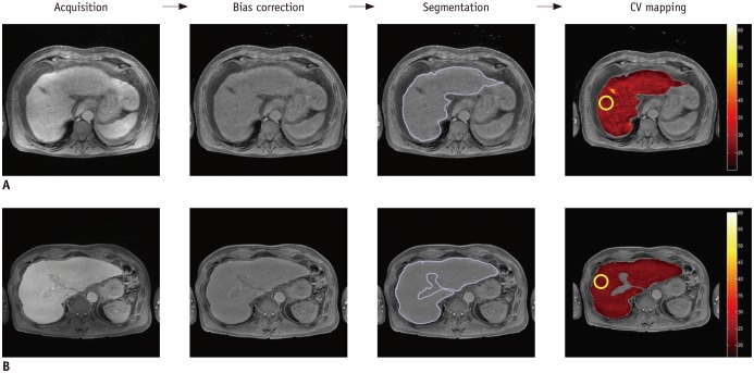
Fig. 1 Workflow for calculation of coefficients of variation (CV) in control subject (A) and in patient with chronic hepatitis B (B) in hepatobiliary phase of Gd-EOB-DTPA-enhanced MRI.To measure hepatic fibrosis quantitatively, MATLAB-based software was used to calculate CV map. After acquisition of MR images, bias correction and segmentation of liver parenchyma were performed, and then mean value of CV map on region of interest at anterior segment of right lobe of liver was calculated. Gd-EOB-DTPA = gadolinium ethoxybenzyl diethylenetriaminepentaacetic acid or gadoxetic acid, MRI = magnetic resonance imaging
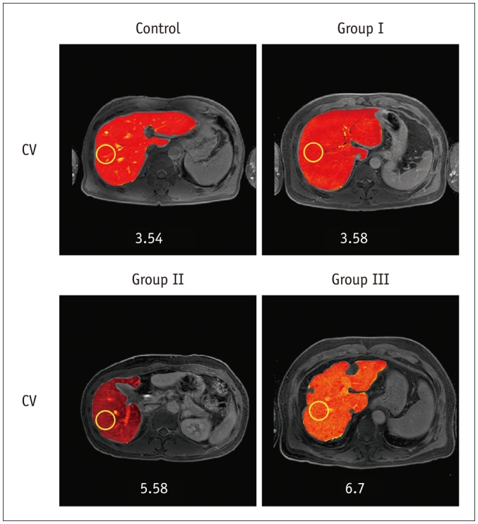
Fig. 2 Images show representative CV maps on control and patients with chronic hepatitis B of group I, II, and III on HB phases of Gd-EOB-DTPA-enhanced MRI.CV values are 3.54, 3.58, 5.58, and 6.7 in HB phase, respectively. CV = coefficients of variation, Gd-EOB-DTPA = gadolinium ethoxybenzyl diethylenetriaminepentaacetic acid or gadoxetic acid, HB = hepatobiliary, MRI = magnetic resonance imaging
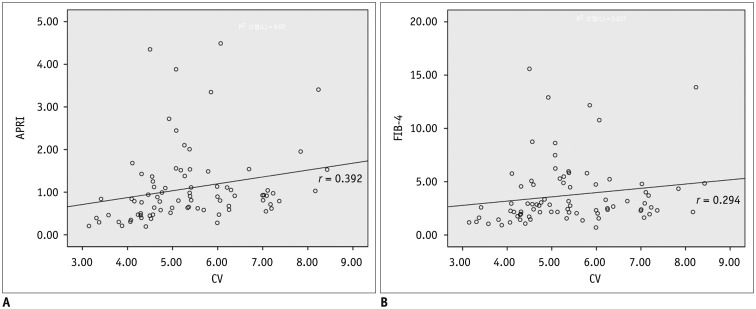
Fig. 3 Correlation of CV with APRI and FIB-4.A. Shows correlation between aspartate aminotransferase-to-platelet ratio and coefficient of variation (CV) (r = 0.392, p < 0.001). B. Shows correlation between FIB-4 and CV values (r = 0.294, p < 0.001) in HB phases of Gd-EOB-DTPA-enhanced MRI. FIB-4 = fibrosis-4, Gd-EOB-DTPA = gadolinium ethoxybenzyl diethylenetriaminepentaacetic acid or gadoxetic acid, HB = hepatobiliary, MRI = magnetic resonance imaging
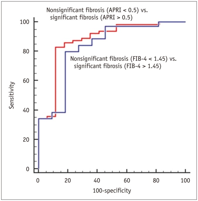
Fig. 4 Receiver operating characteristic curves of coefficient of variation used for diagnosis in patient with significant fibrosis (aspartate aminotransferase-to-platelet ratio > 0.5 and FIB-4 score > 1.45).AUC was 0.875 in APRI version with optimal cut-off values of 4.1866 (red line). AUC was 0.831 in FIB-4 version with optimal cut-off values of 4.5642 (blue line). APRI = aspartate aminotransferase to platelet ratio index, AUC = area under the curve, FIB-4 = fibrosis-4
Cited by 3 articles
-
Age of Data in Contemporary Research Articles Published in Representative General Radiology Journals
Ji Hun Kang, Dong Hwan Kim, Seong Ho Park, Jung Hwan Baek
Korean J Radiol. 2018;19(6):1172-1178. doi: 10.3348/kjr.2018.19.6.1172.The Diagnostic Performance of Liver MRI without Intravenous Contrast for Detecting Hepatocellular Carcinoma: A Case-Controlled Feasibility Study
Seunghee Han, Joon-Il Choi, Michael Yong Park, Moon Hyung Choi, Sung Eun Rha, Young Joon Lee
Korean J Radiol. 2018;19(4):568-577. doi: 10.3348/kjr.2018.19.4.568.Histogram Analysis of Diffusion Kurtosis Magnetic Resonance Imaging for Diagnosis of Hepatic Fibrosis
Ruo-Fan Sheng, Kai-Pu Jin, Li Yang, He-Qing Wang, Hao Liu, Yuan Ji, Cai-Xia Fu, Meng-Su Zeng
Korean J Radiol. 2018;19(5):916-922. doi: 10.3348/kjr.2018.19.5.916.
Reference
-
1. Korean Liver Cancer Study Group (KLCSG); National Cancer Center, Korea (NCC). 2014 Korean Liver Cancer Study Group-National Cancer Center Korea practice guideline for the management of hepatocellular carcinoma. Korean J Radiol. 2015; 16:465–522. PMID: 25995680.2. Ganem D, Prince AM. Hepatitis B virus infection--natural history and clinical consequences. N Engl J Med. 2004; 350:1118–1129. PMID: 15014185.3. Afdhal NH, Nunes D. Evaluation of liver fibrosis: a concise review. Am J Gastroenterol. 2004; 99:1160–1174. PMID: 15180741.
Article4. Yoon JH, Park JW, Lee JM. Noninvasive diagnosis of hepatocellular carcinoma: elaboration on Korean Liver Cancer Study Group-National Cancer Center Korea Practice Guidelines compared with other guidelines and remaining issues. Korean J Radiol. 2016; 17:7–24. PMID: 26798212.
Article5. Bedossa P, Dargère D, Paradis V. Sampling variability of liver fibrosis in chronic hepatitis C. Hepatology. 2003; 38:1449–1457. PMID: 14647056.
Article6. Cadranel JF, Rufat P, Degos F. Practices of liver biopsy in France: results of a prospective nationwide survey. For the Group of Epidemiology of the French Association for the Study of the Liver (AFEF). Hepatology. 2000; 32:477–481. PMID: 10960438.7. Kim BK, Kim SA, Park YN, Cheong JY, Kim HS, Park JY, et al. Noninvasive models to predict liver cirrhosis in patients with chronic hepatitis B. Liver Int. 2007; 27:969–976. PMID: 17696936.
Article8. Motosugi U, Ichikawa T, Sou H, Sano K, Tominaga L, Kitamura T, et al. Liver parenchymal enhancement of hepatocyte-phase images in Gd-EOB-DTPA-enhanced MR imaging: which biological markers of the liver function affect the enhancement? J Magn Reson Imaging. 2009; 30:1042–1046. PMID: 19856436.
Article9. Wai CT, Greenson JK, Fontana RJ, Kalbfleisch JD, Marrero JA, Conjeevaram HS, et al. A simple noninvasive index can predict both significant fibrosis and cirrhosis in patients with chronic hepatitis C. Hepatology. 2003; 38:518–526. PMID: 12883497.
Article10. Shin WG, Park SH, Jang MK, Hahn TH, Kim JB, Lee MS, et al. Aspartate aminotransferase to platelet ratio index (APRI) can predict liver fibrosis in chronic hepatitis B. Dig Liver Dis. 2008; 40:267–274. PMID: 18055281.
Article11. Xiao G, Yang J, Yan L. Comparison of diagnostic accuracy of aspartate aminotransferase to platelet ratio index and fibrosis-4 index for detecting liver fibrosis in adult patients with chronic hepatitis B virus infection: a systemic review and meta-analysis. Hepatology. 2015; 61:292–302. PMID: 25132233.
Article12. Geier A, Dietrich CG, Voigt S, Kim SK, Gerloff T, Kullak-Ublick GA, et al. Effects of proinflammatory cytokines on rat organic anion transporters during toxic liver injury and cholestasis. Hepatology. 2003; 38:345–354. PMID: 12883478.
Article13. Kim SM, Heo SH, Kim JW, Lim HS, Shin SS, Jeong YY, et al. Hepatic arterial phase on gadoxetic acid-enhanced liver MR imaging: a randomized comparison of 0.5 mL/s and 1 mL/s injection rates. Korean J Radiol. 2014; 15:605–612. PMID: 25246821.
Article14. Tsuboyama T, Onishi H, Kim T, Akita H, Hori M, Tatsumi M, et al. Hepatocellular carcinoma: hepatocyte-selective enhancement at gadoxetic acid-enhanced MR imaging--correlation with expression of sinusoidal and canalicular transporters and bile accumulation. Radiology. 2010; 255:824–833. PMID: 20501720.
Article15. Van Beers BE, Pastor CM, Hussain HK. Primovist, Eovist: what to expect? J Hepatol. 2012; 57:421–429. PMID: 22504332.
Article16. Kim H, Park SH, Kim EK, Kim MJ, Park YN, Park HJ, et al. Histogram analysis of gadoxetic acid-enhanced MRI for quantitative hepatic fibrosis measurement. PLoS One. 2014; 9:e114224. PMID: 25460180.
Article17. Kanki A, Tamada T, Higaki A, Noda Y, Tanimoto D, Sato T, et al. Hepatic parenchymal enhancement at Gd-EOB-DTPA-enhanced MR imaging: correlation with morphological grading of severity in cirrhosis and chronic hepatitis. Magn Reson Imaging. 2012; 30:356–360. PMID: 22227353.
Article18. Tamada T, Ito K, Higaki A, Yoshida K, Kanki A, Sato T, et al. Gd-EOB-DTPA-enhanced MR imaging: evaluation of hepatic enhancement effects in normal and cirrhotic livers. Eur J Radiol. 2011; 80:e311–e316. PMID: 21315529.
Article19. Sato Y, Matsushima S, Inaba Y, Sano T, Yamaura H, Kato M, et al. Preoperative estimation of future remnant liver function following portal vein embolization using relative enhancement on gadoxetic acid disodium-enhanced magnetic resonance imaging. Korean J Radiol. 2015; 16:523–530. PMID: 25995681.
Article20. Huh J, Kim SY, Yeh BM, Lee SS, Kim KW, Wu EH, et al. Troubleshooting arterial-phase MR images of gadoxetate disodium-enhanced liver. Korean J Radiol. 2015; 16:1207–1215. PMID: 26576109.
Article21. Li C, Huang R, Ding Z, Gatenby JC, Metaxas DN, Gore JC. A level set method for image segmentation in the presence of intensity inhomogeneities with application to MRI. IEEE Trans Image Process. 2011; 20:2007–2016. PMID: 21518662.22. Roth M, Obaidat A, Hagenbuch B. OATPs, OATs and OCTs: the organic anion and cation transporters of the SLCO and SLC22A gene superfamilies. Br J Pharmacol. 2012; 165:1260–1287. PMID: 22013971.
Article23. Chen ZS, Tiwari AK. Multidrug resistance proteins (MRPs/ABCCs) in cancer chemotherapy and genetic diseases. FEBS J. 2011; 278:3226–3245. PMID: 21740521.
Article24. Leonhardt M, Keiser M, Oswald S, Kühn J, Jia J, Grube M, et al. Hepatic uptake of the magnetic resonance imaging contrast agent Gd-EOB-DTPA: role of human organic anion transporters. Drug Metab Dispos. 2010; 38:1024–1028. PMID: 20406852.
Article25. Schmitt M, Kubitz R, Wettstein M, vom Dahl S, Häussinger D. Retrieval of the MRP2 gene encoded conjugate export pump from the canalicular membrane contributes to cholestasis induced by tert-butyl hydroperoxide and chloro-dinitrobenzene. Biol Chem. 2000; 381:487–495. PMID: 10937881.
Article26. Beuers U, Bilzer M, Chittattu A, Kullak-Ublick GA, Keppler D, Paumgartner G, et al. Tauroursodeoxycholic acid inserts the apical conjugate export pump, MRP2, into canalicular membranes and stimulates organic anion secretion by protein kinase C-dependent mechanisms in cholestatic rat liver. Hepatology. 2001; 33:1206–1216. PMID: 11343250.
Article27. Atzori L, Poli G, Perra A. Hepatic stellate cell: a star cell in the liver. Int J Biochem Cell Biol. 2009; 41:1639–1642. PMID: 19433304.
Article28. Chen BB, Hsu CY, Yu CW, Wei SY, Kao JH, Lee HS, et al. Dynamic contrast-enhanced magnetic resonance imaging with Gd-EOB-DTPA for the evaluation of liver fibrosis in chronic hepatitis patients. Eur Radiol. 2012; 22:171–180. PMID: 21879400.
Article29. Tsuda N, Matsui O. Cirrhotic rat liver: reference to transporter activity and morphologic changes in bile canaliculi--gadoxetic acid-enhanced MR imaging. Radiology. 2010; 256:767–773. PMID: 20663976.
Article30. Chon YE, Choi EH, Song KJ, Park JY, Kim DY, Han KH, et al. Performance of transient elastography for the staging of liver fibrosis in patients with chronic hepatitis B: a meta-analysis. PLoS One. 2012; 7:e44930. PMID: 23049764.
Article31. Shi Y, Guo Q, Xia F, Dzyubak B, Glaser KJ, Li Q, et al. MR elastography for the assessment of hepatic fibrosis in patients with chronic hepatitis B infection: does histologic necroinflammation influence the measurement of hepatic stiffness? Radiology. 2014; 273:88–98. PMID: 24893048.
Article
- Full Text Links
-
- Actions
-
Cited
- CITED
-
- Close
- Share
-
- Similar articles
-
- The Clinical Impact of AST-Platelet Ratio Index for Non-invasive Hepatic Fibrosis in the Patients with Chronic Hepatitis B
- Efficacy of AST to Platelet Ratio Index in Predicting Severe Hepatic Fibrosis and Cirrhosis in Chronic Hepatitis B Virus Infection
- Simple Tests to Predict Hepatic Fibrosis in Nonalcoholic Chronic Liver Diseases
- The Performance of Serum Biomarkers for Predicting Fibrosis in Patients with Chronic Viral Hepatitis
- Evaluation of Non-invasive Fibrosis Markers in Predicting Esophageal Variceal Bleeding
