Korean J Neurotrauma.
2018 Oct;14(2):129-133. 10.13004/kjnt.2018.14.2.129.
Delayed Trochlear Nerve Palsy Following Traumatic Subarachnoid Hemorrhage: Usefulness of High-Resolution Three Dimensional Magnetic Resonance Imaging and Unusual Course of the Nerve
- Affiliations
-
- 1Department of Neurosurgery, Seoul National University College of Medicine, Seoul, Korea.
- 2Department of Neurosurgery, Seoul National University Boramae Medical Center, Seoul, Korea. nsyangdr@gmail.com
- KMID: 2424325
- DOI: http://doi.org/10.13004/kjnt.2018.14.2.129
Abstract
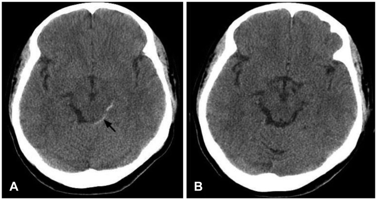
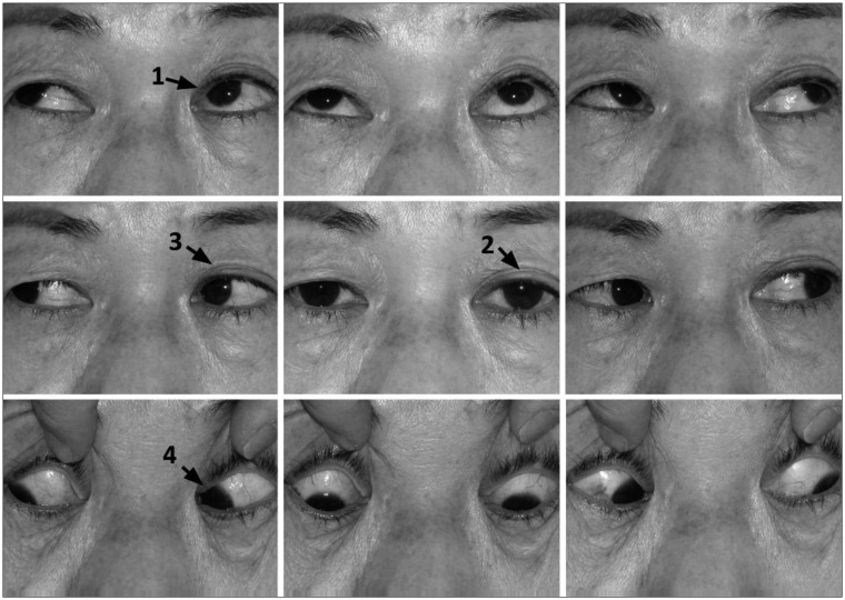
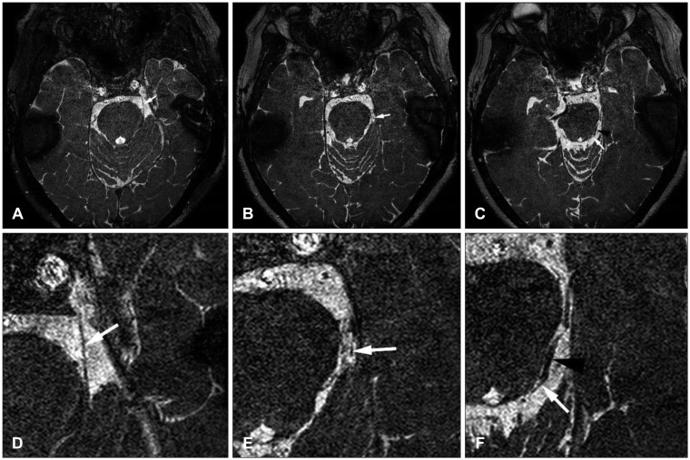
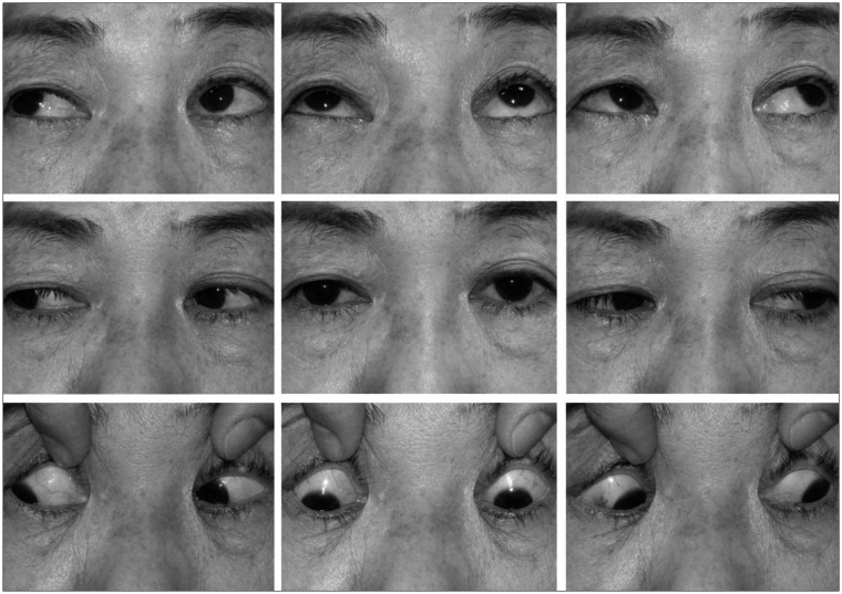
- Cranial nerve palsies are relatively common after trauma, but trochlear nerve palsy is relatively uncommon. Although traumatic trochlear nerve palsy is easy to diagnose clinically because of extraocular movement disturbances, radiologic evaluations of this condition are difficult to perform because of the nerve's small size. Here, we report the case of a patient with delayed traumatic trochlear nerve palsy associated with a traumatic subarachnoid hemorrhage (SAH) and the related radiological findings, as obtained with high-resolution three-dimensional (3D) magnetic resonance imaging (MRI). A 63-year-old woman was brought to the emergency room after a minor head trauma. Neurologic examinations did not reveal any focal neurologic deficits. Brain computed tomography showed a traumatic SAH at the left ambient cistern. The patient complained of vertical diplopia at 3 days post-trauma. Ophthalmologic evaluations revealed trochlear nerve palsy on the left side. High-resolution 3D MRI, performed 20 days post-trauma, revealed continuity of the trochlear nerve and its abutted course by the posterior cerebral artery branch at the brain stem. Chemical irritation due to the SAH and the abutting nerve course were considered causative factors. The trochlear nerve palsy completely resolved during follow-up. This case shows the usefulness of high-resolution 3D MRI for evaluating trochlear nerve palsy.
Keyword
MeSH Terms
-
Brain
Brain Stem
Cranial Nerve Diseases
Craniocerebral Trauma
Diplopia
Emergency Service, Hospital
Female
Follow-Up Studies
Humans
Imaging, Three-Dimensional
Magnetic Resonance Imaging*
Middle Aged
Neurologic Examination
Neurologic Manifestations
Posterior Cerebral Artery
Subarachnoid Hemorrhage, Traumatic*
Trochlear Nerve Diseases*
Trochlear Nerve*
Figure
Reference
-
1. Ammirati M, Musumeci A, Bernardo A, Bricolo A. The microsurgical anatomy of the cisternal segment of the trochlear nerve, as seen through different neurosurgical operative windows. Acta Neurochir (Wien). 2002; 144:1323–1327. PMID: 12478346.2. Borges A, Casselman J. Imaging the cranial nerves: Part I: methodology, infectious and inflammatory, traumatic and congenital lesions. Eur Radiol. 2007; 17:2112–2125. PMID: 17323090.
Article3. Burger LJ, Kalvin NH, Smith JL. Acquired lesions of the fourth cranial nerve. Brain. 1970; 93:567–574. PMID: 4319184.
Article4. Choi BS, Kim JH, Jung C, Hwang JM. High-resolution 3D MR imaging of the trochlear nerve. AJNR Am J Neuroradiol. 2010; 31:1076–1079. PMID: 20093316.
Article5. Coello AF, Canals AG, Gonzalez JM, Martín JJ. Cranial nerve injury after minor head trauma. J Neurosurg. 2010; 113:547–555. PMID: 20635856.
Article6. Eisenkraft B, Ortiz AO. Imaging evaluation of cranial nerves 3, 4, and 6. Semin Ultrasound CT MR. 2001; 22:488–501. PMID: 11770928.
Article7. Hara N, Kan S, Simizu K. Localization of post-traumatic trochlear nerve palsy associated with hemorrhage at the subarachnoid space by magnetic resonance imaging. Am J Ophthalmol. 2001; 132:443–445. PMID: 11530077.
Article8. Hoya K, Kirino T. Traumatic trochlear nerve palsy following minor occipital impact-four case reports. Neurol Med Chir (Tokyo). 2000; 40:358–360. PMID: 10927902.9. Kim JH, Hwang JM. Hypoplastic oculomotor nerve and absent abducens nerve in congenital fibrosis syndrome and synergistic divergence with magnetic resonance imaging. Ophthalmology. 2005; 112:728–732. PMID: 15808269.
Article10. Laine FJ. Cranial nerves III, IV, and VI. Top Magn Reson Imaging. 1996; 8:111–130. PMID: 8784967.
Article11. Li G, Zhu X, Gu X, Sun Y, Gao X, Zhang Y, et al. Ocular movement nerve palsy after mild head trauma. World Neurosurg. 2016; 94:296–302. PMID: 27422684.
Article12. Macdonald RL, Pluta RM, Zhang JH. Cerebral vasospasm after subarachnoid hemorrhage: the emerging revolution. Nat Clin Pract Neurol. 2007; 3:256–263. PMID: 17479073.
Article13. Macdonald RL, Weir BK. A review of hemoglobin and the pathogenesis of cerebral vasospasm. Stroke. 1991; 22:971–982. PMID: 1866764.
Article14. Mansour AM, Reinecke RD. Central trochlear palsy. Surv Ophthalmol. 1986; 30:279–297. PMID: 3520909.
Article15. Marinković S, Gibo H, Zelić O, Nikodijević I. The neurovascular relationships and the blood supply of the trochlear nerve: surgical anatomy of its cisternal segment. Neurosurgery. 1996; 38:161–169. PMID: 8747965.
Article16. Richards BW, Jones FR Jr, Younge BR. Causes and prognosis in 4,278 cases of paralysis of the oculomotor, trochlear, and abducens cranial nerves. Am J Ophthalmol. 1992; 113:489–496. PMID: 1575221.
Article17. Villain M, Segnarbieux F, Bonnel F, Aubry I, Arnaud B. The trochlear nerve: anatomy by microdissection. Surg Radiol Anat. 1993; 15:169–173. PMID: 8235957.
Article
- Full Text Links
- Actions
-
Cited
- CITED
-
- Close
- Share
- Similar articles
-
- Direct Oculomotor Nerve Palsy in Association with A Mild Head Injury: A Case Report
- Combined Facial and Contralateral Trochlear Nerve Palsy in a Patient with Diabetes Mellitus
- Isolated, Contralateral Trochlear Nerve Palsy Associated with a Ruptured Right Posterior Communicating Artery Aneurysm
- A Case of Trochlear Nerve Schwannoma Presenting with Binocular Diplopia
- Imaging of Cranial Nerves III, IV, VI in Congenital Cranial Dysinnervation Disorders


