Imaging of Cranial Nerves III, IV, VI in Congenital Cranial Dysinnervation Disorders
- Affiliations
-
- 1Department of Radiology, Seoul National University Bundang Hospital, Seoul National University College of Medicine, Seongnam, Korea.
- 2Department of Ophthalmology, Seoul National University Bundang Hospital, Seoul National University College of Medicine, Seongnam, Korea. hjm@snu.ac.kr
- KMID: 2379874
- DOI: http://doi.org/10.3341/kjo.2017.0024
Abstract
- Congenital cranial dysinnervation disorders are a group of diseases caused by abnormal development of cranial nerve nuclei or their axonal connections, resulting in aberrant innervation of the ocular and facial musculature. Its diagnosis could be facilitated by the development of high resolution thin-section magnetic resonance imaging. The purpose of this review is to describe the method to visualize cranial nerves III, IV, and VI and to present the imaging findings of congenital cranial dysinnervation disorders including congenital oculomotor nerve palsy, congenital trochlear nerve palsy, Duane retraction syndrome, Möbius syndrome, congenital fibrosis of the extraocular muscles, synergistic divergence, and synergistic convergence.
Keyword
MeSH Terms
Figure
-
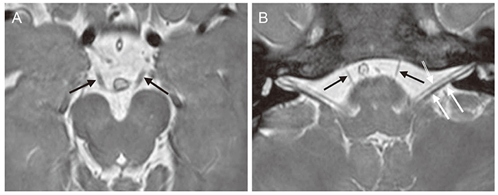
Fig. 1 Normal cranial nerve (CN) III and VI. (A) Axial magnetic resonance image at the level of the lower midbrain shows the cisternal segments of CN III (arrows) as linear dark structures coursing in the anterolateral direction toward the cavernous sinus. (B) Axial magnetic resonance image at the pontomedullary junction shows the cisternal segments of CN VI (black arrows) coursing in the anterosuperior direction toward Dorello's canal. CN VII (double arrow) and CN VIII (white arrows) are also well identified.

Fig. 2 Normal cranial nerve IV. Axial magnetic resonance image at the level of the inferior colliculus shows the cisternal segments of cranial nerve IV (thin arrows) exiting from the posterior aspect of the brainstem and coursing in the anterolateral direction toward the tentorium (thick arrows).
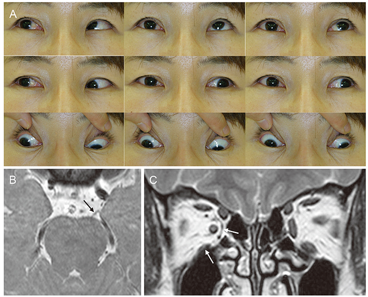
Fig. 3 Congenital cranial nerve (CN) III agenesis. (A) Ocular versions show limited elevation, adduction, and depression of the right eye. (B) Axial magnetic resonance image shows normal left CN III (arrow). Right CN III is not observed. (C) Orbital coronal magnetic resonance image shows atrophy of the right medial rectus and right inferior rectus (arrows).

Fig. 4 Congenital cranial nerve (CN) IV agenesis. (A) Ocular versions show right hypertropia and limited depression both in downgaze and adduction in the right eye. (B) Axial magnetic resonance image shows normal left CN IV (arrows). Right CN IV is not identified. (C) Orbital coronal magnetic resonance image shows atrophy of the right superior oblique muscle (arrow). Compare with the left superior oblique (double arrow).

Fig. 5 Duane's retraction syndrome type 1. (A) Ocular versions show limitations of adduction and abduction and fissure narrowing on adduction in the left eye. (B) Axial magnetic resonance image at the pontomedullary junction shows normal right cranial nerve VI (arrow). Left cranial nerve VI is not identified.

Fig. 6 Bilateral Möbius syndrome. (A) Ocular versions show limitation on abduction in both eyes. (B) Axial magnetic resonance image at the lower pons shows normal cranial nerve VIII bilaterally (white and black arrows). Cranial nerve VI and VII are not observed bilaterally. Refer to normal VI and VII in Fig. 1B.

Fig. 7 Congenital fibrosis of the extraocular muscles in both eyes. (A) Ocular versions show limited ductions of both eyes. (B) Axial magnetic resonance image shows bilateral small-sized cranial nerve III (arrows), suggesting hypoplasia. (C) Orbital coronal magnetic resonance image shows mild atrophy of the right superior rectus and right medial rectus and severe atrophy of the left superior rectus and left medial rectus (arrows).
Reference
-
1. Gutowski NJ, Bosley TM, Engle EC. 110th ENMC International Workshop: the congenital cranial dysinnervation disorders (CCDDs). Naarden, The Netherlands, 25–27 October, 2002. Neuromuscul Disord. 2003; 13:573–578.2. Kim JH, Hwang JM. Presence of the abducens nerve according to the type of Duane's retraction syndrome. Ophthalmology. 2005; 112:109–113.3. Ciftci E, Anik Y, Arslan A, et al. Driven equilibrium (drive) MR imaging of the cranial nerves V-VIII: comparison with the T2-weighted 3D TSE sequence. Eur J Radiol. 2004; 51:234–240.4. Kim JH, Hwang JM. Hypoplastic oculomotor nerve and absent abducens nerve in congenital fibrosis syndrome and synergistic divergence with magnetic resonance imaging. Ophthalmology. 2005; 112:728–732.5. Choi BS, Kim JH, Jung C, Hwang JM. High-resolution 3D MR imaging of the trochlear nerve. AJNR Am J Neuroradiol. 2010; 31:1076–1079.6. Yang HK, Kim JH, Hwang JM. Congenital superior oblique palsy and trochlear nerve absence: a clinical and radiological study. Ophthalmology. 2012; 119:170–177.7. Demer JL, Ortube MC, Engle EC, Thacker N. High-resolution magnetic resonance imaging demonstrates abnormalities of motor nerves and extraocular muscles in patients with neuropathic strabismus. J AAPOS. 2006; 10:135–142.8. Lim KH, Engle EC, Demer JL. Abnormalities of the oculomotor nerve in congenital fibrosis of the extraocular muscles and congenital oculomotor palsy. Invest Ophthalmol Vis Sci. 2007; 48:1601–1606.9. Kim JH, Hwang JM. Magnetic resonance imaging in three patients with congenital oculomotor nerve palsy. Br J Ophthalmol. 2009; 93:1266–1267.10. Hamed LM. Associated neurologic and ophthalmologic findings in congenital oculomotor nerve palsy. Ophthalmology. 1991; 98:708–714.11. Good WV, Barkovich AJ, Nickel BL, Hoyt CS. Bilateral congenital oculomotor nerve palsy in a child with brain anomalies. Am J Ophthalmol. 1991; 111:555–558.12. Sun CC, Kao LY. Unilateral congenital third cranial nerve palsy with central nervous system anomalies: report of two cases. Chang Gung Med J. 2000; 23:776–781.13. Schulz E, Jung H. Unilateral congenital oculomotor nerve palsy, optic nerve hypoplasia and pituitary malformation: a preliminary report. Strabismus. 2001; 9:33–35.14. Kau HC, Tsai CC, Ortube MC, Demer JL. High-resolution magnetic resonance imaging of the extraocular muscles and nerves demonstrates various etiologies of third nerve palsy. Am J Ophthalmol. 2007; 143:280–287.15. Sato M. Magnetic resonance imaging and tendon anomaly associated with congenital superior oblique palsy. Am J Ophthalmol. 1999; 127:379–387.16. Demer JL, Clark RA, Kono R, et al. A 12-year, prospective study of extraocular muscle imaging in complex strabismus. J AAPOS. 2002; 6:337–347.17. Kim JH, Hwang JM. MR imaging of familial superior oblique hypoplasia. Br J Ophthalmol. 2010; 94:346–350.18. Kim JH, Hwang JM. Absence of the trochlear nerve in patients with superior oblique hypoplasia. Ophthalmology. 2010; 117:2208–2213. 2213.e1–2213.e2.19. Lee DS, Yang HK, Kim JH, Hwang JM. Morphometry of the trochlear nerve and superior oblique muscle volume in congenital superior oblique palsy. Invest Ophthalmol Vis Sci. 2014; 55:8571–8575.20. Yang HK, Lee DS, Kim JH, Hwang JM. Association of superior oblique muscle volumes with the presence or absence of the trochlear nerve on high-resolution MR imaging in congenital superior oblique palsy. AJNR Am J Neuroradiol. 2015; 36:774–778.21. Kim JH, Hwang JM. Usefulness of magnetic resonance imaging in a patient with diplopia after cataract surgery. Graefes Arch Clin Exp Ophthalmol. 2012; 250:151–153.22. Lee S, Kim SH, Yang HK, et al. Imaging demonstration of trochlear nerve agenesis in superior oblique palsy emerging during the later life. Clin Neurol Neurosurg. 2015; 139:269–271.23. Yang HK, Kim JH, Kim JS, Hwang JM. Absent trochlear nerve with transient diplopia. Neurol Sci. 2014; 35:935–937.24. Yang HK, Kim JH, Hwang JM. Absent trochlear nerve with contralateral superior oblique underaction. Graefes Arch Clin Exp Ophthalmol. 2013; 251:2297–2298.25. Okanobu H, Kono R, Miyake K, Ohtsuki H. Splitting of the extraocular horizontal rectus muscle in congenital cranial dysinnervation disorders. Am J Ophthalmol. 2009; 147:550–556.e1.26. Duane A. Congenital deficiency of abduction associated with impairment of adduction, retraction movements, contraction of palpebral fissure and oblique movements of the eye. Arch Ophthalmol. 1996; 114:1255–1256.27. Huber A. Electrophysiology of the retraction syndromes. Br J Ophthalmol. 1974; 58:293–300.28. Hotchkiss MG, Miller NR, Clark AW, Green WR. Bilateral Duane's retraction syndrome: a clinical-pathologic case report. Arch Ophthalmol. 1980; 98:870–874.29. Mulhern M, Keohane C, O'Connor G. Bilateral abducens nerve lesions in unilateral type 3 Duane's retraction syndrome. Br J Ophthalmol. 1994; 78:588–591.30. Parsa CF, Grant PE, Dillon WP Jr, et al. Absence of the abducens nerve in Duane syndrome verified by magnetic resonance imaging. Am J Ophthalmol. 1998; 125:399–401.31. Kim JH, Hwang JM. Abducens nerve is present in patients with type 2 Duane's retraction syndrome. Ophthalmology. 2012; 119:403–406.32. Denis D, Dauletbekov D, Alessi G, et al. Duane retraction syndrome: MRI features in two cases. J Neuroradiol. 2007; 34:137–140.33. Denis D, Dauletbekov D, Girard N. Duane retraction syndrome: type II with severe abducens nerve hypoplasia on magnetic resonance imaging. J AAPOS. 2008; 12:91–93.34. Yonghong J, Kanxing Z, Zhenchang W, et al. Detailed magnetic resonance imaging findings of the ocular motor nerves in Duane's retraction syndrome. J Pediatr Ophthalmol Strabismus. 2009; 46:278–285.35. Yang HK, Kim JH, Hwang JM. Abducens nerve in patients with type 3 Duane's retraction syndrome. PLoS One. 2016; 11:e0150670.36. Kim JH, Hwang JM. MR imaging diagnosis of familial Duane's retraction syndrome by documentation of the absence of the abducens nerves. Eye (Lond). 2007; 21:1431–1433.37. Kim JH, Hwang JM. Magnetic resonance imaging in patients with abduction deficit found after head trauma. J Neurol. 2005; 252:224–226.38. Kim JH, Hwang JM. Usefulness of MR imaging in children without characteristic clinical findings of Duane's retraction syndrome. AJNR Am J Neuroradiol. 2005; 26:702–705.39. Kim JH, Kim JS, Hwang JM. Duane's retraction syndrome as a cause of diplopia in adults. Eur J Neurol. 2008; 15:e25–e27.40. Kim JH, Hwang JM. Abducens nerve in a patient with Duane retraction syndrome. Can J Ophthalmol. 2014; 49:e52–e54.41. Kim JH, Hwang JM. A discrepant abduction as an additional characteristic finding in a patient with Duane's retraction syndrome. J Neurol. 2009; 256:1920–1921.42. Kim JH, Kim JS, Hwang JM. Coexistence of different types of Duane's retraction syndrome manifesting as a congenital gaze palsy. J Neurol. 2006; 253:390–391.43. Demer JL, Clark RA, Lim KH, Engle EC. Magnetic resonance imaging of innervational and extraocular muscle abnormalities in Duane-radial ray syndrome. Invest Ophthalmol Vis Sci. 2007; 48:5505–5511.44. Verzijl HT, van der Zwaag B, Cruysberg JR, Padberg GW. Mobius syndrome redefined: a syndrome of rhombencephalic maldevelopment. Neurology. 2003; 61:327–333.45. Ouanounou S, Saigal G, Birchansky S. Mobius syndrome. AJNR Am J Neuroradiol. 2005; 26:430–432.46. Pedraza S, Gamez J, Rovira A, et al. MRI findings in Mobius syndrome: correlation with clinical features. Neurology. 2000; 55:1058–1060.47. Dooley JM, Stewart WA, Hayden JD, Therrien A. Brainstem calcification in Mobius syndrome. Pediatr Neurol. 2004; 30:39–41.48. Pandey PK, Shroff D, Kapoor S, et al. Bilateral incyclotorsion, absent facial nerve, and anotia: fellow travelers in Mobius sequence or oculoauriculovertebral spectrum? J AAPOS. 2007; 11:310–312.49. Verzijl HT, Valk J, de Vries R, Padberg GW. Radiologic evidence for absence of the facial nerve in Mobius syndrome. Neurology. 2005; 64:849–855.50. Fons-Estupina MC, Poo P, Colomer J, Campistol J. Moebius sequence: clinico-radiological f indings. Rev Neurol. 2007; 44:583–588.51. Dumars S, Andrews C, Chan WM, et al. Magnetic resonance imaging of the endophenotype of a novel familial Mobius-like syndrome. J AAPOS. 2008; 12:381–389.52. Jiao YH, Zhao KX, Wang ZC, et al. Magnetic resonance imaging of the extraocular muscles and corresponding cranial nerves in patients with special forms of strabismus. Chin Med J (Engl). 2009; 122:2998–3002.53. Park C, Kim JH, Hwang JM. Coexistence of Mobius syndrome and Duane's retraction syndrome. Graefes Arch Clin Exp Ophthalmol. 2012; 250:1707–1709.54. Engle EC, Goumnerov BC, McKeown CA, et al. Oculomotor nerve and muscle abnormalities in congenital fibrosis of the extraocular muscles. Ann Neurol. 1997; 41:314–325.55. Heidary G, Engle EC, Hunter DG. Congenital fibrosis of the extraocular muscles. Semin Ophthalmol. 2008; 23:3–8.56. Engle EC, Wang SM, Zwaan JT, et al. Linkage and homozygosity mapping of a variant of congenital fibrosis of the extraocular muscles to chromosome 11q13. 1. Am J Hum Genet. 1997; 61:30.57. Yamada K, Andrews C, Chan WM, et al. Heterozygous mutations of the kinesin KIF21A in congenital fibrosis of the extraocular muscles type 1 (CFEOM1). Nat Genet. 2003; 35:318–321.58. Bosley TM, Salih MA, Alorainy IA, et al. Clinical characterization of the HOXA1 syndrome BSAS variant. Neurology. 2007; 69:1245–1253.59. Demer JL, Clark RA, Engle EC. Magnetic resonance imaging evidence for widespread orbital dysinnervation in congenital fibrosis of extraocular muscles due to mutations in KIF21A. Invest Ophthalmol Vis Sci. 2005; 46:530–539.60. Pieh C, Goebel HH, Engle EC, Gottlob I. Congenital fibrosis syndrome associated with central nervous system abnormalities. Graefes Arch Clin Exp Ophthalmol. 2003; 241:546–553.61. Harissi-Dagher M, Dagher JH, Aroichane M. Congenital fibrosis of the extraocular muscles with brain-stem abnormalities: a novel finding. Can J Ophthalmol. 2004; 39:540–545.62. Park KA, Kim HJ, Kim YD. Ectopic brain in the orbit with congenital adduction deficit and simultaneous abduction. Ophthal Plast Reconstr Surg. 2007; 23:244–246.63. Oystreck DT, Khan AO, Vila-Coro AA, et al. Synergistic divergence: a distinct ocular motility dysinnervation pattern. Invest Ophthalmol Vis Sci. 2009; 50:5213–5216.64. Kim JH, Hwang JM. Variable synergistic divergence. Optom Vis Sci. 2009; 86:1386–1388.65. Kim JH, Hwang JM. Adduction on attempted abduction: the opposite of synergistic divergence. Arch Ophthalmol. 2006; 124:918–920.66. Pieh C, Berlis A, Lagreze WA. Synergistic convergence in congenital extraocular muscle misinnervation. Arch Ophthalmol. 2008; 126:574–576.67. Khan AO, Oystreck DT, Al-Tassan N, et al. Bilateral synergistic convergence associated with homozygous ROB03 mutation (p.Pro771Leu). Ophthalmology. 2008; 115:2262–2265.68. Jain NR, Jethani J, Narendran K, Kanth L. Synergistic convergence and split pons in horizontal gaze palsy and progressive scoliosis in two sisters. Indian J Ophthalmol. 2011; 59:162–165.
- Full Text Links
-
- Actions
-
Cited
- CITED
-
- Close
- Share
-
- Similar articles
-
- Morphometric Aspect of Juxta-Clinoidal Cranial Nerves
- Normal Anatomy of Cranial Nerves III–XII on Magnetic Resonance Imaging
- Cranial Nerve Disorders: Clinical Application of High-Resolution Magnetic Resonance Imaging Techniques
- A Case of Ramsay Hunt Syndrome Complicated with Multiple Cranial Nerve Palsy Followed by a Brain Stem Lesion
- Neuromuscular Ultrasound of Cranial Nerves
