A Case of Trochlear Nerve Schwannoma Presenting with Binocular Diplopia
- Affiliations
-
- 1Department of Ophthalmology, Seoul National University Bundang Hospital, Seoul National University College of Medicine, Seongnam, Korea. nan282@snu.ac.kr
- 2Department of Radiology, Seoul National University Bundang Hospital, Seoul National University College of Medicine, Seongnam, Korea.
- KMID: 2357742
- DOI: http://doi.org/10.3341/jkos.2016.57.11.1812
Abstract
- PURPOSE
To report a case of unilateral trochlear nerve schwannoma in a patient without neurofibromatosis.
CASE SUMMARY
A 58-year-old male presented with acute onset of diplopia which developed 10 days prior. Alternate prism cover test, ductions and versions and Bielschowsky three-step test were compatible with left superior oblique muscle palsy. High-resolution magnetic resonance imaging showed a 6-mm-sized lobulated mass in the cisternal segment of the left trochlear nerve passing lateral to the brainstem. An additional thin-section gadolinium-enhanced orbit magnetic resonance imaging showed definite enhancement in the entire portion of the lobulated mass, compatible with a trochlear nerve schwannoma. Diplopia was managed conservatively with prism glasses and regular follow-up examinations were recommended without further treatment.
CONCLUSIONS
A trochlear nerve tumor should be considered in adults who develop diplopia associated with acquired superior oblique muscle palsy.
MeSH Terms
Figure
-

Figure 1. Photographs of the nine cardinal positions of gaze. The left superior oblique muscle shows underaction, without definite overaction of the left inferior oblique muscle.
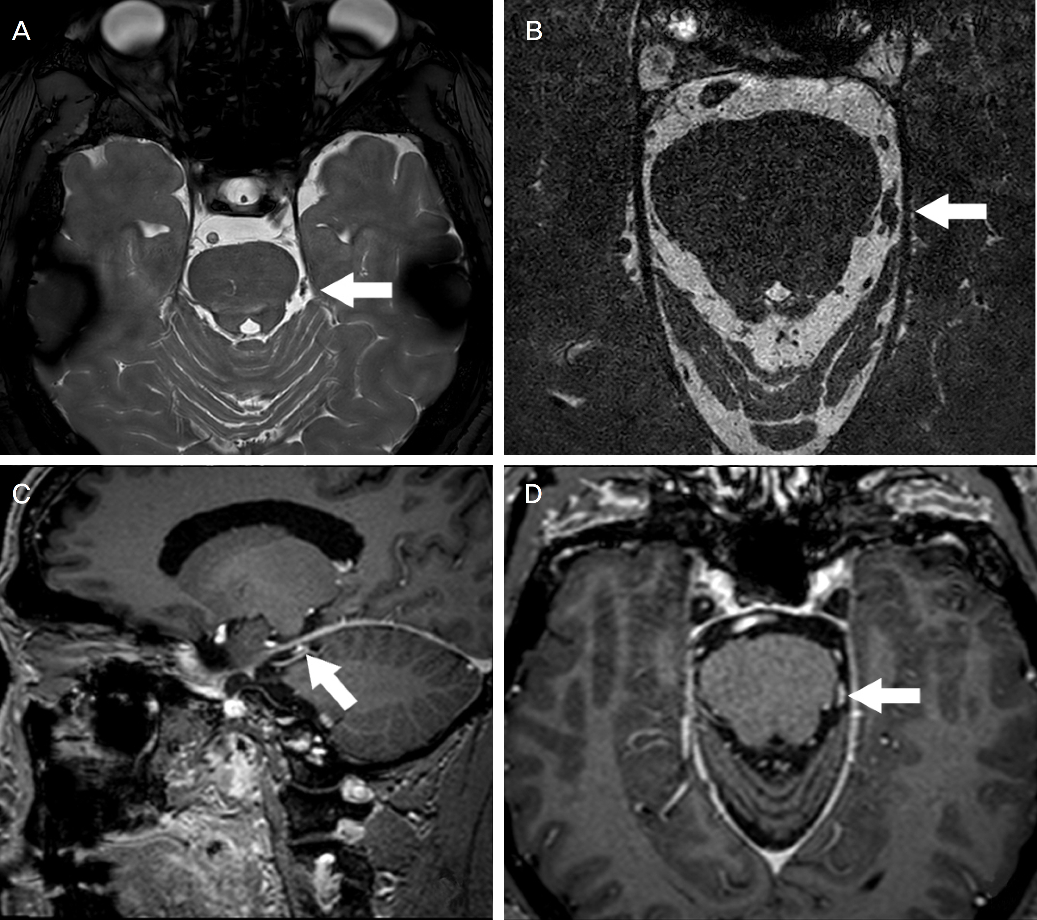
Figure 2. High-resolution magnetic resonance imaging of the trochlear nerve. (A, B) Axial T2-weighted images at the lower midbrain and upper pons reveal a 6 mm-sized lobulated trochlear nerve mass with low signal intensity (arrows). Sagittal (C) and axial (D) planes of gadolinium-enhanced T1-weighted images show a well circumscribed, homogeneously enhancing lesion (arrows) originating from the trochlear nerve in the left ambient cistern.
Cited by 1 articles
-
Trochlear Nerve Palsy Caused by Quadrigeminal Cistern Lipoma
Nam Hyeon Choi, Won Jae Kim, Myung-Mi Kim
J Korean Ophthalmol Soc. 2018;59(11):1087-1090. doi: 10.3341/jkos.2018.59.11.1087.
Reference
-
References
1. Esiri M. Russell and Rubinstein's pathology of tumors of the abdominal system. Sixth edition. J Neurol Neurosurg Psychiatry. 2000; 68:538D.2. Elsharkawy M, Xu Z, Schlesinger D, Sheehan JP. Gamma Knife surgery for nonvestibular schwannomas: radiological and clinical outcomes. J Neurosurg. 2012; 116:66–72.
Article3. Ho KL. Schwannoma of the trochlear nerve. Case report. J Neurosurg. 1981; 55:132–5.4. Celli P, Ferrante L, Acqui M, et al. Neurinoma of the third, fourth, and sixth cranial nerves: a survey and report of a new fourth nerve case. Surg Neurol. 1992; 38:216–24.
Article5. Neurofibromatosis. Conference statement. National Institutes of Health Consensus Development Conference. Arch Neurol. 1988; 45:575–8.6. Aoki S, Barkovich AJ, Nishimura K, et al. Neurofibromatosis types 1 and 2: cranial MR findings. Radiology. 1989; 172:527–34.
Article7. Boucher AB, Michael LM 2nd. The middle fossa approach for the removal of a trochlear schwannoma. Case Rep Neurol Med. 2014; 2014:672314.
Article8. Elmalem VI, Younge BR, Biousse V, et al. Clinical course and prognosis of trochlear nerve schwannomas. Ophthalmology. 2009; 116:2011–6.
Article9. Hatae R, Miyazono M, Kohri R, et al. Trochlear nerve abdominal with intratumoral hemorrhage presenting with persistent hic-cups: a case report. J Neurol Surg Rep. 2014; 75:e183–8.10. Elflein HM, Thömke F, Müller-Forell W, Pitz S. Trochlear palsies caused by isolated trochlear schwannomas. Strabismus. 2010; 18:83–6.
Article11. Du R, Dhoot J, McDermott MW, Gupta N. Cystic schwannoma of the anterior tentorial hiatus. Case report and review of the literature. Pediatr Neurosurg. 2003; 38:167–73.12. Gerganov V, Amir S, Koerbel A, et al. Cystic trochlear nerve schwannoma. Case report. Surg Neurol. 2007; 68:221–5.
Article13. Richards BW, Jones FR Jr, Younge BR. Causes and prognosis in 4,278 cases of paralysis of the oculomotor, trochlear, and abducens cranial nerves. Am J Ophthalmol. 1992; 113:489–96.
Article14. Jackowski A, Weiner G, O'Reilly G. Trochlear nerve abdominals: a case report and literature review. Br J Neurosurg. 1994; 8:219–23.15. Santoreneos S, Hanieh A, Jorgensen RE. Trochlear nerve schwannomas occurring in patients without neurofibromatosis: case report and review of the literature. Neurosurgery. 1997; 41:282–7.
Article16. Pollock BE, Foote RL, Stafford SL. Stereotactic radiosurgery: the preferred management for patients with nonvestibular abdominals? Int J Radiat Oncol Biol Phys. 2002; 52:1002–7.17. Yamamoto M, Jimbo M, Ide M, Kubo O. Trochlear neurinoma. Surg Neurol. 1987; 28:287–90.
Article18. Abe T, Iwata T, Shimazu M, Matsumoto K. Trochlear nerve abdominal associated with a giant thrombosed dissecting aneurysm of the contralateral vertebral artery. Surg Neurol. 1994; 42:438–41.19. Garen PD, Harper CG, Teo C, Johnston IH. Cystic schwannoma of the trochlear nerve mimicking a abdominal tumor. Case report. J Neurosurg. 1987; 67:928–30.20. Boggan JE, Rosenblum ML, Wilson CB. Neurilemmoma of the fourth cranial nerve. Case report. J Neurosurg. 1979; 50:519–21.
- Full Text Links
-
- Actions
-
Cited
- CITED
-
- Close
- Share
-
- Similar articles
-
- A Case of Herpes Zoster Ophthalmicus with Isolated Trochlear Nerve Involvement
- Clinical Features for Patients Presenting with Diplopia
- Transient Isolated Trochlear Nerve Palsy Associated with Rathke's Cleft Cyst
- A Clinical Study of Diplopia Due to Neurologic Disorder
- A Case with Bilateral Dural Arteriovenous Fistulae Manifesting as Sequential Trochlear, Oculomotor Nerve Palsies
