Comparison of Filtered Back Projection, Hybrid Iterative Reconstruction, Model-Based Iterative Reconstruction, and Virtual Monoenergetic Reconstruction Images at Both Low- and Standard-Dose Settings in Measurement of Emphysema Volume and Airway Wall Thickness: A CT Phantom Study
- Affiliations
-
- 1Department of Radiology, Korea University Ansan Hospital, Korea University College of Medicine, Ansan 15355, Korea. kiylee@korea.ac.kr
- 2Department of Pulmonology, Korea University Ansan Hospital, Korea University College of Medicine, Ansan 15355, Korea.
- 3Department of Radiology, Korea University Guro Hospital, Korea University College of Medicine, Seoul 08308, Korea.
- 4Department of Radiology, Korea University Anam Hospital, Korea University College of Medicine, Seoul 02841, Korea.
- 5Medical Science Research Center, Korea University Ansan Hospital, Korea University College of Medicine, Ansan 15355, Korea.
- KMID: 2413711
- DOI: http://doi.org/10.3348/kjr.2018.19.4.809
Abstract
OBJECTIVE
To evaluate the accuracy of emphysema volume (EV) and airway measurements (AMs) produced by various iterative reconstruction (IR) algorithms and virtual monoenergetic images (VME) at both low- and standard-dose settings.
MATERIALS AND METHODS
Computed tomography (CT) images were obtained on phantom at both low- (30 mAs at 120 kVp) and standard-doses (100 mAs at 120 kVp). Each CT scan was reconstructed using filtered back projection, hybrid IR (iDose4; Philips Healthcare), model-based IR (IMR-R1, IMR-ST1, IMR-SP1; Philips Healthcare), and VME at 70 keV (VME70). The EV of each air column and wall area percentage (WA%) of each airway tube were measured in all algorithms. Absolute percentage measurement errors of EV (APEvol) and AM (APEWA%) were then calculated.
RESULTS
Emphysema volume was most accurately measured in IMR-R1 (APEvol in low-dose, 0.053 ± 0.002; APEvol in standard-dose, 0.047 ± 0.003; all p < 0.001) and AM was the most accurate in IMR-SP1 on both low- and standard-doses CT (APEWA% in low-dose, 0.067 ± 0.002; APEWA% in standard-dose, 0.06 ± 0.003; all p < 0.001). There were no significant differences in the APEvol of IMR-R1 between low- and standard-doses (all p > 0.05). VME70 showed a significantly higher APEvol than iDose4, IMR-R1, and IMR-ST1 (all p < 0.004). VME70 also showed a significantly higher APEWA% compared with the other algorithms (all p < 0.001).
CONCLUSION
IMR was the most accurate technique for measurement of both EV and airway wall thickness. However, VME70 did not show a significantly better accuracy compared with other algorithms.
Keyword
MeSH Terms
Figure
-
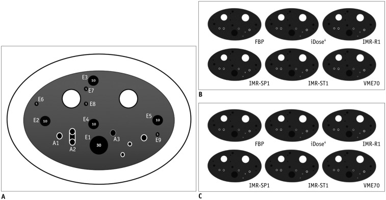
Fig. 1 Illustration and CT images of CTP674 lung phantom (The Phantom Laboratory).A. Illustration of CTP674 lung phantom. Nine air columns with three different diameters and volumes were chosen for EV measurements (E1–E9), and three polycarbonate tubes (airway tubes) with different wall thicknesses and angles were analyzed for airway wall measurements (A1–A3). Two airway tubes (A1 and A2) were placed at different angle of 30° from z-axis of CT couch. B. CT scans with various algorithms at low-dose setting. C. CT scans with various algorithms at standard-dose setting. CT = computed tomography, EV = emphysema volume, FBP = filtered back projection, IMR-R1 = MIR with image definition of body routine level 1, IMR-SP1 = MIR with image definition of sharp plus level, IMR-ST1 = MIR with image definition of body soft tissue level 1, MIR = model-based iterative reconstruction, VME70 = standard virtual monoenergetic dataset at 70 keV
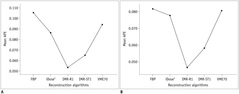
Fig. 2 APEvol of across all air columns (all of 5-, 10-, and 30-mm air columns) on (A) low-dose CT and (B) standard-dose CT.Mean APEvol in all air columns was lowest in IMR-R1 in both low-dose and standard-dose settings. APE = absolute percentage measurement error, APEvol = APE of EV
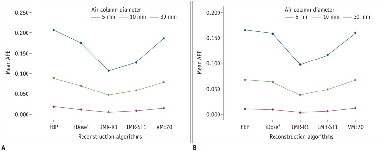
Fig. 3 APEvol of each column (each 5-, 10-, and 30-mm) on (A) low-dose CT and (B) standard-dose CT.Mean APEvol in all air columns was lowest in IMR-R1 in both low-dose and standard-dose settings. Mean APEvol decreased as volume of air columns increased.
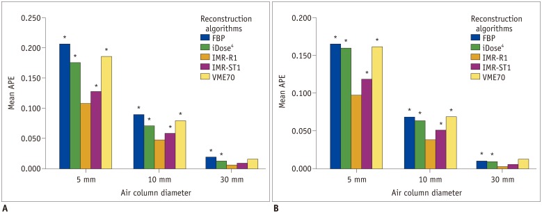
Fig. 4 Mean APEvol in each air column on (A) low-dose CT and (B) standard-dose CT.Significant differences in APEvol from IMR-R1 are marked with an asterisk (*).

Fig. 5 APEWA% of across all airway tubes (all of A1, A2, and A3 airway tubes) on (A) low-dose CT and (B) standard-dose CT.Mean APEWA% of across all airway tubes was lowest in IMR-SP1 in both low-dose and standard-dose settings. Mean APEWA% decreased as WA% increased. APEWA% = absolute percentage measurement error of wall area percentage, WA% = wall area percentage
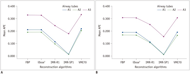
Fig. 6 Absolute percentage measurement error of each airway tube (each A1, A2, and A3 airway tube) on (A) low-dose CT and (B) standard-dose CT.Mean APEWA% decreased as WA% increased.

Fig. 7 Mean APEWA% in each airway tube on (A) low-dose and (B) standard-dose CT.Significant differences in APEWA% from IMR-SP1 are marked with asterisk (*).
Cited by 2 articles
-
Accuracy of Model-Based Iterative Reconstruction for CT Volumetry of Part-Solid Nodules and Solid Nodules in Comparison with Filtered Back Projection and Hybrid Iterative Reconstruction at Various Dose Settings: An Anthropomorphic Chest Phantom Study
Seung Kwan Kim, Cherry Kim, Ki Yeol Lee, Jaehyung Cha, Hyun-ju Lim, Eun-Young Kang, Yu-Whan Oh
Korean J Radiol. 2019;20(7):1195-1206. doi: 10.3348/kjr.2018.0893.Prediction of Pulmonary Function in Patients with Chronic Obstructive Pulmonary Disease: Correlation with Quantitative CT Parameters
Hyun Jung Koo, Sang Min Lee, Joon Beom Seo, Sang Min Lee, Namkug Kim, Sang Young Oh, Jae Seung Lee, Yeon-Mok Oh
Korean J Radiol. 2019;20(4):683-692. doi: 10.3348/kjr.2018.0391.
Reference
-
1. Lynch DA, Al-Qaisi MA. Quantitative computed tomography in chronic obstructive pulmonary disease. J Thorac Imaging. 2013; 28:284–290. PMID: 23748651.
Article2. Kim SS, Jin GY, Li YZ, Lee JE, Shin HS. CT Quantification of Lungs and Airways in Normal Korean Subjects. Korean J Radiol. 2017; 18:739–748. PMID: 28670169.
Article3. Brooks RA, Di Chiro G. Theory of image reconstruction in computed tomography. Radiology. 1975; 117(3 Pt 1):561–572. PMID: 1188102.
Article4. Baumueller S, Winklehner A, Karlo C, Goetti R, Flohr T, Russi EW, et al. Low-dose CT of the lung: potential value of iterative reconstructions. Eur Radiol. 2012; 22:2597–2606. PMID: 22699873.
Article5. Yang WJ, Yan FH, Liu B, Pang LF, Hou L, Zhang H, et al. Can sinogram-affirmed iterative (SAFIRE) reconstruction improve imaging quality on low-dose lung CT screening compared with traditional filtered back projection (FBP) reconstruction? J Comput Assist Tomogr. 2013; 37:301–305. PMID: 23493224.
Article6. Lim HJ, Chung MJ, Shin KE, Hwang HS, Lee KS. The impact of iterative reconstruction in low-dose computed tomography on the evaluation of diffuse interstitial lung disease. Korean J Radiol. 2016; 17:950–960. PMID: 27833411.
Article7. Katsura M, Matsuda I, Akahane M, Sato J, Akai H, Yasaka K, et al. Model-based iterative reconstruction technique for radiation dose reduction in chest CT: comparison with the adaptive statistical iterative reconstruction technique. Eur Radiol. 2012; 22:1613–1623. PMID: 22538629.
Article8. Yuki H, Oda S, Utsunomiya D, Funama Y, Kidoh M, Namimoto T, et al. Clinical impact of model-based type iterative reconstruction with fast reconstruction time on image quality of low-dose screening chest CT. Acta Radiol. 2016; 57:295–302. PMID: 25817455.
Article9. Choo JY, Goo JM, Lee CH, Park CM, Park SJ, Shim MS. Quantitative analysis of emphysema and airway measurements according to iterative reconstruction algorithms: comparison of filtered back projection, adaptive statistical iterative reconstruction and model-based iterative reconstruction. Eur Radiol. 2014; 24:799–806. PMID: 24275806.
Article10. Mets OM, Willemink MJ, de Kort FP, Mol CP, Leiner T, Oudkerk M, et al. The effect of iterative reconstruction on computed tomography assessment of emphysema, air trapping and airway dimensions. Eur Radiol. 2012; 22:2103–2109. PMID: 22618522.
Article11. Nishio M, Matsumoto S, Ohno Y, Sugihara N, Inokawa H, Yoshikawa T, et al. Emphysema quantification by low-dose CT: potential impact of adaptive iterative dose reduction using 3D processing. AJR Am J Roentgenol. 2012; 199:595–601. PMID: 22915399.
Article12. Leng S, Yu L, Fletcher JG, McCollough CH. Maximizing iodine contrast-to-noise ratios in abdominal CT imaging through use of energy domain noise reduction and virtual monoenergetic dual-energy CT. Radiology. 2015; 276:562–570. PMID: 25860839.
Article13. Goo HW, Goo JM. Dual-energy CT: new horizon in medical imaging. Korean J Radiol. 2017; 18:555–569. PMID: 28670151.
Article14. Kaup M, Scholtz JE, Engler A, Albrecht MH, Bauer RW, Kerl JM, et al. Dual-energy computed tomography virtual monoenergetic imaging of lung cancer: assessment of optimal energy levels. J Comput Assist Tomogr. 2016; 40:80–85. PMID: 26466115.15. Frellesen C, Kaup M, Wichmann JL, Hüsers K, Scholtz JE, Albrecht MH, et al. Noise-optimized advanced image-based virtual monoenergetic imaging for improved visualization of lung cancer: comparison with traditional virtual monoenergetic imaging. Eur J Radiol. 2016; 85:665–672. PMID: 26860682.
Article16. Gomez-Cardona D, Nagle SK, Li K, Robinson TE, Chen GH. Influence of radiation dose and reconstruction algorithm in MDCT assessment of airway wall thickness: a phantom study. Med Phys. 2015; 42:5919–5927. PMID: 26429266.
Article17. de Margerie-Mellon C, de Bazelaire C, Montlahuc C, Lambert J, Martineau A, Coulon P, et al. Reducing radiation dose at chest CT: comparison among model-based type iterative reconstruction, hybrid iterative reconstruction, and filtered back projection. Acad Radiol. 2016; 23:1246–1254. PMID: 27346234.18. Nakajo C, Heinzer S, Montandon S, Dunet V, Bize P, Feldman A, et al. Chest CT at a dose below 0.3 mSv: impact of iterative reconstruction on image quality and lung analysis. Acta Radiol. 2016; 57:311–317. PMID: 25838452.
Article19. Blechschmidt RA, Werthschützky R, Lörcher U. Automated CT image evaluation of the lung: a morphology-based concept. IEEE Trans Med Imaging. 2001; 20:434–442. PMID: 11403202.
Article
- Full Text Links
-
- Actions
-
Cited
- CITED
-
- Close
- Share
-
- Similar articles
-
- Effects of Iterative Reconstruction Algorithm, Automatic Exposure Control on Image Quality, and Radiation Dose: Phantom Experiments with Coronary CT Angiography Protocols
- Contrast-Enhanced CT with Knowledge-Based Iterative Model Reconstruction for the Evaluation of Parotid Gland Tumors: A Feasibility Study
- Accuracy of Model-Based Iterative Reconstruction for CT Volumetry of Part-Solid Nodules and Solid Nodules in Comparison with Filtered Back Projection and Hybrid Iterative Reconstruction at Various Dose Settings: An Anthropomorphic Chest Phantom Study
- Impact of Model-Based Iterative Reconstruction on the Correlation between Computed Tomography Quantification of a Low Lung Attenuation Area and Airway Measurements and Pulmonary Function Test Results in Normal Subjects
- Low-Dose Abdominal CT Using a Deep Learning-Based Denoising Algorithm: A Comparison with CT Reconstructed with Filtered Back Projection or Iterative Reconstruction Algorithm
