Association of Insulin Resistance with Lower Bone Volume and Strength Index of the Proximal Femur in Nondiabetic Postmenopausal Women
- Affiliations
-
- 1Department of Public Health, Yonsei University Graduate School, Seoul, Korea.
- 2Department of Internal Medicine, Severance Hospital, Endocrine Research Institute, Yonsei University College of Medicine, Seoul, Korea.
- 3Cardiovascular and Metabolic Diseases Etiology Research Center, Yonsei University College of Medicine, Seoul, Korea. hckim@yuhs.ac
- 4Department of Preventive Medicine, Yonsei University College of Medicine, Seoul, Korea.
- KMID: 2413374
- DOI: http://doi.org/10.11005/jbm.2018.25.2.123
Abstract
- BACKGROUND
Type 2 diabetes mellitus is associated with an increased risk of osteoporotic fracture despite relatively preserved bone mineral density (BMD). Although this paradox might be attributed to the influence of insulin resistance (IR) on bone structure and material properties, the association of IR with femur bone geometry and strength indices remains unclear.
METHODS
Using data from the Cardiovascular and Metabolic Disease Etiology Research Center cohort study, we conducted a cross-sectional analysis among nondiabetic postmenopausal women. IR was estimated using the homeostasis model assessment of IR (HOMA-IR). Compartment-specific volumetric BMD (vBMD) and bone volume of proximal femur were measured using quantitative computed tomography. The compressive strength index (CSI), section modulus (Z), and buckling ratio of the femoral neck were calculated as bone strength indices.
RESULTS
Among 1,008 subjects (mean age, 57.3 years; body mass index [BMI], 23.6 kg/m²), BMI, waist circumference, and vBMD of the femoral neck and total hip increased in a linear trend from the lowest ( < 1.37) to highest (≥2.27) HOMA-IR quartile (P < 0.05 for all). The HOMA-IR showed an independent negative association with total bone volume (standardized β=−0.12), cortical volume (β=−0.05), CSI (β=−0.013), and Z (β=−0.017; P < 0.05 for all) of the femoral neck after adjustment for age, weight, height, physical activity, and vitamin D and high-sensitivity C-reactive protein levels. However, the association between HOMA-IR and vBMD was attenuated in the adjusted model (femoral neck, β=0.94; P=0.548).
CONCLUSIONS
Elevated HOMA-IR was associated with lower cortical bone volume and bone strength indices in nondiabetic postmenopausal women, independent of age and body size.
Keyword
MeSH Terms
-
Body Mass Index
Body Size
Bone Density
C-Reactive Protein
Cohort Studies
Compressive Strength
Cross-Sectional Studies
Diabetes Mellitus, Type 2
Female
Femur Neck
Femur*
Hip
Homeostasis
Humans
Insulin Resistance*
Insulin*
Metabolic Diseases
Motor Activity
Neck
Osteoporosis
Osteoporotic Fractures
Postmenopause
Vitamin D
Waist Circumference
C-Reactive Protein
Insulin
Vitamin D
Figure
-
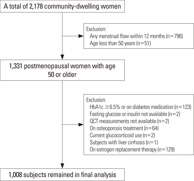
Fig. 1 Study flow diagram. HbA1c, hemoglobin A1c; QCT, quantitative computed tomography.
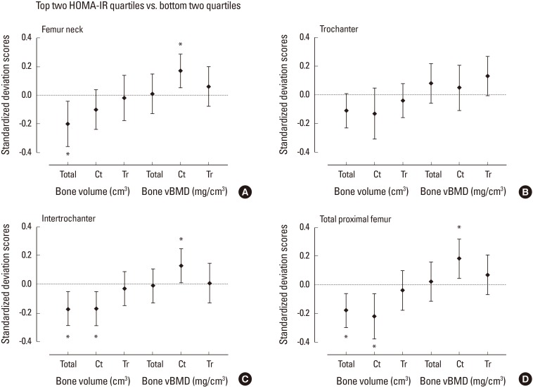
Fig. 2 Standardized difference (in standard deviation [SD] units±95% confidence interval) in quantitative computed tomography-derived bone volume and density at proximal femur for subjects in the top quartiles of homeostasis model assessment of insulin resistance (HOMA-IR) vs. lower quartiles after adjustment for age, weight, and height. Dashed line indicates the mean of the bottom two quartiles. The mean difference between the groups were expressed in SD scores as divided by the SD of the bottom two quartile group. Asterisk (*) indicated P<0.05 for the difference between the top and bottom two quartiles. vBMD, volumetric bone mineral density.
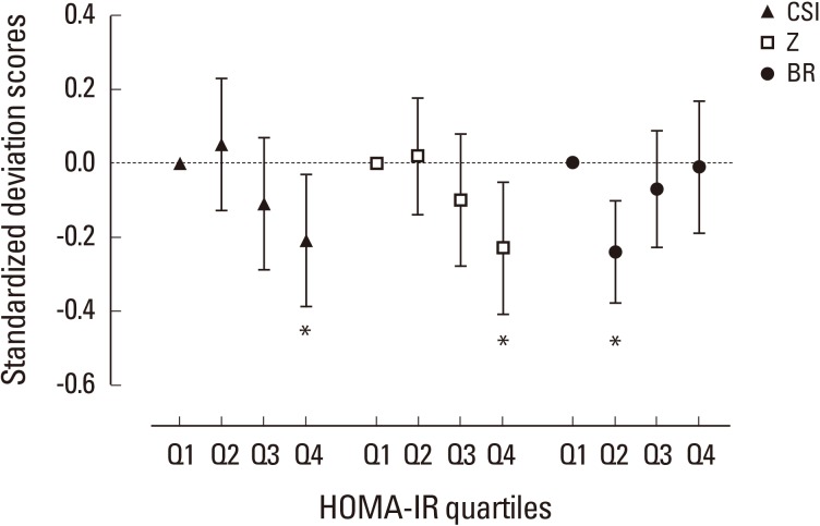
Fig. 3 Standardized difference (in standard deviation [SD] units±95% confidence interval) in quantitative computed tomography-derived bone geometry parameters at femur according to homeostasis model assessment of insulin resistance (HOMA-IR) quartiles. Dashed line indicates the mean value of the subjects in lowest quartile after adjustment for age, weight, and height. The mean difference between the groups were expressed in SD scores as divided by the SD of the lowest quartile group. Asterisk (*) indicated P<0.05 as compared with the lowest quartile as reference group. Q, quartile; CSI, compressive strength index; Z, section modulus; BR, buckling ratio.
Cited by 2 articles
-
Skeletal Fragility in Type 2 Diabetes Mellitus
Jakob Starup-Linde, Katrine Hygum, Bente Lomholt Langdahl
Endocrinol Metab. 2018;33(3):339-351. doi: 10.3803/EnM.2018.33.3.339.Cohort Profile: The Cardiovascular and Metabolic Diseases Etiology Research Center Cohort in Korea
Jee-Seon Shim, Bo Mi Song, Jung Hyun Lee, Seung Won Lee, Ji Hye Park, Dong Phil Choi, Myung Ha Lee, Kyoung Hwa Ha, Dae Jung Kim, Sungha Park, Won-Woo Lee, Yoosik Youm, Eui-Cheol Shin, Hyeon Chang Kim
Yonsei Med J. 2019;60(8):804-810. doi: 10.3349/ymj.2019.60.8.804.
Reference
-
1. Russo GT, Giandalia A, Romeo EL, et al. Fracture risk in type 2 diabetes: current perspectives and gender differences. Int J Endocrinol. 2016; 2016:1615735. PMID: 28044077.
Article2. Nicodemus KK, Folsom AR. Type 1 and type 2 diabetes and incident hip fractures in postmenopausal women. Diabetes Care. 2001; 24:1192–1197. PMID: 11423501.
Article3. Schwartz AV, Sellmeyer DE, Ensrud KE, et al. Older women with diabetes have an increased risk of fracture: a prospective study. J Clin Endocrinol Metab. 2001; 86:32–38. PMID: 11231974.
Article4. Bonds DE, Larson JC, Schwartz AV, et al. Risk of fracture in women with type 2 diabetes: the Women’s Health Initiative Observational Study. J Clin Endocrinol Metab. 2006; 91:3404–3410. PMID: 16804043.
Article5. Shull RL, Guilkey M, Witty W. Changing the response unit from a single peck to a fixed number of pecks in fixed-interval schedules. J Exp Anal Behav. 1972; 17:193–200. PMID: 16811581.6. Schwartz AV, Sellmeyer DE, Strotmeyer ES, et al. Diabetes and bone loss at the hip in older black and white adults. J Bone Miner Res. 2005; 20:596–603. PMID: 15765178.
Article7. Melton LJ 3rd, Riggs BL, Leibson CL, et al. A bone structural basis for fracture risk in diabetes. J Clin Endocrinol Metab. 2008; 93:4804–4809. PMID: 18796521.
Article8. Shanbhogue VV, Finkelstein JS, Bouxsein ML, et al. Association between insulin resistance and bone structure in nondiabetic postmenopausal women. J Clin Endocrinol Metab. 2016; 101:3114–3122. PMID: 27243136.
Article9. Srikanthan P, Crandall CJ, Miller-Martinez D, et al. Insulin resistance and bone strength: findings from the study of midlife in the United States. J Bone Miner Res. 2014; 29:796–803. PMID: 23983216.
Article10. Shim JS, Song BM, Lee JH, et al. Cardiovascular and metabolic diseases etiology research center (CMERC) cohort: study protocol and results of the first 3 years of enrollment. Epidemiol Health. 2017; 39:e2017016. PMID: 28395401.
Article11. Chun MY. Validity and reliability of Korean version of international physical activity questionnaire short form in the elderly. Korean J Fam Med. 2012; 33:144–151. PMID: 22787536.
Article12. Matthews DR, Hosker JP, Rudenski AS, et al. Homeostasis model assessment: insulin resistance and beta-cell function from fasting plasma glucose and insulin concentrations in man. Diabetologia. 1985; 28:412–419. PMID: 3899825.13. Levey AS, Stevens LA, Schmid CH, et al. A new equation to estimate glomerular filtration rate. Ann Intern Med. 2009; 150:604–612. PMID: 19414839.
Article14. Danielson ME, Beck TJ, Karlamangla AS, et al. A comparison of DXA and CT based methods for estimating the strength of the femoral neck in post-menopausal women. Osteoporos Int. 2013; 24:1379–1388. PMID: 22810918.
Article15. Khoo BC, Brown K, Zhu K, et al. Effects of the assessment of 4 determinants of structural geometry on QCT- and DXA-derived hip structural analysis measurements in elderly women. J Clin Densitom. 2014; 17:38–46. PMID: 23578719.
Article16. Lee SJ, Kim KM, Brown JK, et al. Negative impact of aromatase inhibitors on proximal femoral bone mass and geometry in postmenopausal women with breast cancer. Calcif Tissue Int. 2015; 97:551–559. PMID: 26232103.
Article18. Strotmeyer ES, Cauley JA, Schwartz AV, et al. Diabetes is associated independently of body composition with BMD and bone volume in older white and black men and women: the Health, Aging, and Body Composition Study. J Bone Miner Res. 2004; 19:1084–1091. PMID: 15176990.
Article19. Napoli N, Chandran M, Pierroz DD, et al. Mechanisms of diabetes mellitus-induced bone fragility. Nat Rev Endocrinol. 2017; 13:208–219. PMID: 27658727.
Article20. Reid IR, Evans MC, Cooper GJ, et al. Circulating insulin levels are related to bone density in normal postmenopausal women. Am J Physiol. 1993; 265:E655–E659. PMID: 8238341.
Article21. Verroken C, Zmierczak HG, Goemaere S, et al. Insulin resistance is associated with smaller cortical bone size in nondiabetic men at the age of peak bone mass. J Clin Endocrinol Metab. 2017; 102:1807–1815. PMID: 28001453.
Article22. Petit MA, Paudel ML, Taylor BC, et al. Bone mass and strength in older men with type 2 diabetes: the Osteoporotic Fractures in Men Study. J Bone Miner Res. 2010; 25:285–291. PMID: 19594301.
Article23. Gilsanz V, Chalfant J, Mo AO, et al. Reciprocal relations of subcutaneous and visceral fat to bone structure and strength. J Clin Endocrinol Metab. 2009; 94:3387–3393. PMID: 19531595.
Article24. Farr JN, Chen Z, Lisse JR, et al. Relationship of total body fat mass to weight-bearing bone volumetric density, geometry, and strength in young girls. Bone. 2010; 46:977–984. PMID: 20060079.
Article25. Bredella MA, Lin E, Gerweck AV, et al. Determinants of bone microarchitecture and mechanical properties in obese men. J Clin Endocrinol Metab. 2012; 97:4115–4122. PMID: 22933540.
Article26. Frost HM. Bone “mass” and the “mechanostat”: a proposal. Anat Rec. 1987; 219:1–9. PMID: 3688455.
Article27. Lebrasseur NK, Achenbach SJ, Melton LJ 3rd, et al. Skeletal muscle mass is associated with bone geometry and microstructure and serum insulin-like growth factor binding protein-2 levels in adult women and men. J Bone Miner Res. 2012; 27:2159–2169. PMID: 22623219.
Article28. Lee S, Kim Y, White DA, et al. Relationships between insulin sensitivity, skeletal muscle mass and muscle quality in obese adolescent boys. Eur J Clin Nutr. 2012; 66:1366–1368. PMID: 23073260.
Article29. Ferron M, Wei J, Yoshizawa T, et al. Insulin signaling in osteoblasts integrates bone remodeling and energy metabolism. Cell. 2010; 142:296–308. PMID: 20655470.
Article30. Hahn TJ, Westbrook SL, Sullivan TL, et al. Glucose transport in osteoblast-enriched bone explants: characterization and insulin regulation. J Bone Miner Res. 1988; 3:359–365. PMID: 2463740.
Article31. Karlsson MK, Weigall SJ, Duan Y, et al. Bone size and volumetric density in women with anorexia nervosa receiving estrogen replacement therapy and in women recovered from anorexia nervosa. J Clin Endocrinol Metab. 2000; 85:3177–3182. PMID: 10999805.
Article32. Szulc P, Seeman E, Duboeuf F, et al. Bone fragility: failure of periosteal apposition to compensate for increased endocortical resorption in postmenopausal women. J Bone Miner Res. 2006; 21:1856–1863. PMID: 17002580.
Article33. Kindler JM, Pollock NK, Laing EM, et al. Insulin resistance negatively influences the muscle-dependent IGF-1-bone mass relationship in premenarcheal girls. J Clin Endocrinol Metab. 2016; 101:199–205. PMID: 26574958.
Article34. Kindler JM, Pollock NK, Laing EM, et al. Insulin resistance and the IGF-I-cortical bone relationship in children ages 9 to 13 years. J Bone Miner Res. 2017; 32:1537–1545. PMID: 28300329.
Article35. Yakar S, Canalis E, Sun H, et al. Serum IGF-1 determines skeletal strength by regulating subperiosteal expansion and trait interactions. J Bone Miner Res. 2009; 24:1481–1492. PMID: 19257833.
Article
- Full Text Links
-
- Actions
-
Cited
- CITED
-
- Close
- Share
-
- Similar articles
-
- The relationship between grip strength and femoral and vertebral bone mineral density in peri-and postmenopausal women
- Comparative Study of Serum Leptin and Insulin Resistance Levels Between Korean Postmenopausal Vegetarian and Non-vegetarian Women
- Determinants of Bone Mass and Insulin Resistance in Korean Postmenopausal Women: Muscle Area, Strength, or Composition?
- Low Handgrip Strength is Associated with Low Bone Mineral Density and Fragility Fractures in Postmenopausal Healthy Korean Women
- Effect of Reproductive Factors on Bone Mineral Density in Postmenopausal Women
