Protective effects of an ethanol extract of Angelica keiskei against acetaminophen-induced hepatotoxicity in HepG2 and HepaRG cells
- Affiliations
-
- 1Department of Food Science and Nutrition, Hallym University, 1 Hallymdaehak-gil, Chuncheon, Gangwon 24252, Korea. ijkang@hallym.ac.kr
- 2Department of Food Science and Nutrition, Dongseo University, Busan 47011, Korea.
- 3Center for Efficacy Assessment and Development of Functional Food and Drugs, Hallym University, 1 Hallymdaehak-gil, Chuncheon, Gangwon 24252, Korea. myej4@hallym.ac.kr
- KMID: 2412021
- DOI: http://doi.org/10.4162/nrp.2017.11.2.97
Abstract
- BACKGROUND
/OBJECTIVE: Although Angelica keiskei (AK) has widely been utilized for the purpose of general health improvement among Asian, its functionality and mechanism of action. The aim of this study was to determine the protective effect of ethanol extract of AK (AK-Ex) on acute hepatotoxicity induced by acetaminophen (AAP) in HepG2 human hepatocellular liver carcinoma cells and HepaRG human hepatic progenitor cells.
MATERIALS/METHODS
AK-Ex was prepared HepG2 and HepaRG cells were cultured with various concentrations and 30 mM AAP. The protective effects of AK-Ex against AAP-induced hepatotoxicity in HepG2 and HepaRG cells were evaluated using 3-[4,5-dimethylthiazol-2-yl]-2,5-diphenyltetrazolium bromide, lactate dehydrogenase (LDH) assay, flow cytometry, and Western blotting.
RESULTS
AK-Ex, when administered prior to AAP, increased cell growth and decreased leakage of LDH in a dose-dependent manner in HepG2 and HepaRG cells against AAP-induced hepatotoxicity. AK-Ex increased the level of Bcl-2 and decreased the levels of Bax, Bok and Bik decreased the permeability of the mitochondrial membrane in HepG2 cells intoxicated with AAP. AK-Ex decreased the cleavage of poly (ADP-ribose) polymerase (PARP) and the activation of caspase-9, -7, and -3.
CONCLUSIONS
These results demonstrate that AK-Ex downregulates apoptosis via intrinsic and extrinsic pathways against AAP-induced hepatotoxicity. We suggest that AK could be a useful preventive agent against AAP-induced apoptosis in hepatocytes.
Keyword
MeSH Terms
Figure
-
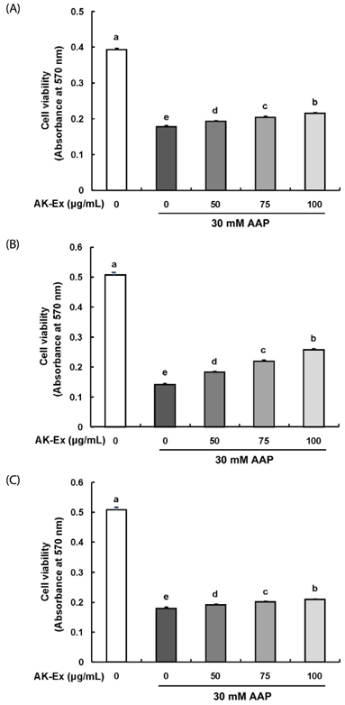
Fig. 1 Effects of an ethanol extract of Angelica keiskei (AK-Ex) on viable cell numbers in HepG2 cells. Cells were plated at a density of 5 × 10⁴ cells/well in 24-well plates. One day later, (A) cells were co-treated with the indicated concentration of AK-Ex and 30 mM AAP for 48 h. (B) Cells were treated with various concentration of AK-Ex for 24 h and then treated with 30 mM AAP for 24 h. (C) Cells were treated with 30 mM AAP for 24 h and then treated with various concentration of AK-Ex for 24 h. Cell numbers were estimated by using the MTT assay. Each bar represents mean ± SEM values (n = 4). Means without a common letter differ significantly, P < 0.05.
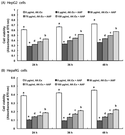
Fig. 2 Effect of AK-Ex pretreatment period on viable cell numbers in HepG2 and HepaRG cells. (A) HepG2 cells and (B) HepaRG cells were plated at a density of 5 × 10⁴ cells/well in 24-well plates. After 24 h, cells were pretreated with the indicated concentration of AK-Ex for 24, 36, or 48 h, respectively. Subsequently, cells were incubated with 30 mM AAP for 24 h. Cell numbers were estimated by using the MTT assay. Each bar represents mean ± SEM values (n = 4). Means without a common letter differ significantly, P < 0.05.
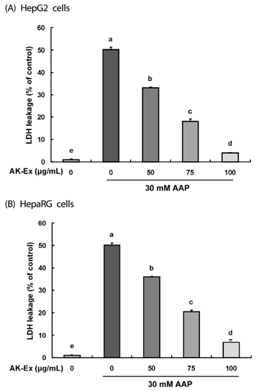
Fig. 3 Effect of AK-Ex on LDH leakage in HepG2 and HepaRG cells. (A) HepG2 cells were plated at a density of 5 × 10⁴ cells/well in a 24-well plate and incubated for 24 h. (B) HepaRG cells were plated at a density of 1.2 × 10⁵ cells/well in a 24-well plate and incubated for 72 h. Cells were treated for 48 h with various concentration of AK-Ex and then treated with 30 mM AAP for an additional 24 h. The 24 h-conditioned media were collected for LDH activity assay. LDH activity was measured by using an LDH cytotoxicity detection assay kit. Each bar represents the mean ± SEM values (n = 4). Means without a common letter differ significantly, P < 0.05.
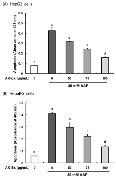
Fig. 4 Effect of AK-Ex on apoptosis in HepG2 and HepaRG cells. (A) HepG2 and (B) HepaRG cells were treated with AK-Ex and/or AAP, as described in Fig. 3. The number of apoptotic cells was assayed by using a cell death detection ELISAPLUS assay kit. Each bar represents mean ± SEM values (n = 4). Means without a common letter differ significantly, P < 0.05.

Fig. 5 Effect of AK-Ex on mitochondrial membrane potential in HepG2 and HepaRG cells. (A) HepG2 and (B) HepaRG cells were treated with AK-Ex and/or AAP, as described in Fig. 3. Cells were loaded with JC-1 and then analyzed by performing flow cytometry. The numbers of cells with normally polarized mitochondrial membranes (red) or with depolarized mitochondrial membranes (green) are expressed as percentages of the total cell number. Each bar represents the mean ± SEM values (n = 4). Means without a common letter differ significantly, P < 0.05.
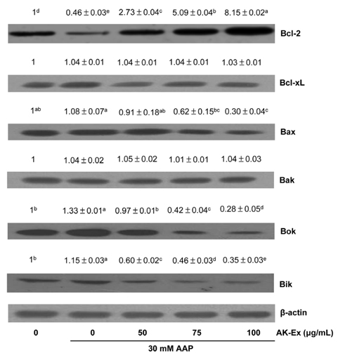
Fig. 6 Effect of AK-Ex on the protein levels of Bal-2 family proteins in HepG2 cells. Cells were plated at a density of 1 × 10⁶ cells/dish in 100 mm dishes and treated with AK-Ex and/or AAP, as described in Fig. 3. Cell lysates were analyzed by western blotting with the indicated antibodies. Photographs of the chemiluminescent detection of the blots, which are representative of three independent experiments, are shown. Relative abundance of each band to their own β-actin level was quantified and the control levels were set at 1. The adjusted mean ± SEM values (n = 3) of each band are shown above each blot. Means without a common letter differ significantly, P < 0.05.

Fig. 7 Effect of AK-Ex on the protein levels of cleaved caspases and cleaved PARP in HepG2 and HepaRG cells. HepG2 and HepaRG cells were plated at a density of 1 × 10⁶ cells/dish in 100 mm dishes and treated with AK-Ex and/or AAP, respectively, as described in Fig. 3. Cell lysates were analyzed by western blotting with the indicated antibodies. Photographs of the chemiluminescent detection of the blots, which are representative of three independent experiments, are shown. Relative abundance of each band to their own β-actin level was quantified and the control levels were set at 1. The adjusted mean ± SEM values (n = 3) of each band are shown above each blot. Means without a common letter differ significantly, P < 0.05.
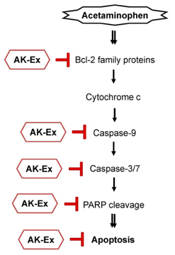
Fig. 8 Schematic representation of a possible mechanism for the hepatoprotective effect of AK-Ex in HepG2 and HepaRG cells.
Reference
-
1. Maronpot RR. Toxicological assessment of Ashitaba Chalcone. Food Chem Toxicol. 2015; 77:111–119.
Article2. Chang HR, Lee HJ, Ryu JH. Chalcones from Angelica keiskei attenuate the inflammatory responses by suppressing nuclear translocation of NF-kappaB. J Med Food. 2014; 17:1306–1313.
Article3. Yasuda M, Kawabata K, Miyashita M, Okumura M, Yamamoto N, Takahashi M, Ashida H, Ohigashi H. Inhibitory effects of 4-hydroxyderricin and xanthoangelol on lipopolysaccharide-induced inflammatory responses in RAW264 macrophages. J Agric Food Chem. 2014; 62:462–467.
Article4. Zhang T, Yamashita Y, Yasuda M, Yamamoto N, Ashida H. Ashitaba (Angelica keiskei) extract prevents adiposity in high-fat diet-fed C57BL/6 mice. Food Funct. 2015; 6:135–145.
Article5. Ogawa H, Okada Y, Kamisako T, Baba K. Beneficial effect of xanthoangelol, a chalcone compound from Angelica keiskei, on lipid metabolism in stroke-prone spontaneously hypertensive rats. Clin Exp Pharmacol Physiol. 2007; 34:238–243.
Article6. Kim E, Choi J, Yeo I. The effects of Angelica keiskei Koidz on the expression of antioxidant enzymes related to lipid profiles in rats fed a high fat diet. Nutr Res Pract. 2012; 6:9–15.
Article7. Akihisa T, Kikuchi T, Nagai H, Ishii K, Tabata K, Suzuki T. 4-Hydroxyderricin from Angelica keiskei roots induces caspasedependent apoptotic cell death in HL60 human leukemia cells. J Oleo Sci. 2011; 60:71–77.
Article8. Noh HM, Ahn EM, Yun JM, Cho BL, Paek YJ. Angelica keiskei Koidzumi extracts improve some markers of liver function in habitual alcohol drinkers: a randomized double-blind clinical trial. J Med Food. 2015; 18:166–172.
Article9. Ni HM, Bockus A, Boggess N, Jaeschke H, Ding WX. Activation of autophagy protects against acetaminophen-induced hepatotoxicity. Hepatology. 2012; 55:222–232.
Article10. Labbe G, Pessayre D, Fromenty B. Drug-induced liver injury through mitochondrial dysfunction: mechanisms and detection during preclinical safety studies. Fundam Clin Pharmacol. 2008; 22:335–353.
Article11. Larson AM, Polson J, Fontana RJ, Davern TJ, Lalani E, Hynan LS, Reisch JS, Schiødt FV, Ostapowicz G, Shakil AO, Lee WM. Acute Liver Failure Study Group. Acetaminophen-induced acute liver failure: results of a United States multicenter, prospective study. Hepatology. 2005; 42:1364–1372.
Article12. Jaeschke H. Glutathione disulfide formation and oxidant stress during acetaminophen-induced hepatotoxicity in mice in vivo: the protective effect of allopurinol. J Pharmacol Exp Ther. 1990; 255:935–941.13. Holownia A, Braszko JJ. Acetaminophen alters microsomal ryanodine Ca2+ channel in HepG2 cells overexpressing CYP2E1. Biochem Pharmacol. 2004; 68:513–521.
Article14. Kon K, Kim JS, Jaeschke H, Lemasters JJ. Mitochondrial permeability transition in acetaminophen-induced necrosis and apoptosis of cultured mouse hepatocytes. Hepatology. 2004; 40:1170–1179.
Article15. Kim EJ, Holthuizen PE, Park HS, Ha YL, Jung KC, Park JH. Trans-10, cis-12-conjugated linoleic acid inhibits Caco-2 colon cancer cell growth. Am J Physiol Gastrointest Liver Physiol. 2002; 283:G357–G367.16. Thomas JP, Geiger PG, Girotti AW. Lethal damage to endothelial cells by oxidized low density lipoprotein: role of selenoperoxidases in cytoprotection against lipid hydroperoxide- and iron-mediated reactions. J Lipid Res. 1993; 34:479–490.
Article17. Jung JI, Lim SS, Choi HJ, Cho HJ, Shin HK, Kim EJ, Chung WY, Park KK, Park JH. Isoliquiritigenin induces apoptosis by depolarizing mitochondrial membranes in prostate cancer cells. J Nutr Biochem. 2006; 17:689–696.
Article18. Cho HJ, Kim WK, Kim EJ, Jung KC, Park S, Lee HS, Tyner AL, Park JH. Conjugated linoleic acid inhibits cell proliferation and ErbB3 signaling in HT-29 human colon cell line. Am J Physiol Gastrointest Liver Physiol. 2003; 284:G996–1005.19. Jetten MJ, Kleinjans JC, Claessen SM, Chesne C, van Delft JH. Baseline and genotoxic compound induced gene expression profiles in HepG2 and HepaRG compared to primary human hepatocytes. Toxicol In Vitro. 2013; 27:2031–2040.
Article20. Jennen DG, Magkoufopoulou C, Ketelslegers HB, van Herwijnen MH, Kleinjans JC, van Delft JH. Comparison of HepG2 and HepaRG by whole-genome gene expression analysis for the purpose of chemical hazard identification. Toxicol Sci. 2010; 115:66–79.
Article21. Liguori MJ, Blomme EA, Waring JF. Trovafloxacin-induced gene expression changes in liver-derived in vitro systems: comparison of primary human hepatocytes to HepG2 cells. Drug Metab Dispos. 2008; 36:223–233.
Article22. Guo L, Dial S, Shi L, Branham W, Liu J, Fang JL, Green B, Deng H, Kaput J, Ning B. Similarities and differences in the expression of drug-metabolizing enzymes between human hepatic cell lines and primary human hepatocytes. Drug Metab Dispos. 2011; 39:528–538.
Article23. Aninat C, Piton A, Glaise D, Le Charpentier T, Langouët S, Morel F, Guguen-Guillouzo C, Guillouzo A. Expression of cytochromes P450, conjugating enzymes and nuclear receptors in human hepatoma HepaRG cells. Drug Metab Dispos. 2006; 34:75–83.
Article24. Andersson TB. The application of HepRG cells in evaluation of cytochrome P450 induction properties of drug compounds. Methods Mol Biol. 2010; 640:375–387.
Article25. Ferreira A, Rodrigues M, Silvestre S, Falcão A, Alves G. HepaRG cell line as an in vitro model for screening drug-drug interactions mediated by metabolic induction: amiodarone used as a model substance. Toxicol In Vitro. 2014; 28:1531–1535.
Article26. Danpure CJ. Lactate dehydrogenase and cell injury. Cell Biochem Funct. 1984; 2:144–148.
Article27. Cory S, Huang DC, Adams JM. The Bcl-2 family: roles in cell survival and oncogenesis. Oncogene. 2003; 22:8590–8607.
Article28. Ohkura N, Nakakuki Y, Taniguchi M, Kanai S, Nakayama A, Ohnishi K, Sakata T, Nohira T, Matsuda J, Baba K, Atsumi G. Xanthoangelols isolated from Angelica keiskei inhibit inflammatory-induced plasminogen activator inhibitor 1 (PAI-1) production. Biofactors. 2011; 37:455–461.
Article29. Nagata J, Morino T, Saito M. Effects of dietary Angelica keiskei on serum and liver lipid profiles, and body fat accumulations in rats. J Nutr Sci Vitaminol (Tokyo). 2007; 53:133–137.
Article30. Aoki N, Muko M, Ohta E, Ohta S. C-geranylated chalcones from the stems of Angelica keiskei with superoxide-scavenging activity. J Nat Prod. 2008; 71:1308–1310.
Article31. Ohnogi H, Hayami S, Kudo Y, Deguchi S, Mizutani S, Enoki T, Tanimura Y, Aoi W, Naito Y, Kato I, Yoshikawa T. Angelica keiskei extract improves insulin resistance and hypertriglyceridemia in rats fed a high-fructose drink. Biosci Biotechnol Biochem. 2012; 76:928–932.
Article32. Kawabata K, Sawada K, Ikeda K, Fukuda I, Kawasaki K, Yamamoto N, Ashida H. Prenylated chalcones 4-hydroxyderricin and xanthoangelol stimulate glucose uptake in skeletal muscle cells by inducing GLUT4 translocation. Mol Nutr Food Res. 2011; 55:467–475.
Article33. Oh SR, Kim SJ, Kim DH, Ryu JH, Ahn EM, Jung JW. Angelica keiskei ameliorates scopolamine-induced memory impairments in mice. Biol Pharm Bull. 2013; 36:82–88.
Article34. Choi SH, Park KH. Protective effects of Angelica keiskei extracts against D-galactosamine(GalN)-induced hepatotoxicity in rats. J Food Hyg Saf. 2011; 26:235–241.35. Huerta S, Goulet EJ, Huerta-Yepez S, Livingston EH. Screening and detection of apoptosis. J Surg Res. 2007; 139:143–156.
Article36. Thomadaki H, Scorilas A. BCL2 family of apoptosis-related genes: functions and clinical implications in cancer. Crit Rev Clin Lab Sci. 2006; 43:1–67.
Article37. Boise LH, González-García M, Postema CE, Ding L, Lindsten T, Turka LA, Mao X, Nuñez G, Thompson CB. bcl-x, a bcl-2-related gene that functions as a dominant regulator of apoptotic cell death. Cell. 1993; 74:597–608.
Article38. Nicholson DW. Caspase structure, proteolytic substrates, and function during apoptotic cell death. Cell Death Differ. 1999; 6:1028–1042.
Article39. Bürkle A, Brabeck C, Diefenbach J, Beneke S. The emerging role of poly(ADP-ribose) polymerase-1 in longevity. Int J Biochem Cell Biol. 2005; 37:1043–1053.
Article40. Andrabi SA, Kim NS, Yu SW, Wang H, Koh DW, Sasaki M, Klaus JA, Otsuka T, Zhang Z, Koehler RC, Hurn PD, Poirier GG, Dawson VL, Dawson TM. Poly(ADP-ribose) (PAR) polymer is a death signal. Proc Natl Acad Sci U S A. 2006; 103:18308–18313.
Article41. Isabelle M, Moreel X, Gagné JP, Rouleau M, Ethier C, Gagné P, Hendzel MJ, Poirier GG. Investigation of PARP-1, PARP-2, and PARG interactomes by affinity-purification mass spectrometry. Proteome Sci. 2010; 8:22.
Article
- Full Text Links
-
- Actions
-
Cited
- CITED
-
- Close
- Share
-
- Similar articles
-
- The Evaluation of Metabolizable Energy of Angelica Keiskei (Angelica utilis Makino) Products
- Comparison of in vitro models for drug-induced liver injury assessment
- Two Faces of "Green Juice"
- Protective effect of chlorophyllremoved ethanol extract of Lycium barbarum leaves against nonalcoholic fatty liver disease
- Ibuprofen Increases the Hepatotoxicity of Ethanol through Potentiating Oxidative Stress
