Cellular Effect of Curcumin and Citral Combination on Breast Cancer Cells: Induction of Apoptosis and Cell Cycle Arrest
- Affiliations
-
- 1Biochemistry Division, B. R. Doshi School of Biosciences, Sardar Patel University, Vallabh Vidyanagar, India. patel_pinaki@yahoo.com
- 2P. D. Patel Institute of Applied Science, Charotar University of Science and Technology (CHARUSAT), Changa, India.
- KMID: 2407569
- DOI: http://doi.org/10.4048/jbc.2015.18.3.225
Abstract
- PURPOSE
The unmanageable side effects caused by current chemotherapy regimens to treat cancer are an unresolved problem. Although many phytonutrients are useful as chemoprevention without side effects, their effects are slower and smaller than conventional chemotherapy. In the present work, we examined the cumulative effect of two phytonutrients, curcumin and citral, on breast cancer cell lines and compared their effect with the known chemotherapy regimen of cyclophosphamide, methotrexate, and 5-fluorouracil.
METHODS
Using cultured breast cancer and normal epithelial cells, the cytotoxic and apoptotic effect of curcumin and citral was evaluated in vitro. The synergistic effect of curcumin and citral was calculated by a combination index study using the method by Chou and Talalay. Cell death pathways and mechanisms were analyzed by measuring intracellular reactive oxygen species (ROS) and apoptotic protein levels.
RESULTS
Curcumin and citral caused dose and time dependent cell death and showed a synergistic effect at effective concentration EC50 and above concentrations in breast cancer cells without disturbing normal breast epithelial cells. With combination curcumin and citral treatment, apoptosis induction and cell cycle arrest at G0/G1 phase in breast cancer cells were observed. Curcumin and citral generated ROS and activated p53 and poly (ADP-ribose) polymerase-1 mediated apoptotic pathways.
CONCLUSION
The results of this study suggest that curcumin and citral in combination may be a useful therapeutic intervention for breast cancer.
MeSH Terms
Figure
-

Figure 1 Survival of MCF 7 and MDA MB 231 cells after exposure to chemotherapeutic drugs. Dose dependent cell viability of MCF 7, MDA MB 231, and MCF 10A cells, after combination treatment with cyclophosphamide, methotrexate, and 5-fluorouracil (CMF) was assessed by MTT assay. The concentration of each combination is shown below the graph. Data were plotted as percent viability (% control). At a zero concentration of drugs, % viability was considered 100%. The data represent the mean±standard deviation of one of the three similar experiments each performed in triplicate.
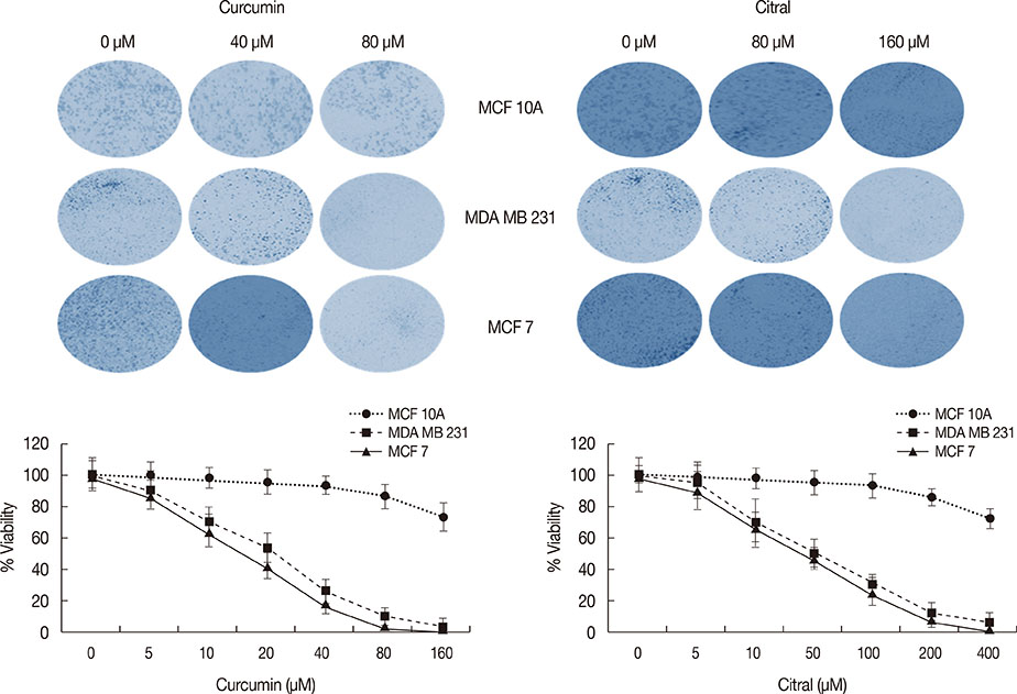
Figure 2 Clonogenic survival of breast cancer (MCF 7 and MDA MB 231) and breast epithelial (MCF 10A) cell lines treated with various concentrations of curcumin and citral for 24 hours. Data were plotted as percent viability (% control). At a zero concentration of curcumin and citral, the % viability was considered 100%. The data represent the mean±standard deviation of one of the three similar experiments each performed in triplicate.
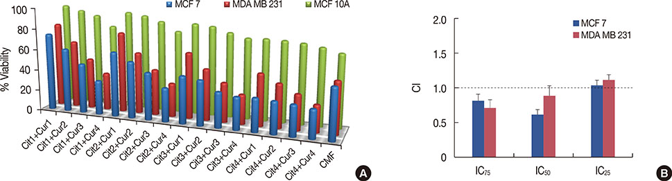
Figure 3 Combined effect of curcumin (Cur) and citral (Cit) on the survival of MCF 7 and MDA MB 231 cells. (A) The concentration used of Cur1, Cur2, Cur3, and Cur4 was 10, 20, 40, and 80 µM, respectively. Whereas, the concentration used of Cit1, Cit2, Cit3, and Cit4 was 20, 40, 80, and 160 µM, respectively. The concentration of cyclophosphamide, methotrexate, and 5-fluorouracil (CMF) was 3.5, 0.75, and 1.5 mM, respectively. Data were plotted as percent viability (% control). At a zero concentration of drugs, curcumin and citral, the % viability was considered 100%. The data represent the mean±standard deviation of one of the three similar experiments each performed in triplicate. (B) Combination Index (CI) of curcumin and citral. The graph shows the mean values of the combination index at the affected fractions of 25.0% (IC25), 50.0% (IC50), and 75.0% (IC75), when curcumin and citral were used in combination in MCF 7 and MDA MB 231 cells. A CI value less than 1 indicates synergism, a CI not different from 1 indicates an addition, and a CI higher than 1 indicates antagonism. The data represent the mean±standard deviation of one of the three similar experiments each performed in triplicate.
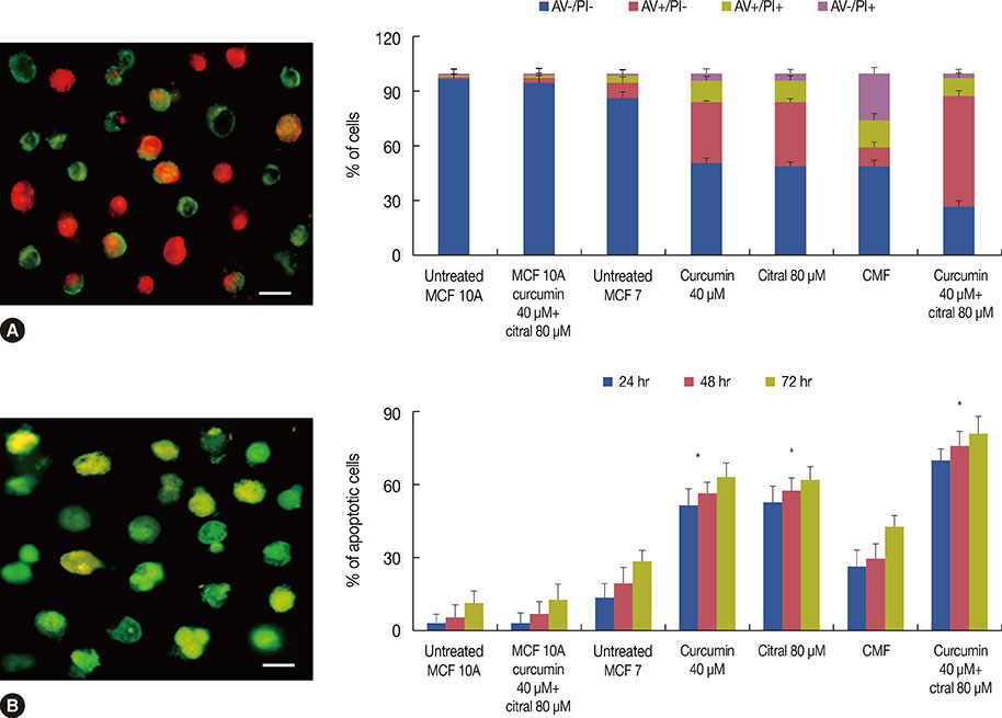
Figure 4 Induction of apoptosis in MCF 7 cells by curcumin and citral treatment. (A) Annexin V-fluorescein isothiocyanate/propidium iodide (PI) dual staining of the MCF 7 cells exposed to cyclophosphamide, methotrexate, and 5-fluorouracil (CMF), curcumin or citral and to the combination of curcumin and citral. Graph represents the distribution of apoptotic cells based on annexin V/PI staining. The apoptotic cells are stained with annexin V (green fluorescence), and the necrotic cells are stained with PI (red fluorescence). The image showed the stained cells treated with curcuminn (40 µM) and citral (80 µM). Scale bar, 20 µm. (B) TUNEL assay. Apoptosis detection in MCF 7 cells treated with CMF, curcumin or citral and the combination of curcumin and citral for 24, 48, and 72 hours. Cells were observed under microscope and % apoptotic cells were counted out of total 100 cells. The image showed the stained cells treated with curcumin (40 µM) and citral (80 µM). Scale bar, 10 µm. The data represent the means of three independent experiments performed in triplicate, with standard deviations represented by vertical bars. *p< 0.05.

Figure 5 Measurement of DNA damage in MCF 7 cells caused by curcumin and citral treatment. (A) The DNA damage in MCF 7 cells exposed to CMF, curcumin or citral and the combination of curcumin and citral were assessed by COMET assay. A1, untreated MCF 10 A cells; A2, untreated MCF 7 cells; A3, CMF treated MCF 7 cells, and A4, curcumun (40 µM) and citral (80 µM) treated MCF 7 cells. (B) Graph showed the measure of COMET parameters. The data represent the means of three independent experiments performed in triplicate.
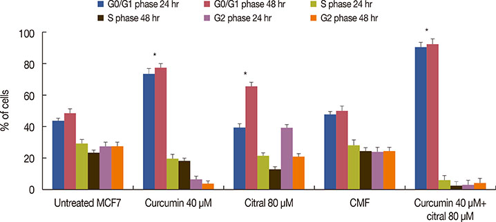
Figure 6 Distribution of MCF 7 cells in various phases of cell cycle; exposed to cyclophosphamide, methotrexate, and 5-fluorouracil (CMF), curcumin or citral and the combination of curcumin and citral; analyzed by flow cytometry. The data represent the means of three independent experiments performed in triplicate, with standard deviations represented by vertical bars. *p<0.05.
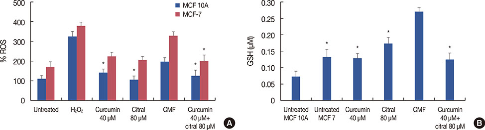
Figure 7 Intracellular reactive oxygen species (ROS) generation and total glutathione (GSH) content. (A) Effect of curcumin and citral on intracellular ROS. The graph shows the generation of ROS in MCF7 and MCF 10A cells, treated with cyclophosphamide, methotrexate, and 5-fluorouracil (CMF), curcumin or citral and the combination of curcumin and citral. The data represent the means of three independent experiments performed in triplicate, with standard deviations represented by vertical bars. (B) Effect of curcumin and citral on the total GSH content. The total GSH content of MCF 7 cells exposed to cyclophosphamide, methotrexate, and 5-fluorouracil (CMF), curcumin or citral and the combination of curcumin and citral was measured. Values in graph represent the total GSH content in µM. The data represent the means of three independent experiments performed in triplicate, with standard deviations represented by vertical bars. *p<0.05.

Figure 8 Measurement of levels of apoptotic proteins. The apoptotic protein levels in the MCF 7cells treated with cyclophosphamide, methotrexate, and 5-fluorouracil (CMF), curcumin or citral and the combination of curcumin and citral in MCF 7 and untreated MCF 10A cells were measured using the Sandwich Enzyme-Linked Immunosorbent Assay kit. Values of all protein represented their absorbance at 450 nm. The data represent the means of three independent experiments performed in triplicate, with standard deviations represented by vertical bars. *p<0.05.
Reference
-
1. Pan MH, Ho CT. Chemopreventive effects of natural dietary compounds on cancer development. Chem Soc Rev. 2008; 37:2558–2574.
Article2. Lee SJ, Krauthauser C, Maduskuie V, Fawcett PT, Olson JM, Rajasekaran SA. Curcumin-induced HDAC inhibition and attenuation of medulloblastoma growth in vitro and in vivo. BMC Cancer. 2011; 11:144.
Article3. Ravindran J, Prasad S, Aggarwal BB. Curcumin and cancer cells: how many ways can curry kill tumor cells selectively? AAPS J. 2009; 11:495–510.
Article4. Ramachandran C, You W. Differential sensitivity of human mammary epithelial and breast carcinoma cell lines to curcumin. Breast Cancer Res Treat. 1999; 54:269–278.
Article5. Shah G, Shri R, Panchal V, Sharma N, Singh B, Mann AS. Scientific basis for the therapeutic use of Cymbopogon citratus, stapf (Lemon grass). J Adv Pharm Technol Res. 2011; 2:3–8.
Article6. Rabbani SI, Devi K, Shivananda TN. Studies on antimutagenic effects of citral in mice. J Food Agric Environ. 2004; 2:62–64.7. Ozben T. Oxidative stress and apoptosis: impact on cancer therapy. J Pharm Sci. 2007; 96:2181–2196.
Article8. Lau AT, Wang Y, Chiu JF. Reactive oxygen species: current knowledge and applications in cancer research and therapeutic. J Cell Biochem. 2008; 104:657–667.
Article9. Bhaumik S, Anjum R, Rangaraj N, Pardhasaradhi BV, Khar A. Curcumin mediated apoptosis in AK-5 tumor cells involves the production of reactive oxygen intermediates. FEBS Lett. 1999; 456:311–314.
Article10. Somasundaram S, Edmund NA, Moore DT, Small GW, Shi YY, Orlowski RZ. Dietary curcumin inhibits chemotherapy-induced apoptosis in models of human breast cancer. Cancer Res. 2002; 62:3868–3875.11. Dudai N, Weinstein Y, Krup M, Rabinski T, Ofir R. Citral is a new inducer of caspase-3 in tumor cell lines. Planta Med. 2005; 71:484–488.
Article12. Xia H, Liang W, Song Q, Chen X, Chen X, Hong J. The in vitro study of apoptosis in NB4 cell induced by citral. Cytotechnology. 2013; 65:49–57.
Article13. Zhang T, Zhang Q, Chen D, Jiang J, Zhou Q. Growth inhibition of human breast cancer cell line MDA-MB-231 by rosiglitazone through activation of PPARgamma. Chin J Clin Oncol. 2008; 5:407–412.
Article14. Reynolds CP, Maurer BJ. Evaluating response to antineoplastic drug combinations in tissue culture models. Methods Mol Med. 2005; 110:173–183.
Article15. Tice RR, Agurell E, Anderson D, Burlinson B, Hartmann A, Kobayashi H, et al. Single cell gel/comet assay: guidelines for in vitro and in vivo genetic toxicology testing. Environ Mol Mutagen. 2000; 35:206–221.
Article16. Sies H, Akerboom TP. Glutathione disulfide (GSSG) efflux from cells and tissues. Methods Enzymol. 1984; 105:445–451.17. Singh RP, Dhanalakshmi S, Agarwal R. Phytochemicals as cell cycle modulators: a less toxic approach in halting human cancers. Cell Cycle. 2002; 1:156–161.
Article18. Chaouki W, Leger DY, Liagre B, Beneytout JL, Hmamouchi M. Citral inhibits cell proliferation and induces apoptosis and cell cycle arrest in MCF-7 cells. Fundam Clin Pharmacol. 2009; 23:549–556.
Article19. Shen L, Ji HF. Theoretical study on physicochemical properties of curcumin. Spectrochim Acta A Mol Biomol Spectrosc. 2007; 67:619–623.
Article20. Liu Q, Loo WT, Sze SC, Tong Y. Curcumin inhibits cell proliferation of MDA-MB-231 and BT-483 breast cancer cells mediated by down-regulation of NFkappaB, cyclinD and MMP-1 transcription. Phytomedicine. 2009; 16:916–922.
Article21. Syng-Ai C, Kumari AL, Khar A. Effect of curcumin on normal and tumor cells: role of glutathione and bcl-2. Mol Cancer Ther. 2004; 3:1101–1108.22. Fang HY, Chen SB, Guo DJ, Pan SY, Yu ZL. Proteomic identification of differentially expressed proteins in curcumin-treated MCF-7 cells. Phytomedicine. 2011; 18:697–703.
Article23. Shyur LF, Lee SH, Chang ST, Lo CP, Kuo YH, Wang SY. Taiwanin A inhibits MCF-7 cancer cell activity through induction of oxidative stress, upregulation of DNA damage checkpoint kinases, and activation of p53 and FasL/Fas signaling pathways. Phytomedicine. 2010; 18:16–24.
Article24. Ataee R, Ataie A, Shadifar M, Nasri N, Hagghi H, Hayati E. Synergic effect of curcumin and melatonin on proliferation and apoptosis of HT29 colorectal cancer cell line. Res Pharm Sci. 2012; 7:S117.25. Li H, Jin L, Wu F, Li X, You J, Cao Z, et al. Effect of curcumin on proliferation, cell cycle, and caspases and MCF-7 cells. Afr J Pharm Pharmacol. 2012; 6:864–870.26. Toyokuni S, Okamoto K, Yodoi J, Hiai H. Persistent oxidative stress in cancer. FEBS Lett. 1995; 358:1–3.
Article
- Full Text Links
-
- Actions
-
Cited
- CITED
-
- Close
- Share
-
- Similar articles
-
- Kaempferol induced the apoptosis via cell cycle arrest in human breast cancer MDA-MB-453 cells
- Curcumin Induces Apoptosis and Inhibits Metalloproteinase Activity in Renal Cancer Cell Line
- Mechanism Underlying Curcumin-induced Apoptosis and Cell Cycle Arrest on SCC25 Human Tongue Squamous Cell Carcinoma Cell Line
- Growth inhibition by fusidic acid in cervical, thyroid, and breast carcinoma cell lines
- Kaempferol Synergistically Enhances Cisplatin-induced Apoptosis and Cell Cycle Arrest in Colon Cancer Cells
