Removal efficacy and cytotoxicity of a calcium hydroxide paste using N-2-methyl-pyrrolidone as a vehicle
- Affiliations
-
- 1Department of Conservative Dentistry, Chonbuk National University School of Dentistry and Institute of Oral Bioscience, Jeonju, Korea. mksdd@jbnu.ac.kr
- 2Department of Life Science, College of Natural Sciences, Chonbuk National University, Jeonju, Korea.
- 3Research Institute of Clinical Medicine of Chonbuk National University, Jeonju, Korea.
- 4Biomedical Research Institute of Chonbuk National University Hospital, Jeonju, Korea.
- KMID: 2403070
- DOI: http://doi.org/10.5395/rde.2017.42.4.290
Abstract
OBJECTIVES
This study investigated the removal efficacy and cytotoxicity of a newly developed calcium hydroxide paste (cleaniCal, Maruchi) using N-2-methyl-pyrrolidone (NMP) as a vehicle in comparison with ApexCal (Ivoclar Vivadent) and Calcipex II (Nishika), which use different vehicles such as polyethylene glycol and propylene glycol, respectively.
MATERIALS AND METHODS
Thirty maxillary premolars with oval-shaped canals were divided into 3 groups and the teeth were filled with one of the pastes. After removal of the paste, micro-computed tomographic (μ-CT) imaging was obtained to assess the volume of residual paste in the root canal of each tooth. The teeth were then split longitudinally and the area of the paste-coated surface was evaluated by stereomicroscopy. The cytotoxicity of each product was assessed using an agar overlay assay. The effect of each vehicle on cell viability was evaluated using the 3-(4,5-dimethylthiazol-2-yl)-2,5-diphenyltetrazolium bromide (MTT) assay. The data were analyzed using one-way analysis of variance and Tukey's tests to detect any significance (p < 0.05).
RESULTS
In the μ-CT and stereomicroscopic analysis, cleaniCal exhibited less remnants of medicament than ApexCal and Calcipex. cleaniCal showed a higher cytotoxicity than the other pastes in the agar overlay assay. Furthermore, NMP exhibited lower cell viability compared to the other vehicles.
CONCLUSIONS
cleaniCal showed better removal efficacy compared to the other products. However, clinicians should be aware of the higher cytotoxicity of the NMP-based material and consider its possible adverse effects on periradicular tissue when it is overfilled.
Keyword
MeSH Terms
Figure
-
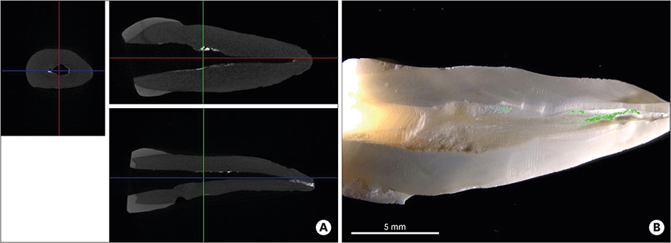
Figure 1 (A) Representative micro-computed tomographic (μ-CT) images show the radiopaque remnants of each material. (B) A stereomicroscopic image shows the coated surface (indicated as the green area) with the remaining material in a split root canal system.
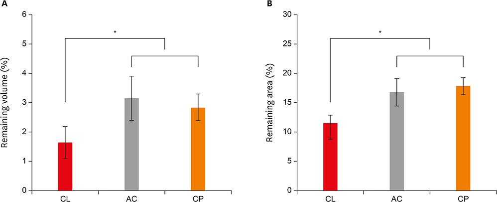
Figure 2 Removal efficacy and radiopacity of the tested materials. Mean percentage and standard deviation of the remaining volume (A) and remaining area (B) of the remnants in micro-computed tomographic (μ-CT) and stereomicroscopic assessments, respectively. *A significant difference was determined at p < 0.05. CL, cleaniCal; AC, ApexCal; CP, Calcipex II.
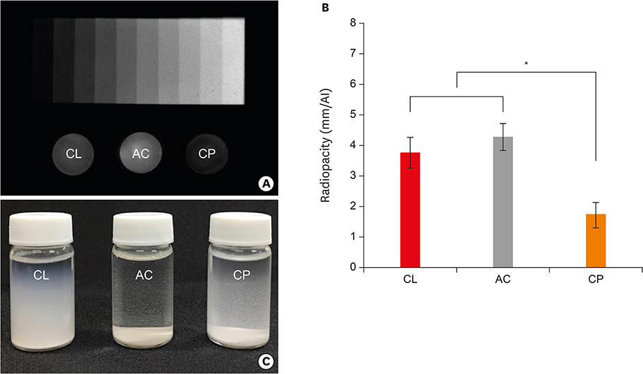
Figure 3 (A) A radiograph showing the radiopacity of each material and its equivalence to that of the aluminum step wedge. (B) Relative radiographic density of each material in comparison with that of a 10-step aluminum step wedge. (C) An image showing the turbidity of the suspension in which the pastes were dissolved. Note that white precipitate formed after sedimentation. *A significant difference was determined at p < 0.05. CL, cleaniCal; AC, ApexCal; CP, Calcipex II.
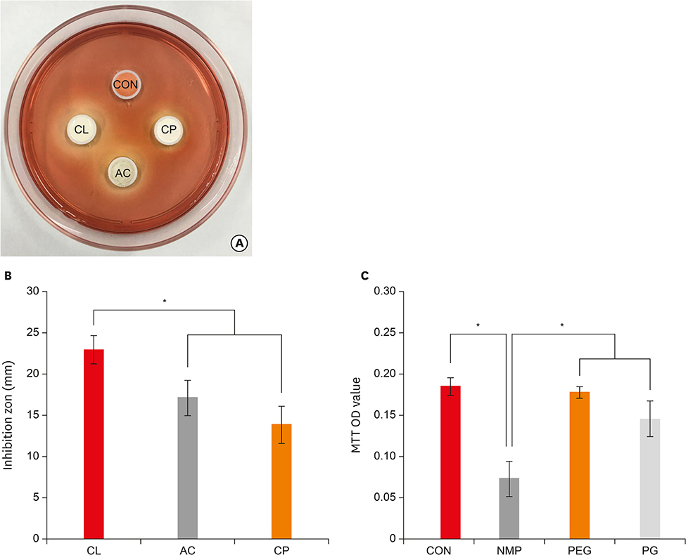
Figure 4 (A) A representative image showing the inhibition zone of each material in the agar overlay assay. (B) Mean and standard deviation of the inhibition zone of each material. (C) Cell viability of the vehicle for each product. *A significant difference was determined at p < 0.05. CON, control; CL, cleaniCal; AC, ApexCal; CP, Calcipex II; NMP, N-2-methyl-pyrrolidone; PEG, polyethylene glycol; PG, propylene glycol.
Reference
-
1. Bystrom A, Claesson R, Sundqvist G. The antibacterial effect of camphorated paramonochlorophenol, camphorated phenol and calcium hydroxide in the treatment of infected root canals. Endod Dent Traumatol. 1985; 1:170–175.
Article2. Calt S, Serper A. Dentinal tubule penetration of root canal sealers after root canal dressing with calcium hydroxide. J Endod. 1999; 25:431–433.
Article3. Hosoya N, Kurayama H, Iino F, Arai T. Effects of calcium hydroxide on physical and sealing properties of canal sealers. Int Endod J. 2004; 37:178–184.
Article4. Guiotti FA, Kuga MC, Duarte MA, Sant'Anna AJ, Faria G. Effect of calcium hydroxide dressing on push-out bond strength of endodontic sealers to root canal dentin. Braz Oral Res. 2014; 28:1–6.
Article5. Grover C, Shetty N. Evaluation of calcium ion release and change in pH on combining calcium hydroxide with different vehicles. Contemp Clin Dent. 2014; 5:434–439.
Article6. Gomes BP, Ferraz CC, Garrido FD, Rosalen PL, Zaia AA, Teixeira FB, de Souza-Filho FJ. Microbial susceptibility to calcium hydroxide pastes and their vehicles. J Endod. 2002; 28:758–761.
Article7. Simi J Junior, Machado R, Souza CJ, Loyola AM, Vansan LP, Antoniazzi JH. Histological analysis of the biocompatibility of calcium hydroxide associated with a new vehicle. Indian J Dent Res. 2015; 26:627–632.
Article8. Simon ST, Bhat KS, Francis R. Effect of four vehicles on the pH of calcium hydroxide and the release of calcium ion. Oral Surg Oral Med Oral Pathol Oral Radiol Endod. 1995; 80:459–464.
Article9. Camargo CH, Bernardineli N, Valera MC, de Carvalho CA, de Oliveira LD, Menezes MM, Afonso SE, Mancini MN. Vehicle influence on calcium hydroxide pastes diffusion in human and bovine teeth. Dent Traumatol. 2006; 22:302–306.
Article10. Deonizio MD, da Silva WJ, Batista A, Sydney GB, do Nascimento FC, Goncalves LM, Rosa ER, Gabardo MC. Efficacy of calcium hydroxide paste prepared with different vehicles against salivary microbial infiltration of root canals. Gen Dent. 2014; 62:e22–e25.11. Cruz EV, Kota K, Huque J, Iwaku M, Hoshino E. Penetration of propylene glycol into dentine. Int Endod J. 2002; 35:330–336.
Article12. Camões IC, Salles MR, Chevitarese O, Gomes GC. Influence on pH of vehicle containing glycerin used with calcium hydroxide. Dent Traumatol. 2003; 19:132–138.
Article13. Balvedi RP, Versiani MA, Manna FF, Biffi JC. A comparison of two techniques for the removal of calcium hydroxide from root canals. Int Endod J. 2010; 43:763–768.
Article14. Lloyd A, Navarrete G, Marchesan MA, Clement D. Removal of calcium hydroxide from Weine Type II systems using photon-induced photoacoustic streaming, passive ultrasonic, and needle irrigation: a microcomputed tomography study. J Appl Oral Sci. 2016; 24:543–548.
Article15. da Silva JM, Andrade CV Junior, Zaia AA, Pessoa OF. Microscopic cleanliness evaluation of the apical root canal after using calcium hydroxide mixed with chlorhexidine, propylene glycol, or antibiotic paste. Oral Surg Oral Med Oral Pathol Oral Radiol Endod. 2011; 111:260–264.
Article16. Hamishehkar H, Khoshbakht M, Jouyban A, Ghanbarzadeh S. The relationship between solubility and transdermal absorption of Tadalafil. Adv Pharm Bull. 2015; 5:411–417.
Article17. Kenee DM, Allemang JD, Johnson JD, Hellstein J, Nichol BK. A quantitative assessment of efficacy of various calcium hydroxide removal techniques. J Endod. 2006; 32:563–565.
Article18. Sanghvi R, Narazaki R, Machatha SG, Yalkowsky SH. Solubility improvement of drugs using N-methyl pyrrolidone. AAPS PharmSciTech. 2008; 9:366–376.
Article19. Jain P, Yalkowsky SH. Solubilization of poorly soluble compounds using 2-pyrrolidone. Int J Pharm. 2007; 342:1–5.
Article20. Li Q, Liu R, Zhao J, Lu Q. N-methyl pyrrolidone (NMP) ameliorates the hypoxia-reduced osteoblast differentiation via inhibiting the NF-κB signaling. J Toxicol Sci. 2016; 41:701–709.
Article21. Aboudzadeh N, Imani M, Shokrgozar MA, Khavandi A, Javadpour J, Shafieyan Y, Farokhi M. Fabrication and characterization of poly(D,L-lactide-co-glycolide)/hydroxyapatite nanocomposite scaffolds for bone tissue regeneration. J Biomed Mater Res A. 2010; 94:137–145.
Article22. Sasaki H, Kojima M, Mori Y, Nakamura J, Shibasaki J. Enhancing effect of pyrrolidone derivatives on transdermal penetration of 5-fluorouracil, triamcinolone acetonide, indomethacin, and flurbiprofen. J Pharm Sci. 1991; 80:533–538.
Article23. Agrawal AG, Kumar A, Gide PS. Toxicity Study of a Self-nanoemulsifying Drug Delivery System Containing N-methyl pyrrolidone. Drug Res (Stuttg). 2015; 65:446–448.
Article24. Gjoksi B, Ruangsawasdi N, Ghayor C, Siegenthaler B, Zenobi-Wong M, Weber FE. Influence of N-methyl pyrrolidone on the activity of the pulp-dentine complex and bone integrity during osteoporosis. Int Endod J. 2017; 50:271–280.
Article25. Karfeld-Sulzer LS, Ghayor C, Siegenthaler B, de Wild M, Leroux JC, Weber FE. N-methyl pyrrolidone/bone morphogenetic protein-2 double delivery with in situ forming implants. J Control Release. 2015; 203:181–188.
Article26. Leira HL, Tiltnes A, Svendsen K, Vetlesen L. Irritant cutaneous reactions to N-methyl-2-pyrrolidone (NMP). Contact Dermat. 1992; 27:148–150.
Article27. Flick B, Talsness CE, Jäckh R, Buesen R, Klug S. Embryotoxic potential of N-methyl-pyrrolidone (NMP) and three of its metabolites using the rat whole embryo culture system. Toxicol Appl Pharmacol. 2009; 237:154–167.
Article28. Kim JW, Cho KM, Park SH, Song SG, Park MS, Jung HR, Song JY, Kim YS, Lee SK. Overfilling of calcium hydroxide-based paste Calcipex II produced a foreign body granuloma without acute inflammatory reaction. Oral Surg Oral Med Oral Pathol Oral Radiol Endod. 2009; 107:e73–e76.
Article29. Kim JW, Cho KM, Park SH, Park SR, Lee SS, Lee SK. Chronic maxillary sinusitis caused by root canal overfilling of Calcipex II. Restor Dent Endod. 2014; 39:63–67.
Article30. Asheibi F, Qualtrough AJ, Mellor A, Withers PJ, Lowe T. Micro-CT evaluation of the effectiveness of the combined use of rotary and hand instrumentation in removal of Resilon. Dent Mater J. 2014; 33:1–6.
Article31. Wiseman A, Cox TC, Paranjpe A, Flake NM, Cohenca N, Johnson JD. Efficacy of sonic and ultrasonic activation for removal of calcium hydroxide from mesial canals of mandibular molars: a microtomographic study. J Endod. 2011; 37:235–238.
Article32. Silva LJ, Pessoa OF, Teixeira MB, Gouveia CH, Braga RR. Micro-CT evaluation of calcium hydroxide removal through passive ultrasonic irrigation associated with or without an additional instrument. Int Endod J. 2015; 48:768–773.
Article33. Ma JZ, Shen Y, Al-Ashaw AJ, Khaleel HY, Yang Y, Wang ZJ, Peng B, Haapasalo M. Micro-computed tomography evaluation of the removal of calcium hydroxide medicament from C-shaped root canals of mandibular second molars. Int Endod J. 2015; 48:333–341.
Article
- Full Text Links
-
- Actions
-
Cited
- CITED
-
- Close
- Share
-
- Similar articles
-
- Effect of two different calcium hydroxide paste removal techniques on apical leakage: an electrochemical study
- The significance of diagnosis and treatment planning in periapical lesion overfilled with calcium hydroxide paste
- Effect of calcium hydroxide application time on dentin
- A Comparison of the irrigation systems in calcium hydroxide removal
- A study of ionic dissociation on various calcium hydroxide pastes using molecular sieving model
