J Dent Rehabil Appl Sci.
2021 Jun;37(2):95-100. 10.14368/jdras.2021.37.2.95.
The significance of diagnosis and treatment planning in periapical lesion overfilled with calcium hydroxide paste
- Affiliations
-
- 1Dental Clinic Center, Pusan National University Hospital, Busan, Republic of Korea
- 2Department of Conservative Dentistry, School of Dentistry, Pusan National University, Yangsan, Republic of Korea
- KMID: 2525983
- DOI: http://doi.org/10.14368/jdras.2021.37.2.95
Abstract
- Calcium hydroxide has been widely used for root canal dressing material in endodontic treatment. This report describes that when the accurate diagnosis and proper nonsurgical endodontic retreatment is applied to periapical lesion with accidentally extruded calcium hydroxide paste, the lesion can be successfully treated. Overfilled calcium hydroxide can affect the healing process, so the overextension of calcium hydroxide agent should be avoided.
Keyword
Figure
-

Fig. 1 (A) Preoperative radiograph of mandibular left second premolar, (B) Photograph of dens evaginatus (arrow) on mandibular right second premolar, (C) Photograph showing a discharge of pus through pulp chamber of mandibular left second premolar.

Fig. 2 Radiographs during endodontic treatment. (A) Preoperative radiograph, (B) Radiograph of the first visit. (C) Radiograph of the second visit, (D) Radiograph of the third visit, (E) Radiograph of the fourth visit. The canal filling was done.
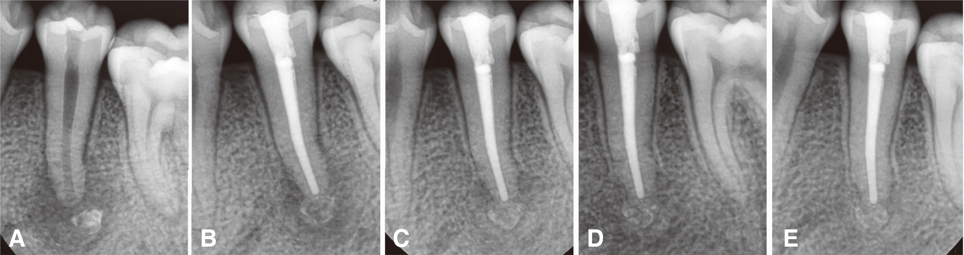
Fig. 3 Radiographs. (A) Preoperative radiograph, (B) 6 months follow up radiograph, (C) 12 months follow up radiograph, (D) 18 months follow up radiograph, (E) 24 months follow up radiograph.
Reference
-
References
1. Kakehashi S, Stanley HR, Fitzgerald RJ. 1965; The effects of surgical exposures of dental pulps in germ-free and conventional laboratory rats. Oral Surg Oral Med Oral Pathol. 20:340–9. DOI: 10.1016/0030-4220(65)90166-0. PMID: 14342926.2. Kim JW, Cho KM, Park SH, Song SG, Park MS, Jung HR, Song JY, Kim YS, Lee SK. 2009; Overfilling of calcium hydroxide based paste Calcipex II produced a foreign body granuloma without acute inflammatory reaction. Oral Surg Oral Med Oral Pathol Oral Radiol Endod. 107:e73–6. DOI: 10.1016/j.tripleo.2008.10.019. PMID: 19168376.3. Hosoya N, Kurayama H, Iino F, Arai T. 2004; Effects of calcium hydroxide on physical and sealing properties of canal sealers. Int Endod J. 37:178–84. DOI: 10.1111/j.0143-2885.2004.00781.x. PMID: 15009407.4. Kim JW, Cho KM, Park SH, Park SR, Lee SS, Lee SK. 2014; Chronic maxillary sinusitis caused by root canal overfilling of Calcipex II. Restor Dent Endod. 39:63–7. DOI: 10.5395/rde.2014.39.1.63. PMID: 24516832. PMCID: PMC3916508.5. Kim SM, Yoo YJ, Lee SK. 2016; Birefringence detection of Calcipex II endodontic material in chronic granulomatous lesion of periapical cyst. Korean J Oral Maxillofac Pathol. 40:775–9. DOI: 10.17779/KAOMP.2016.40.2.005.6. Shin Y, Roh BD, Kim Y, Kim T, Kim H. 2016; Accidental injury of the inferior alveolar nerve due to the extrusion of calcium hydroxide in endodontic treatment: a case report. Restor Dent Endod. 41:63–7. DOI: 10.5395/rde.2016.41.1.63. PMID: 26877992. PMCID: PMC4751209.7. Levitan ME, Himel VT. 2006; Dens evaginatus: literature review, pathophysiology, and comprehensive treatment regimen. J Endod. 32:1–9. DOI: 10.1016/j.joen.2005.10.009. PMID: 16410059.8. Hill FJ, Bellis WJ. 1984; Dens evaginatus and its management. Br Dent J. 156:400–2. DOI: 10.1038/sj.bdj.4805383. PMID: 6587891.9. Chen JW, Huang GT, Bakland LK. 2020; Dens evaginatus: current treatment options. J Am Dent Assoc. 151:358–67. DOI: 10.1016/j.adaj.2020.01.015. PMID: 32209245.10. Stecker S, DiAngelis AJ. 2002; Dens evaginatus: a diagnostic and treatment challenge. J Am Dent Assoc. 133:190–3. DOI: 10.14219/jada.archive.2002.0143. PMID: 11868837.11. Orucoglu H, Cobankara FK. 2008; Effect of unintentionally extruded calcium hydroxide paste including barium sulfate as a radiopaquing agent in treatment of teeth with periapical lesions: report of a case. J Endod. 34:888–91. DOI: 10.1016/j.joen.2008.04.012. PMID: 18571001.12. Song M, Kim HC, Lee W, Kim E. 2011; Analysis of the cause of failure in nonsurgical endodontic treatment by microscopic inspection during endodontic microsurgery. J Endod. 37:1516–9. DOI: 10.1016/j.joen.2011.06.032. PMID: 22000454.13. Tabassum S, Khan FR. 2016; Failure of endodontic treatment: The usual suspects. Eur J Dent. 10:144–7. DOI: 10.4103/1305-7456.175682. PMID: 27011754. PMCID: PMC4784145.14. Lin LM, Skribner JE, Gaengler P. 1992; Factors associated with endodontic treatment failures. J Endod. 18:625–7. DOI: 10.1016/S0099-2399(06)81335-X. PMID: 1298804.15. Siqueira JF Jr. 2001; Aetiology of root canal treatment failure: why well-treated teeth can fail. Int Endod J. 34:1–10. DOI: 10.1046/j.1365-2591.2001.00396.x. PMID: 11307374.16. Torabinejad M, Corr R, Handysides R, Shabahang S. 2009; Outcomes of nonsurgical retreatment and endodontic surgery: a systematic review. J Endod. 35:930–7. DOI: 10.1016/j.joen.2009.04.023. PMID: 19567310.17. Alaçam T, Görgül G, Omürlü H. 1990; Evaluation of diagnostic radiopaque contrast materials used with calcium hydroxide. J Endod. 16:365–8. DOI: 10.1016/S0099-2399(06)81907-2. PMID: 2081953.18. Vernieks AA, Messer LB. 1978; Calcium hydroxide induced healing of periapical lesions: a study of 78 non-vital teeth. J Br Endod Soc. 11:61–9. DOI: 10.1111/j.1365-2591.1978.tb00663.x. PMID: 294435.
- Full Text Links
-
- Actions
-
Cited
- CITED
-
- Close
- Share
-
- Similar articles
-
- Nerve Injury from Overfilled Calcium Hydroxide Root Canal Filling Paste for Maxillary Lateral Incisor Endodontic Treatment
- Removal efficacy and cytotoxicity of a calcium hydroxide paste using N-2-methyl-pyrrolidone as a vehicle
- Effect of two different calcium hydroxide paste removal techniques on apical leakage: an electrochemical study
- Effect of calcium hydroxide application time on dentin
- A study of ionic dissociation on various calcium hydroxide pastes using molecular sieving model
