Caffeic acid phenethyl ester protects against photothrombotic cortical ischemic injury in mice
- Affiliations
-
- 1Department of Pharmacology, Pusan National University School of Medicine, Yangsan 50612, Korea. wonslee@pusan.ac.kr
- KMID: 2398560
- DOI: http://doi.org/10.4196/kjpp.2018.22.1.101
Abstract
- In this study, we aimed to investigate the neuroprotective effects of caffeic acid phenethyl ester (CAPE), an active component of propolis purified from honeybee hives, on photothrombotic cortical ischemic injury in mice. Permanent focal ischemia was achieved in the medial frontal and somatosensory cortices of anesthetized male C57BL/6 mice by irradiation of the skull with cold light laser in combination with systemic administration of rose bengal. The animals were treated with CAPE (0.5-5 mg/kg, i.p.) twice 1 and 6 h after ischemic insult. CAPE significantly reduced the infarct size as well as the expression of tumor necrosis factor-α, hypoxiainducible factor-1α, monocyte chemoattractant protein-1, interleukin-1α, and indoleamine 2,3-dioxygenase in the cerebral cortex ipsilateral to the photothrombosis. Moreover, it induced an increase in heme oxygenase-1 immunoreactivity and interleukin-10 expression. These results suggest that CAPE exerts a remarkable neuroprotective effect on ischemic brain injury via its anti-inflammatory properties, thereby providing a benefit to the therapy of cerebral infarction.
MeSH Terms
-
Animals
Brain Injuries
Brain Ischemia
Cerebral Cortex
Cerebral Infarction
Chemokine CCL2
Heme Oxygenase-1
Humans
Indoleamine-Pyrrole 2,3,-Dioxygenase
Interleukin-10
Ischemia
Male
Mice*
Necrosis
Neuroprotective Agents
Propolis
Rose Bengal
Skull
Urticaria
Chemokine CCL2
Heme Oxygenase-1
Indoleamine-Pyrrole 2,3,-Dioxygenase
Interleukin-10
Neuroprotective Agents
Propolis
Rose Bengal
Figure
-
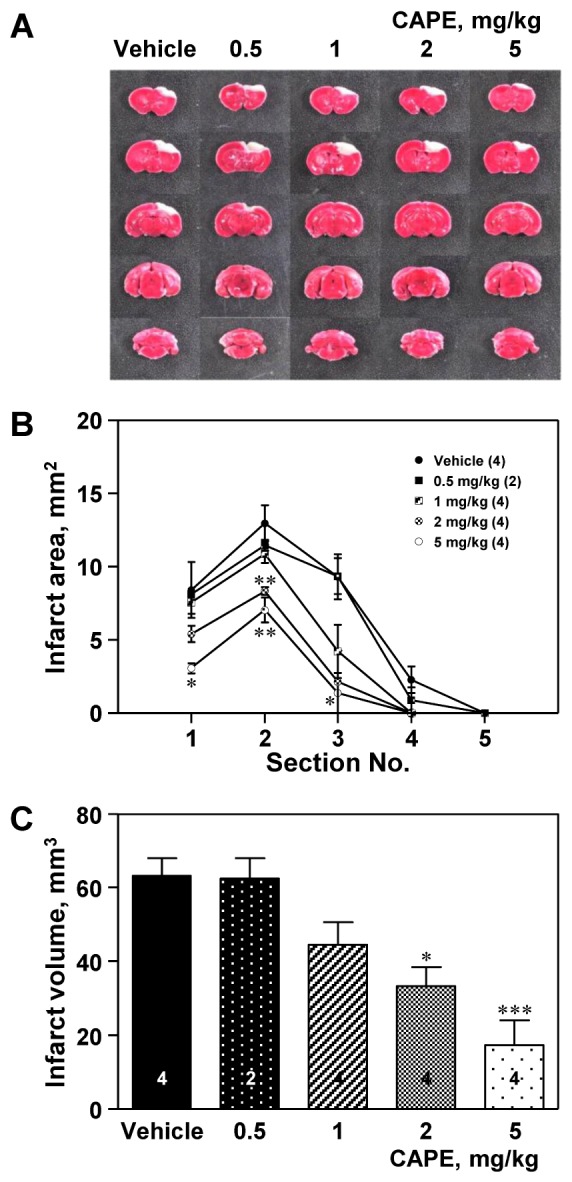
Fig. 1 Effect of caffeic acid phenethyl ester (CAPE) on infarct size.(A) Representative photographs showing the ischemic lesion. Male C57BL/6 mice were treated with CAPE (0.5–5 mg/kg, i.p.) twice 1 and 6 h after ischemic insult, and were sacrificed 24 h after photothrombotic cortical ischemia. The pale non-stained area indicates the lesion of ischemic injury, while the deep red-colored area indicates the viable tissue. CAPE elicited the reduction in the infarct area (B) and infarct volume (C). The numbers in parentheses and columns indicate the numbers of animals. Data are presented as mean±SEM. *p<0.05; **p<0.01; ***p<0.001 vs. vehicle group.
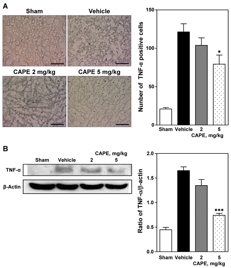
Fig. 2 Immunohistochemical and Western blot analyses of expression of tumor necrosis factor-α (TNF-α).(A) Representative photomicrographs showing TNF-α positive cells (left panel) and number of TNF-α positive cells (right panel) in the cortical ischemic core region 24 h after photothrombosis. Treatment with CAPE 5 mg/kg significantly reduced the immunohistochemical staining of TNF-α positive cells compared to the vehicle group. Five animals were used in each group. Scale bar=100 µm. (B) Western blot analysis revealed that TNF-α expression was reduced by CAPE in a dose-dependent manner. Data are presented as mean±SEM. *p<0.05; ***p<0.001 vs. vehicle group.
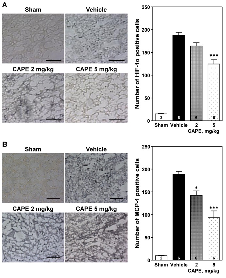
Fig. 3 Immunohistochemical analysis of expression of hypoxia-inducible factor-1α (HIF-1α) and monocyte chemoattractant protein-1 (MCP-1).Representative photomicrographs showing HIF-1α and MCP-1 positive cells (left panels of A and B, respectively) and number of HIF-1α and MCP-1 positive cells (right panel panels of A and B, respectively) in the cortical ischemic core region 24 h after photothrombosis. Treatment with CAPE dose-dependently reduced the immunohistochemical staining of HIF-1α and MCP-1 positive cells compared to the vehicle group. Scale bar=100 µm. The numbers in columns indicate the numbers of animals. Data are presented as mean±SEM. *p<0.05; ***p<0.001 vs. vehicle group.
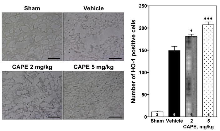
Fig. 4 Immunohistochemical analysis of expression of heme oxygenase-1 (HO-1).Representative photomicrographs showing HO-1 positive cells (left panel) and number of HO-1 positive cells (right panel) in the cortical ischemic core region 24 h after photothrombosis. CAPE dose-dependently increased the number of HO-1 positive cells compared to the vehicle group. Scale bar=100 µm. The numbers in columns indicate the numbers of animals. Data are presented as mean±SEM. *p<0.05; ***p<0.001 vs. vehicle group.
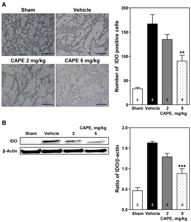
Fig. 5 Immunohistochemical and Western blot analyses of expression of indoleamine 2,3-dioxygenase (IDO).(A) Representative photomicrographs showing IDO positive cells (left panel) and number of IDO positive cells (right panel) in the cortical ischemic core region 24 h after photothrombosis. Treatment with CAPE 5 mg/kg significantly reduced the immunohistochemical staining of IDO positive cells compared to the vehicle group. Scale bar=100 µm. (B) Western blot analysis revealed that IDO expression was significantly reduced by CAPE 5 mg/kg. The numbers in columns indicate the numbers of animals. Data are presented as mean±SEM. **p<0.01; ***p<0.001 vs. vehicle group.
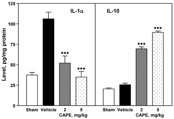
Fig. 6 Effect of CPAE on production of IL-1α and IL-10.The cerebral cortex ipsilateral to the photothrombosis was obtained 24 h after photothrombotic cortical ischemia, and was homogenized in lysis buffer. The supernatant of centrifuged sample was used for measuring the levels of IL-1α and IL-10 using ELISA (left and right panels, respectively). CAPE reduced the production of IL-1α (left panel) and enhanced production of IL-10 (right panel) in the ischemic region in a dose-dependent manner. Four animals were used in each group. ***p<0.001 vs. vehicle group.
Reference
-
1. GBD 2013 Mortality and Causes of Death Collaborators. Global, regional, and national age-sex specific all-cause and cause-specific mortality for 240 causes of death, 1990-2013: a systematic analysis for the Global Burden of Disease Study 2013. Lancet. 2015; 385:117–171. PMID: 25530442.2. Benjamin EJ, Blaha MJ, Chiuve SE, Cushman M, Das SR, Deo R, de Ferranti SD, Floyd J, Fornage M, Gillespie C, Isasi CR, Jiménez MC, Jordan LC, Judd SE, Lackland D, Lichtman JH, Lisabeth L, Liu S, Longenecker CT, Mackey RH, Matsushita K, Mozaffarian D, Mussolino ME, Nasir K, Neumar RW, Palaniappan L, Pandey DK, Thiagarajan RR, Reeves MJ, Ritchey M, Rodriguez CJ, Roth GA, Rosamond WD, Sasson C, Towfighi A, Tsao CW, Turner MB, Virani SS, Voeks JH, Willey JZ, Wilkins JT, Wu JH, Alger HM, Wong SS, Muntner P. American Heart Association Statistics Committee and Stroke Statistics Subcommittee. Heart disease and stroke statistics-2017 update: a report from the American Heart Association. Circulation. 2017; 135:e146–e603. PMID: 28122885.
Article3. National Institute of Neurological Disorders and Stroke rt-PA Stroke Study Group. Tissue plasminogen activator for acute ischemic stroke. N Engl J Med. 1995; 333:1581–1587. PMID: 7477192.4. Lambrinos A, Schaink AK, Dhalla I, Krings T, Casaubon LK, Sikich N, Lum C, Bharatha A, Pereira VM, Stotts G, Saposnik G, Kelloway L, Xie X, Hill MD. Mechanical thrombectomy in acute ischemic stroke: a systematic review. Can J Neurol Sci. 2016; 43:455–460. PMID: 27071728.
Article5. Denes A, Thornton P, Rothwell NJ, Allan SM. Inflammation and brain injury: acute cerebral ischaemia, peripheral and central inflammation. Brain Behav Immun. 2010; 24:708–723. PMID: 19770034.
Article6. Lakhan SE, Kirchgessner A, Hofer M. Inflammatory mechanisms in ischemic stroke: therapeutic approaches. J Transl Med. 2009; 7:97. PMID: 19919699.
Article7. Amantea D, Nappi G, Bernardi G, Bagetta G, Corasaniti MT. Postischemic brain damage: pathophysiology and role of inflammatory mediators. FEBS J. 2009; 276:13–26. PMID: 19087196.
Article8. Schilling M, Besselmann M, Leonhard C, Mueller M, Ringelstein EB, Kiefer R. Microglial activation precedes and predominates over macrophage infiltration in transient focal cerebral ischemia: a study in green fluorescent protein transgenic bone marrow chimeric mice. Exp Neurol. 2003; 183:25–33. PMID: 12957485.
Article9. Tanaka R, Komine-Kobayashi M, Mochizuki H, Yamada M, Furuya T, Migita M, Shimada T, Mizuno Y, Urabe T. Migration of enhanced green fluorescent protein expressing bone marrow-derived microglia/macrophage into the mouse brain following permanent focal ischemia. Neuroscience. 2003; 117:531–539. PMID: 12617960.
Article10. Gerhard A, Neumaier B, Elitok E, Glatting G, Ries V, Tomczak R, Ludolph AC, Reske SN. In vivo imaging of activated microglia using [11C]PK11195 and positron emission tomography in patients after ischemic stroke. Neuroreport. 2000; 11:2957–2960. PMID: 11006973.11. Price CJ, Menon DK, Peters AM, Ballinger JR, Barber RW, Balan KK, Lynch A, Xuereb JH, Fryer T, Guadagno JV, Warburton EA. Cerebral neutrophil recruitment, histology, and outcome in acute ischemic stroke: an imaging-based study. Stroke. 2004; 35:1659–1664. PMID: 15155970.12. Agata I1. Studies on caffeic acid derivatives in medicinal plants. Yakugaku Zasshi. 1999; 119:237–248. PMID: 10228448.
Article13. Sud'ina GF, Mirzoeva OK, Pushkareva MA, Korshunova GA, Sumbatyan NV, Varfolomeev SD. Caffeic acid phenethyl ester as a lipoxygenase inhibitor with antioxidant properties. FEBS Lett. 1993; 329:21–24. PMID: 7689063.14. Altuğ ME, Serarslan Y, Bal R, Kontaş T, Ekici F, Melek IM, Aslan H, Duman T. Caffeic acid phenethyl ester protects rabbit brains against permanent focal ischemia by antioxidant action: a biochemical and planimetric study. Brain Res. 2008; 1201:135–142. PMID: 18308295.
Article15. Ilhan A, Akyol O, Gurel A, Armutcu F, Iraz M, Oztas E. Protective effects of caffeic acid phenethyl ester against experimental allergic encephalomyelitis-induced oxidative stress in rats. Free Radic Biol Med. 2004; 37:386–394. PMID: 15223072.
Article16. Khan M, Elango C, Ansari MA, Singh I, Singh AK. Caffeic acid phenethyl ester reduces neurovascular inflammation and protects rat brain following transient focal cerebral ischemia. J Neurochem. 2007; 102:365–377. PMID: 17437550.
Article17. Park JH, Lee JK, Kim HS, Chung ST, Eom JH, Kim KA, Chung SJ, Paik SY, Oh HY. Immunomodulatory effect of caffeic acid phenethyl ester in Balb/c mice. Int Immunopharmacol. 2004; 4:429–436. PMID: 15037220.
Article18. Sforcin JM. Propolis and the immune system: a review. J Ethnopharmacol. 2007; 113:1–14. PMID: 17580109.
Article19. Shin TK, Kang MS, Lee HY, Seo MS, Kim SG, Kim CD, Lee WS. Fluoxetine and sertraline attenuate postischemic brain injury in mice. Korean J Physiol Pharmacol. 2009; 13:257–263. PMID: 19885045.
Article20. Bederson JB, Pitts LH, Germano SM, Nishimura MC, Davis RL, Bartkowski HM. Evaluation of 2,3,5-triphenyltetrazolium chloride as a stain for detection and quantification of experimental cerebral infarction in rats. Stroke. 1986; 17:1304–1308. PMID: 2433817.
Article21. Park CH, Shin TK, Lee HY, Kim SJ, Lee WS. Matrix metalloproteinase inhibitors attenuate neuroinflammation following focal cerebral ischemia in mice. Korean J Physiol Pharmacol. 2011; 15:115–122. PMID: 21660152.
Article22. Castaldo S, Capasso F. Propolis, an old remedy used in modern medicine. Fitoterapia. 2002; 73(Suppl 1):S1–S6. PMID: 12495704.
Article23. Ilhan A, Koltuksuz U, Ozen S, Uz E, Ciralik H, Akyol O. The effects of caffeic acid phenethyl ester (CAPE) on spinal cord ischemia/reperfusion injury in rabbits. Eur J Cardiothorac Surg. 1999; 16:458–463. PMID: 10571095.
Article24. Fesen MR, Pommier Y, Leteurtre F, Hiroguchi S, Yung J, Kohn KW. Inhibition of HIV-1 integrase by flavones, caffeic acid phenethyl ester (CAPE) and related compounds. Biochem Pharmacol. 1994; 48:595–608. PMID: 7520698.
Article25. Lee SK, Song L, Mata-Greenwood E, Kelloff GJ, Steele VE, Pezzuto JM. Modulation of in vitro biomarkers of the carcinogenic process by chemopreventive agents. Anticancer Res. 1999; 19:35–44. PMID: 10226522.26. Uz E, Söğüt S, Sahin S, Var A, Ozyurt H, Güleç M, Akyol O. The protective role of caffeic acid phenethyl ester (CAPE) on testicular tissue after testicular torsion and detorsion. World J Urol. 2002; 20:264–270. PMID: 12215859.
Article27. Mohamadin AM, Hammad LN, El-Bab MF, Abdel Gawad HS. Attenuation of oxidative stress in plasma and tissues of rats with experimentally induced hyperthyroidism by caffeic acid phenylethyl ester. Basic Clin Pharmacol Toxicol. 2007; 100:84–90. PMID: 17244256.
Article28. Grome JJ, Gojowczyk G, Hofmann W, Graham DI. Quantitation of photochemically induced focal cerebral ischemia in the rat. J Cereb Blood Flow Metab. 1988; 8:89–95. PMID: 3339108.
Article29. Hossmann KA. Pathophysiology and therapy of experimental stroke. Cell Mol Neurobiol. 2006; 26:1057–1083. PMID: 16710759.
Article30. Astrup J, Siesjö BK, Symon L. Thresholds in cerebral ischemia - the ischemic penumbra. Stroke. 1981; 12:723–725. PMID: 6272455.
Article31. Dirnagl U, Iadecola C, Moskowitz MA. Pathobiology of ischaemic stroke: an integrated view. Trends Neurosci. 1999; 22:391–397. PMID: 10441299.
Article32. Taylor RA, Sansing LH. Microglial responses after ischemic stroke and intracerebral hemorrhage. Clin Dev Immunol. 2013; 2013:746068. PMID: 24223607.
Article33. Jin R, Liu L, Zhang S, Nanda A, Li G. Role of inflammation and its mediators in acute ischemic stroke. J Cardiovasc Transl Res. 2013; 6:834–851. PMID: 24006091.
Article34. Mojsilovic-Petrovic J, Callaghan D, Cui H, Dean C, Stanimirovic DB, Zhang W. Hypoxia-inducible factor-1 (HIF-1) is involved in the regulation of hypoxia-stimulated expression of monocyte chemoattractant protein-1 (MCP-1/CCL2) and MCP-5 (Ccl12) in astrocytes. J Neuroinflammation. 2007; 4:12. PMID: 17474992.
Article35. Liu T, Clark RK, McDonnell PC, Young PR, White RF, Barone FC, Feuerstein GZ. Tumor necrosis factor-a expression in ischemic neurons. Stroke. 1994; 25:1481–1488. PMID: 8023366.36. Zaremba J, Losy J. Early TNF-α levels correlate with ischaemic stroke severity. Acta Neurol Scand. 2001; 104:288–295. PMID: 11696023.
Article37. Sriram K, O'Callaghan JP. Divergent roles for tumor necrosis factor-alpha in the brain. J Neuroimmune Pharmacol. 2007; 2:140–153. PMID: 18040839.38. Helton R, Cui J, Scheel JR, Ellison JA, Ames C, Gibson C, Blouw B, Ouyang L, Dragatsis I, Zeitlin S, Johnson RS, Lipton SA, Barlow C. Brain-specific knock-out of hypoxia-inducible factor-1α reduces rather than increases hypoxic-ischemic damage. J Neurosci. 2005; 25:4099–4107. PMID: 15843612.
Article39. Chen C, Hu Q, Yan J, Lei J, Qin L, Shi X, Luan L, Yang L, Wang K, Han J, Nanda A, Zhou C. Multiple effects of 2ME2 and D609 on the cortical expression of HIF-1alpha and apoptotic genes in a middle cerebral artery occlusion-induced focal ischemia rat model. J Neurochem. 2007; 102:1831–1841. PMID: 17532791.40. Jones NM, Bergeron M. Hypoxic preconditioning induces changes in HIF-1 target genes in neonatal rat brain. J Cereb Blood Flow Metab. 2001; 21:1105–1114. PMID: 11524615.
Article41. Mu D, Jiang X, Sheldon RA, Fox CK, Hamrick SE, Vexler ZS, Ferriero DM. Regulation of hypoxia-inducible factor 1alpha and induction of vascular endothelial growth factor in a rat neonatal stroke model. Neurobiol Dis. 2003; 14:524–534. PMID: 14678768.42. Sharp FR, Bergeron M, Bernaudin M. Hypoxia-inducible factor in brain. Adv Exp Med Biol. 2001; 502:273–291. PMID: 11950144.
Article43. Halterman MW, Miller CC, Federoff HJ. Hypoxia-inducible factor-1α mediates hypoxia-induced delayed neuronal death that involves p53. J Neurosci. 1999; 19:6818–6824. PMID: 10436039.
Article44. Chen C, Hu Q, Yan J, Yang X, Shi X, Lei J, Chen L, Huang H, Han J, Zhang JH, Zhou C. Early inhibition of HIF-1α with small interfering RNA reduces ischemic-reperfused brain injury in rats. Neurobiol Dis. 2009; 33:509–517. PMID: 19166937.
Article45. Lee PJ, Jiang BH, Chin BY, Iyer NV, Alam J, Semenza GL, Choi AM. Hypoxia-inducible factor-1 mediates transcriptional activation of the heme oxygenase-1 gene in response to hypoxia. J Biol Chem. 1997; 272:5375–5381. PMID: 9038135.
Article46. Dawn B, Bolli R. HO-1 induction by HIF-1: a new mechanism for delayed cardioprotection? Am J Physiol Heart Circ Physiol. 2005; 289:H522–H524. PMID: 16014614.
Article47. Panahian N, Yoshiura M, Maines MD. Overexpression of heme oxygenase-1 is neuroprotective in a model of permanent middle cerebral artery occlusion in transgenic mice. J Neurochem. 1999; 72:1187–1203. PMID: 10037492.
Article48. Fu R, Zhao ZQ, Zhao HY, Zhao JS, Zhu XL. Expression of heme oxygenase-1 protein and messenger RNA in permanent cerebral ischemia in rats. Neurol Res. 2006; 28:38–45. PMID: 16464361.
Article49. Demougeot C, Van Hoecke M, Bertrand N, Prigent-Tessier A, Mossiat C, Beley A, Marie C. Cytoprotective efficacy and mechanisms of the liposoluble iron chelator 2,2′-dipyridyl in the rat photothrombotic ischemic stroke model. J Pharmacol Exp Ther. 2004; 311:1080–1087. PMID: 15280435.
Article50. Che X, Ye W, Panga L, Wu DC, Yang GY. Monocyte chemoattractant protein-1 expressed in neurons and astrocytes during focal ischemia in mice. Brain Res. 2001; 902:171–177. PMID: 11384610.
Article51. Hughes PM, Allegrini PR, Rudin M, Perry VH, Mir AK, Wiessner C. Monocyte chemoattractant protein-1 deficiency is protective in a murine stroke model. J Cereb Blood Flow Metab. 2002; 22:308–317. PMID: 11891436.
Article52. Chen Y, Hallenbeck JM, Ruetzler C, Bol D, Thomas K, Berman NE, Vogel SN. Overexpression of monocyte chemoattractant protein 1 in the brain exacerbates ischemic brain injury and is associated with recruitment of inflammatory cells. J Cereb Blood Flow Metab. 2003; 23:748–755. PMID: 12796723.
Article53. Konsman JP, Blond D, Vigues S. Neurobiology of interleukin-1 receptors: getting the message. Eur Cytokine Netw. 2000; 11:699–702. PMID: 11125316.54. Harkness KA, Sussman JD, Davies-Jones GA, Greenwood J, Woodroofe MN. Cytokine regulation of MCP-1 expression in brain and retinal microvascular endothelial cells. J Neuroimmunol. 2003; 142:1–9. PMID: 14512159.
Article55. Pérez-De La Cruz V, Königsberg M, Santamaría A. Kynurenine pathway and disease: an overview. CNS Neurol Disord Drug Targets. 2007; 6:398–410. PMID: 18220779.56. Saito K, Nowak TS Jr, Markey SP, Heyes MP. Mechanism of delayed increases in kynurenine pathway metabolism in damaged brain regions following transient cerebral ischemia. J Neurochem. 1993; 60:180–192. PMID: 8417138.
Article57. Saito K, Nowak TS Jr, Suyama K, Quearry BJ, Saito M, Crowley JS, Markey SP, Heyes MP. Kynurenine pathway enzymes in brain: responses to ischemic brain injury versus systemic immune activation. J Neurochem. 1993; 61:2061–2070. PMID: 8245962.
Article58. Spalletta G, Bossù P, Ciaramella A, Bria P, Caltagirone C, Robinson RG. The etiology of poststroke depression: a review of the literature and a new hypothesis involving inflammatory cytokines. Mol Psychiatry. 2006; 11:984–991. PMID: 16894392.
Article59. Touzani O, Boutin H, Chuquet J, Rothwell N. Potential mechanisms of interleukin-1 involvement in cerebral ischaemia. J Neuroimmunol. 1999; 100:203–215. PMID: 10695731.
Article60. Rothwell NJ, Luheshi GN. Interleukin 1 in the brain: biology, pathology and therapeutic target. Trends Neurosci. 2000; 23:618–625. PMID: 11137152.
Article61. Luheshi NM, Kovács KJ, Lopez-Castejon G, Brough D, Denes A. Interleukin-1α expression precedes IL-1β after ischemic brain injury and is localised to areas of focal neuronal loss and penumbral tissues. J Neuroinflammation. 2011; 8:186. PMID: 22206506.
Article
- Full Text Links
-
- Actions
-
Cited
- CITED
-
- Close
- Share
-
- Similar articles
-
- Effects of Propolis and Caffeic Acid Phenethyl Ester on Tumorigenesis, Pulmonary Metastases, and Activities of Splenocytes and Macrophages in Mice
- Effect of Caffeic Acid Phenethyl Ester on Lipopolysaccharide-induced Murine Macrophage Activation
- Caffeic Acid Phenethyl Ester Inhibits the PKC-Induced IL-6 Gene Expression in the Synoviocytes of Rheumatoid Arthritis Patients
- Neuroprotective effect of caffeic acid phenethyl ester in 3-nitropropionic acid-induced striatal neurotoxicity
- Caffeic Acid Phenethyl Ester Increases Radiosensitivity of Estrogen Receptor-Positive and -Negative Breast Cancer Cells by Prolonging Radiation-Induced DNA Damage
