Mammary Carcinoma Arising in Microglandular Adenosis: A Report of Five Cases
- Affiliations
-
- 1Department of Pathology, Seoul National University College of Medicine, Seoul, Korea. sypmd@snu.ac.kr
- 2Department of Pathology, Seoul National University Bundang Hospital, Seongnam, Korea.
- KMID: 2392584
- DOI: http://doi.org/10.4132/jptm.2016.11.11
Abstract
- Mammary carcinoma arising in microglandular adenosis (MGA) is extremely rare, and MGA is regarded as a non-obligate precursor of triple-negative breast cancer. We report five cases of carcinoma arising in MGA of the breast. All cases showed a spectrum of proliferative lesions ranging from MGA to atypical MGA, ductal carcinoma in situ or invasive carcinoma. Immunohistochemically, all cases were triple-negative and expression of S-100 protein gradually decreased as the lesions progressed from MGA to atypical MGA and carcinoma. Three cases showed acinic cell differentiation with reactivity to α1-antitrypsin, and one case was metaplastic carcinoma. During clinical follow-up, one patient developed local recurrence. Carcinoma arising in MGA is a rare but distinct subset of triple-negative breast cancer with characteristic histologic and immunohistochemical findings.
Keyword
MeSH Terms
Figure
-

Fig. 1. Representative histologic features of the five cases. (A, C, E, G, I) Microglandular adenosis (MGA) or atypical MGA component. (B, D, F, H, J) Carcinoma component arising in MGA in each case. (H) Histologic features of matrix forming metaplastic carcinoma. (J) Ductal carcinoma in situ (A, B, case 1; C, D, case 2; E, F, case 3; G, H, case 4; I, J, case 5).
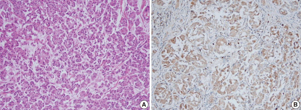
Fig. 2. Acinic cell differentiation in case 3. Tumor cells have eosinophilic granular cytoplasm (A) and show immunoreactivity to α1-antitrypsin (B).

Fig. 3. S-100 protein and p53 expression in case 4. (A, B) Atypical microglandular adenosis (AMGA) shows diffuse strong positivity to S-100 protein (A), while carcinoma arising in microglandular adenosis (MGACA) shows decreased expression to S-100 protein (B). (C, D) p53 staining is evident in MGACA (D), but not in AMGA (C).
Reference
-
1. Rosen PP. Rosen’s breast pathology. 2nd ed. Philadelphia: Lippincott Williams & Wilkins;2001.2. Rosenblum MK, Purrazzella R, Rosen PP. Is microglandular adenosis a precancerous disease? A study of carcinoma arising therein. Am J Surg Pathol. 1986; 10:237–45.3. Koenig C, Dadmanesh F, Bratthauer GL, Tavassoli FA. Carcinoma arising in microglandular adenosis: an immunohistochemical analysis of 20 intraepithelial and invasive neoplasms. Int J Surg Pathol. 2000; 8:303–15.
Article4. Khalifeh IM, Albarracin C, Diaz LK, et al. Clinical, histopathologic, and immunohistochemical features of microglandular adenosis and transition into in situ and invasive carcinoma. Am J Surg Pathol. 2008; 32:544–52.
Article5. Shui R, Bi R, Cheng Y, Lu H, Wang J, Yang W. Matrix-producing carcinoma of the breast in the Chinese population: a clinicopathological study of 13 cases. Pathol Int. 2011; 61:415–22.
Article6. Shui R, Yang W. Invasive breast carcinoma arising in microglandular adenosis: a case report and review of the literature. Breast J. 2009; 15:653–6.
Article7. Marenholz I, Heizmann CW, Fritz G. S100 proteins in mouse and man: from evolution to function and pathology (including an update of the nomenclature). Biochem Biophys Res Commun. 2004; 322:1111–22.
Article8. Lehmann BD, Bauer JA, Chen X, et al. Identification of human triple-negative breast cancer subtypes and preclinical models for selection of targeted therapies. J Clin Invest. 2011; 121:2750–67.
Article9. Geyer FC, Kushner YB, Lambros MB, et al. Microglandular adenosis or microglandular adenoma? A molecular genetic analysis of a case associated with atypia and invasive carcinoma. Histopathology. 2009; 55:732–43.
Article10. Shin SJ, Simpson PT, Da Silva L, et al. Molecular evidence for progression of microglandular adenosis (MGA) to invasive carcinoma. Am J Surg Pathol. 2009; 33:496–504.
Article11. Geyer FC, Lacroix-Triki M, Colombo PE, et al. Molecular evidence in support of the neoplastic and precursor nature of microglandular adenosis. Histopathology. 2012; 60:E115–30.
Article12. Tsang JY, Tse GM. Microglandular adenosis: a prime suspect in triple-negative breast cancer development. J Pathol. 2016; 239:129–32.
Article
- Full Text Links
-
- Actions
-
Cited
- CITED
-
- Close
- Share
-
- Similar articles
-
- Ductal Carcinoma In Situ of the Breast Arising in Microglandular Adenosis
- Invasive Breast Carcinoma Arising in Microglandular Adenosis: Two Case Reports
- Microglandular Adenosis
- Metaplastic Carcinoma with Chondroid Differentiation Arising in Microglandular Adenosis
- Lobular carcinoma in situ in sclerosing adenosis
