Prog Med Phys.
2016 Sep;27(3):162-168. 10.14316/pmp.2016.27.3.162.
Secondary Neutron Dose Measurement for Proton Line Scanning Therapy
- Affiliations
-
- 1Department of Radiological Science, Yonsei University, Wonju, Korea.
- 2Program in Biomedical Radiation Sciences, Department of Transdisciplinary Studies, Graduate School of Convergence Science and Technology, Seoul National University, Seoul, Korea. jinsung@yuhs.ac
- 3Department of Radiation Oncology, Samsung Medical Center, Sungkyunkwan University School of Medicine, Seoul, Korea.
- 4Department of Radiation Oncology, Yonsei Cancer Center, Yonsei University College of Medicine, Seoul, Korea.
- KMID: 2376547
- DOI: http://doi.org/10.14316/pmp.2016.27.3.162
Abstract
- Proton therapy is increasingly being actively used in the treatment of cancer. In contrast to photons, protons have the potential advantage of delivering higher doses to the cancerous tissue and lower doses to the surrounding normal tissue. However, a range shifter is needed to degrade the beam energy in order to apply the pencil beam scanning technique to tumors located close to the minimum range. The secondary neutrons are produced in the beam path including within the patient's body as a result of nuclear interactions. Therefore, unintended side effects may possibly occur. The research related to the secondary neutrons generated during proton therapy has been presented in a variety of studies worldwide, since 2007. In this study, we measured the magnitude of the secondary neutron dose depending on the location of the detector and the use of a range shifter at the beam nozzle of the proton scanning mode, which was recently installed. In addition, the production of secondary neutrons was measured and estimated as a function of the distance between the isocenter and detector. The neutron dose was measured using WENDI-II (Wide Energy Neutron Detection Instruments) and a Plastic Water phantom; a Zebra dosimeter and 4-cm-thick range shifter were also employed as a phantom. In conclusion, we need to consider the secondary neutron dose at proton scanning facilities to employ the range shifter reasonably and effectively.
Keyword
Figure
-
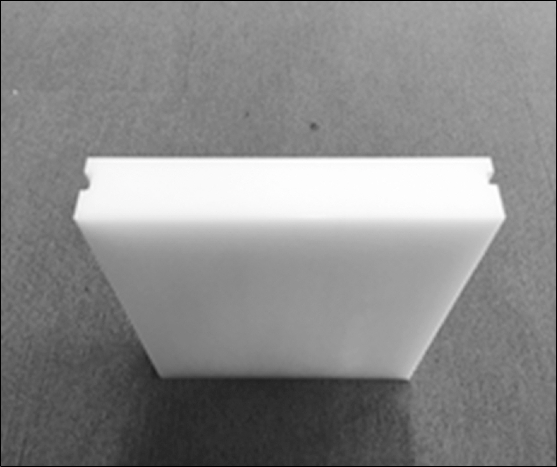
Fig. 1 Range shifter.
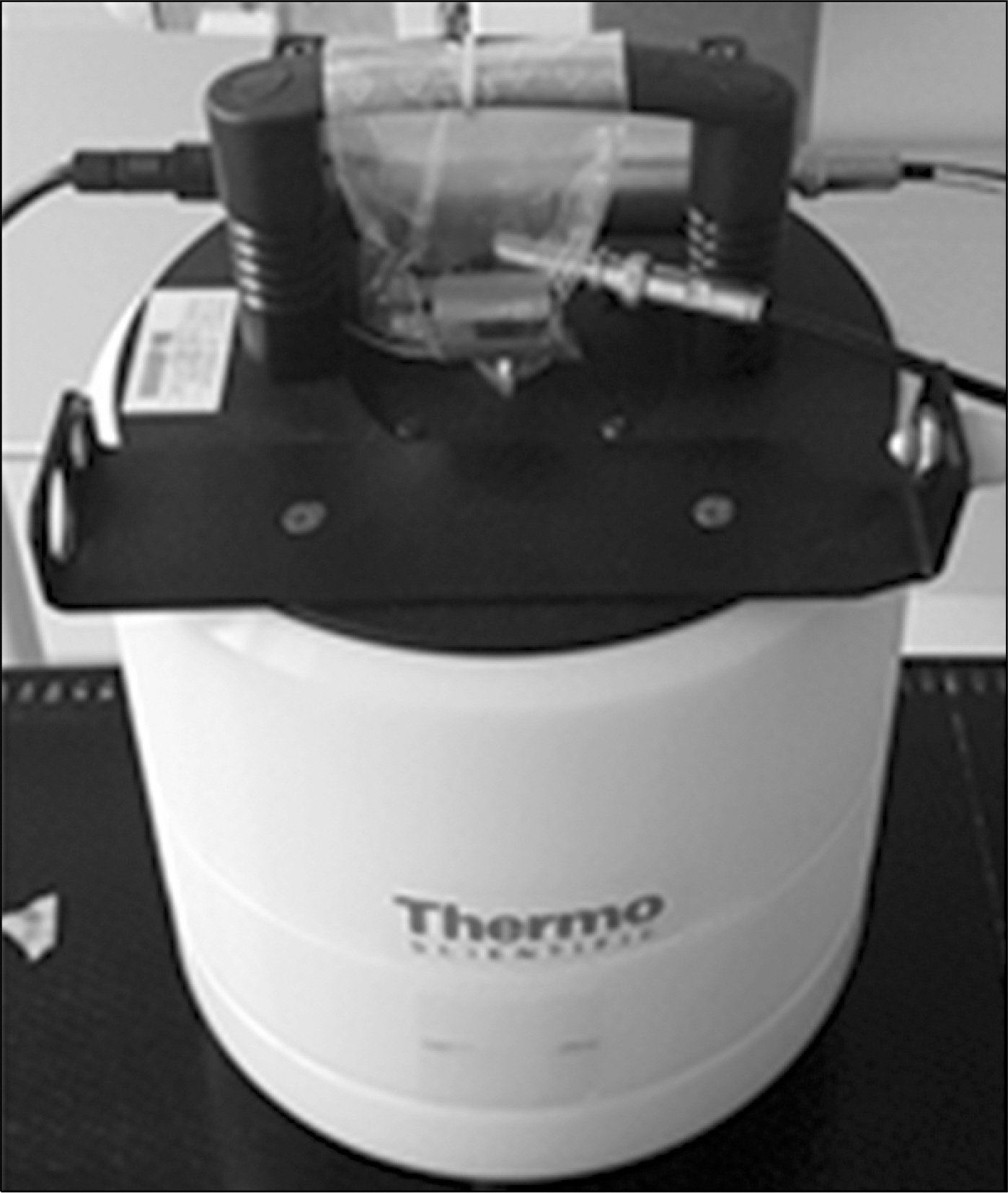
Fig. 2 WENDI-II detector.

Fig. 3 Schematic of first experiment.
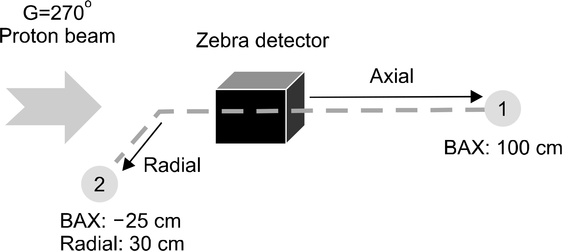
Fig. 4 Schematic of second experiment.
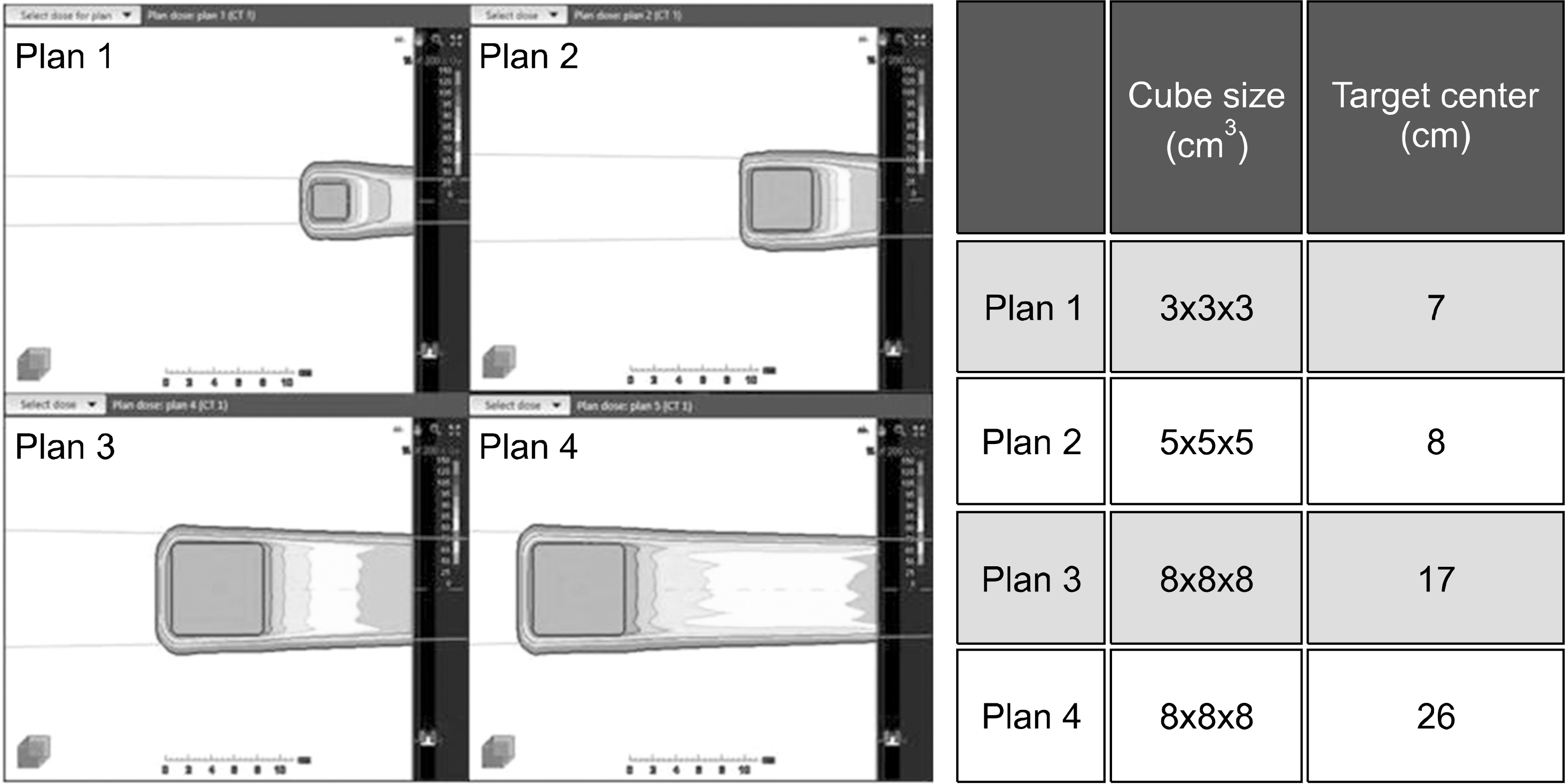
Fig. 5 Two-dimensional dosage distribution within phantom irradiated by four proton scanning methods to assess neutron dose of range shifter.
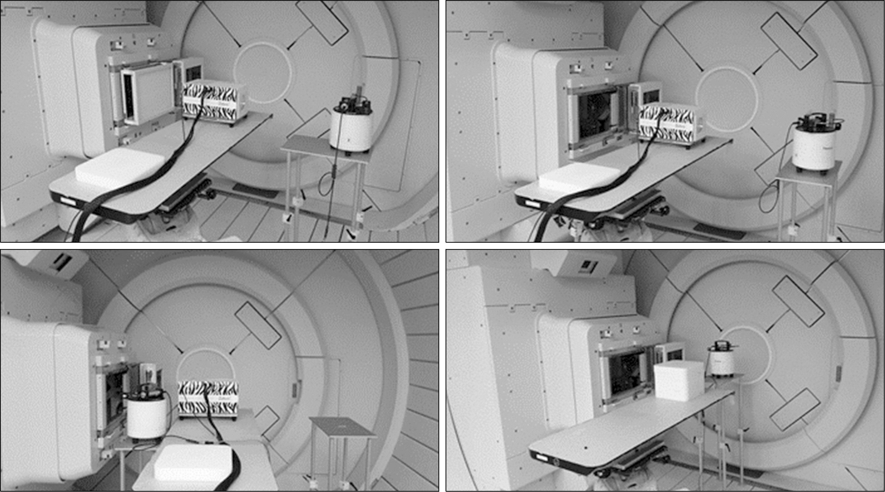
Fig. 6 Measurement of secondary neutron using WENDI-II. Upper left and right: neutron emission measurements taken with a range shifter; lower left: emission measurement taken without a range shifter; lower right: emission measurements taken from a phantom.

Fig. 7 Secondary neutron dose on the radial axis from isocenter.

Fig. 8 Secondary neutron dose on the beam axis from the isocenter.
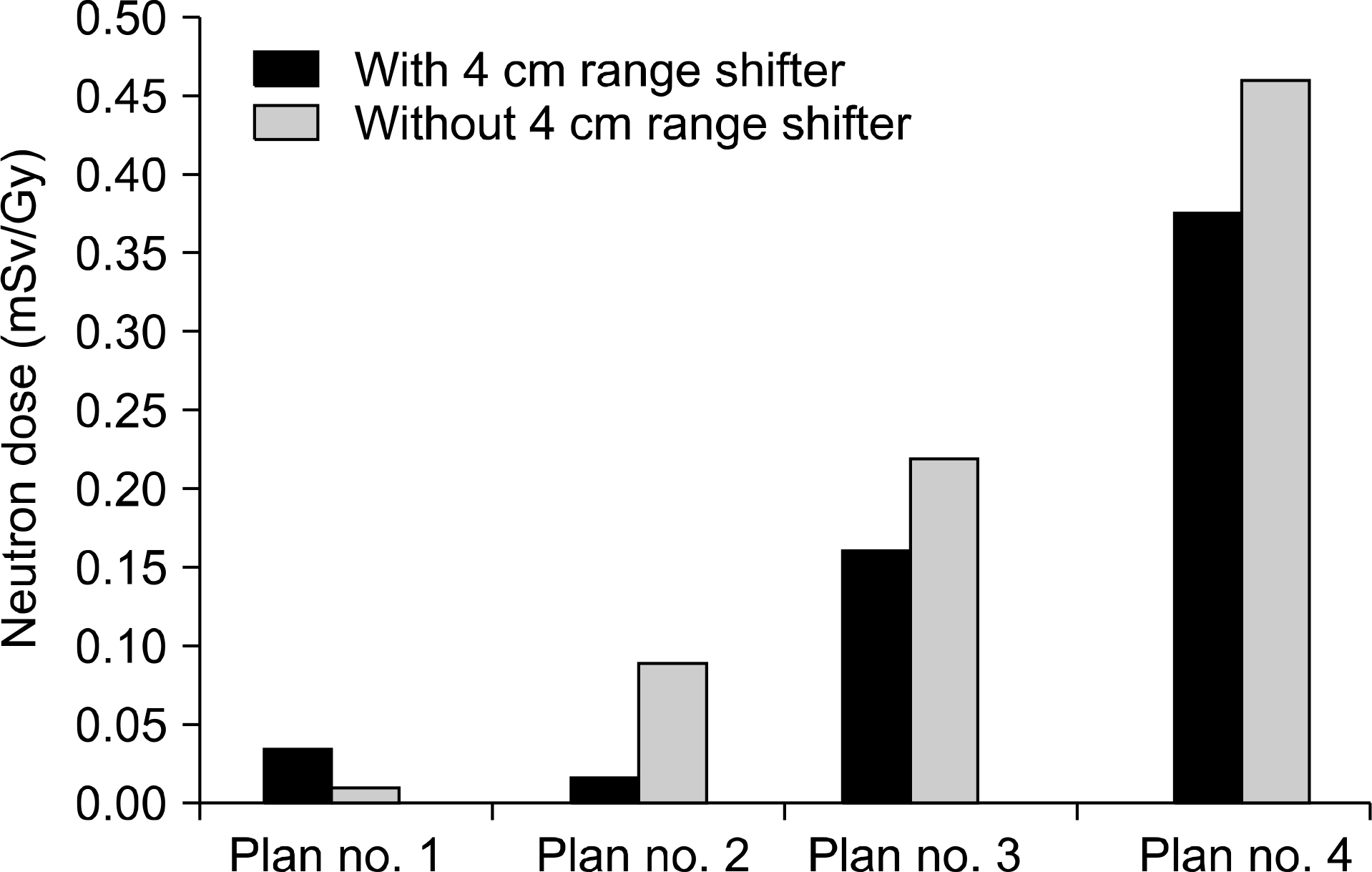
Fig. 9 Value of secondary neutron/Exp_1.
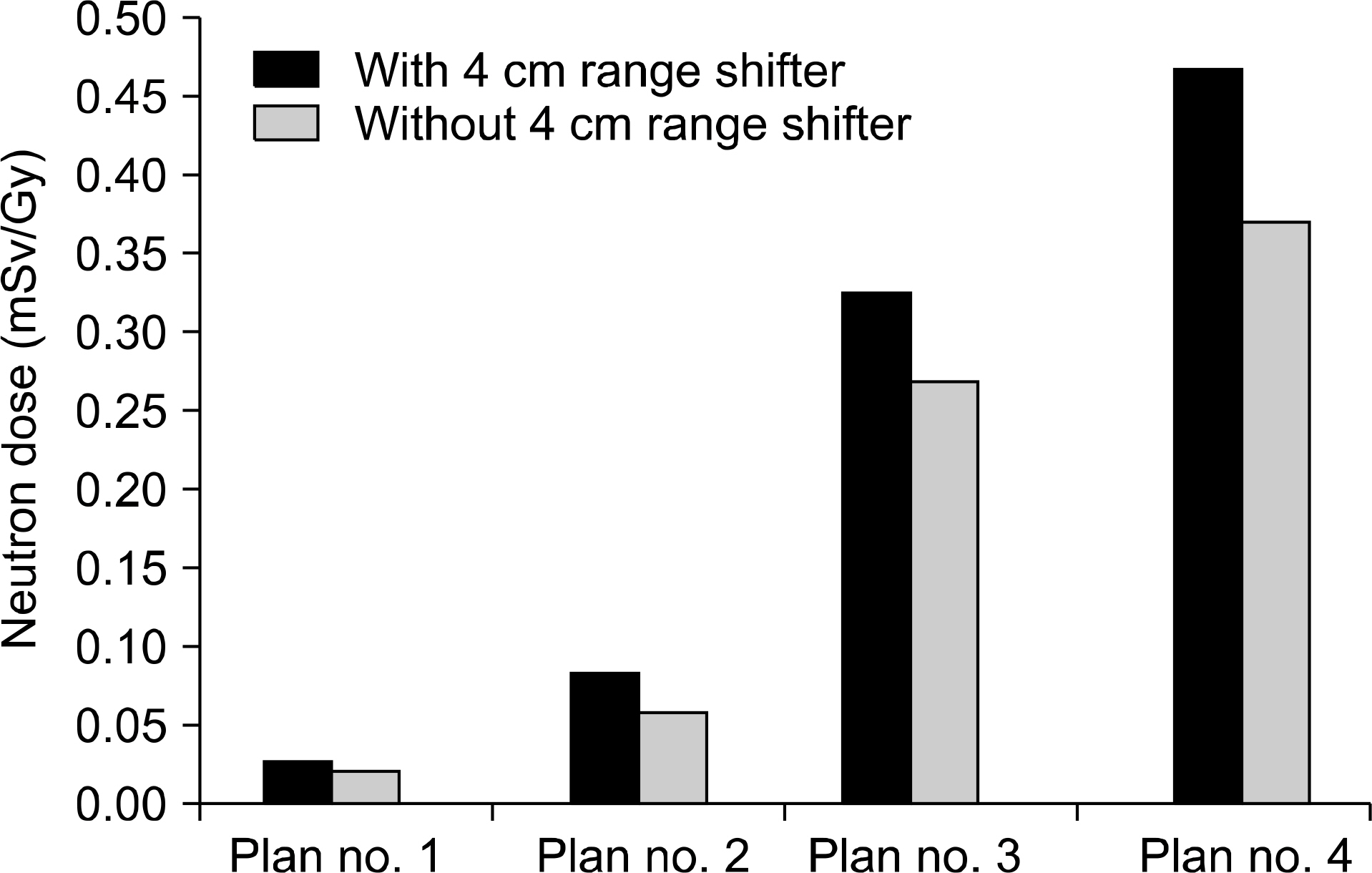
Fig. 10. Value of secondary neutron/Exp_2.
Reference
-
1.Miller DW. A review of proton beam radiation therapy. Medical physics. 22:1943. 1995.
Article2.Olsen DR, et al. Proton therapy –A systematic review of clinical effectiveness. Radiother Oncol 83: 123. 2007.3.Particle Therapy Co-Operative Group. Particle therapy facilities under construction [Internet]. [place un known]: Particle Therapy Co-Operative Group; Available from:. http://www.ptcog.ch/index.php/facilities-under-construction.4.Shin JS, et al. Analysis of changes in dose distribution due to respiration during IMRT. Radiat Oncol J 29: 206. 2011.
Article5.Schneider U., Lomax A., Lombriser N. Comparative risk assessment of secondary cancer incidence after treatment of Hodgkin's disease with photon and proton radiation. Radiat Res. 154(4):): 382–388. 2000.
Article6.Paganetti H. Nuclear interactions in proton therapy: Dose and relative biological effect distributions originating from primary and secondary particles. Phys Med Biol. 47:747–764. 2002.
Article7.Zacharatou JC., Paganetti H. Risk of developing second cancer from neutron dose in proton therapy as function of field characteristics: organ and patient age. Int J Radiat Oncol Biol Phys. 82:228–35. 2008.8.Kim JS, et al. Feasibility study of neutron dose for real-time image-guided proton therapy: a Monte Carlo study. JKPS. 67(1):142–146. 2015.9.Yonai S, et al. Measurement of neutron ambient dose equivalent in passive carbon-ion and proton radiotherapies. Med Phys. 35:4782–4792. 2008.
Article10.Hall EJ. Intensity modulated radiation therapy, protons, and the risk of second cancers Int J Radiat Oncol Biol Phys. 65:1–7. 2006.11.Chung KZ, et al. The first private-hospital based proton therapy center in Korea; Status of the Proton Therapy Center at Samsung Medical Center. Radiat Oncol J. 33(4):1–7. 2015. . DOI: doi: 10.3857/roj.2015.33.4.337.
Article12.https://www.thermofisher.com/order/catalog/product/FHT762WENDI2.13.ICRP. 1977. Recommendations of the ICRP. ICRP Publication 26. Ann. ICRP. 1(3):
- Full Text Links
-
- Actions
-
Cited
- CITED
-
- Close
- Share
-
- Similar articles
-
- A Pilot Study of the Scanning Beam Quality Assurance Using Machine Log Files in Proton Beam Therapy
- Initial Experience of Patient-Specific QA for Wobbling and Line-Scanning Proton Therapy at Samsung Medical Center
- Preliminary Study for Dosimetry of Boron Neutron Capture Therapy with Polymer Gel
- Feasibility Test of Flat-Type Faraday Cup for UltrahighDose-Rate Transmission Proton Beam Therapy
- Proton Therapy Review: Proton Therapy from a Medical
