Morphological and Microstructural Changes of the Hippocampus in Early MCI: A Study Utilizing the Alzheimer's Disease Neuroimaging Initiative Database
- Affiliations
-
- 1Department of Bio and Brain Engineering, Korea Advanced Institute of Science and Technology (KAIST) Daejeon, Korea. yong@kaist.ac.kr
- 2School of Computing, Korea Advanced Institute of Science and Technology (KAIST) Daejeon, Korea. jinahpark@kaist.ac.kr
- 3KI for Health Science and Technology, Korea Advanced Institute of Science and Technology (KAIST) Daejeon, Korea.
- KMID: 2376015
- DOI: http://doi.org/10.3988/jcn.2017.13.2.144
Abstract
- BACKGROUND AND PURPOSE
With the aim of facilitating the early detection of Alzheimer's disease, the Alzheimer's Disease Neuroimaging Initiative proposed two stages based on the memory performance: early mild cognitive impairment (EMCI) and late mild cognitive impairment (LMCI). The current study was designed to investigate structural differences in terms of surface atrophy and microstructural changes of the hippocampus in EMCI and LMCI.
METHODS
Hippocampal shape modeling based on progressive template surface deformation was performed on T1-weighted MRI images obtained from 20 cognitive normal (CN) subjects, 17 EMCI patients, and 20 LMCI patients. A template surface in CN was used as a region of interest for diffusion-tensor imaging (DTI) voxel-based morphometry (VBM) analysis. Cluster-wise group comparison was performed based on DTI indices within the hippocampus. Linear regression was performed to identify correlations between DTI metrics and clinical scores.
RESULTS
The hippocampal surface analysis showed significant atrophies in bilateral CA1 regions and the right ventral subiculum in EMCI, in contrast to widespread atrophy in LMCI. DTI VBM analysis showed increased diffusivity in the CA2-CA4 regions in EMCI and additionally in the subiculum region in LMCI. Hippocampal diffusivity was significantly correlated with scores both for the Mini Mental State Examination and on the Modified Alzheimer Disease Assessment Scale cognitive subscale. However, the hippocampal diffusivity did not vary significantly with the fractional anisotropy.
CONCLUSIONS
EMCI showed hippocampal surface changes mainly in the CA1 region and ventral subiculum. Diffusivity increased mainly in the CA2-CA4 regions in EMCI, while it decreased throughout the hippocampus in LMCI. Although axial diffusivity showed prominent changes in the right hippocampus in EMCI, future studies need to confirm the presence of this laterality difference. In addition, diffusivity is strongly correlated with the cognitive performance, indicating the possibility of using diffusivity as a biomarker for disease progression.
Keyword
MeSH Terms
Figure
-
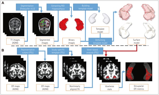
Fig. 1 Overall scheme of the analysis. A: T1-weighted images were segmented using Freesurfer to extract the hippocampus. The template model from the normal group was used to construct the group surface model and determine intergroup morphometry differences. B: DTI maps were processed using the TBSS pipeline up to the nonlinear register in an MNI space. Using a surface model of the hippocampus generated from T1-weighted images as a mask, we compared hippocampal diffusivity differences among groups. ADNI: Alzheimer's Disease Neuroimaging Initiative, DTI: diffusion-tensor imaging, FA: fractional anisotropy, FSL: functional MRI of the brain software library, ROI: region of interest, TBSS: Tract-Based Spatial Statistics.
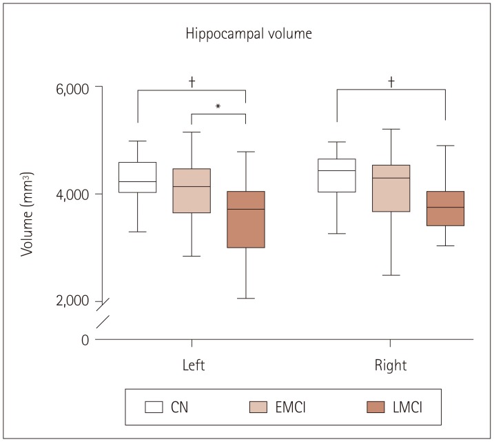
Fig. 2 Hippocampal volumes in each group. Bilateral hippocampal volumes in EMCI were not significantly smaller than those in CN. While the left hippocampal volumes in EMCI were significantly smaller than those in LMCI, they did not differ significantly from those in CN (*p<0.05, †p<0.001). CN: cognitive normal, EMCI: early mild cognitive impairment, LMCI: late mild cognitive impairment.
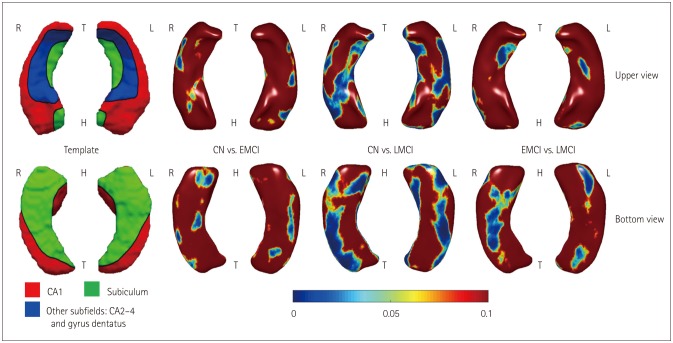
Fig. 3 Morphometry changes. Delineation of hippocampal subfields in the average template from CN (leftmost). Hippocampal surface analysis showed significant atrophy in bilateral CA1 regions and the right ventral subiculum in EMCI compared to CN, and in bilateral CA1 and CA2–CA4 regions and the subiculum in LMCI (uncorrected p<0.05). CN: cognitive normal, EMCI: early mild cognitive impairment, H: head, LMCI: late mild cognitive impairment, T: tail.
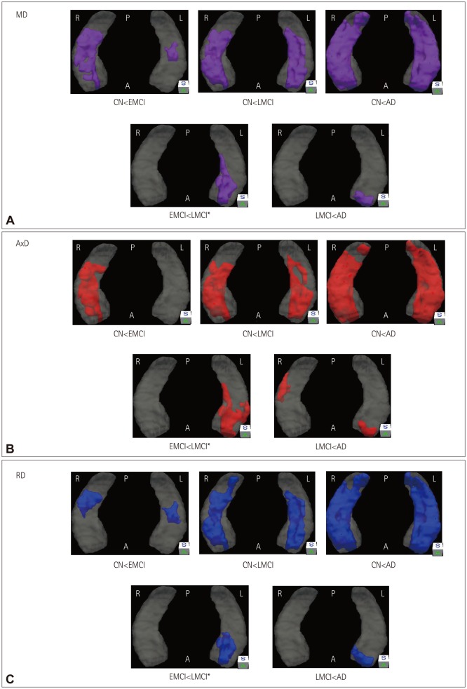
Fig. 4 Microstructural changes in each group. Clusters showing changes in DTI indices: MD (A), AxD (B), and RD (C). All comparisons except those in EMCI and LMCI were corrected for multiple comparisons. A and C: MD and RD were higher in EMCI than in CN in the bilateral CA2–CA4 regions, and higher in LMCI in the bilateral CA2–CA4 and subiculum regions. B: AxD was higher in EMCI than in CN in the right CA2–CA4 regions, and higher in LMCI in the bilateral CA2–CA4 and subiculum regions (purple, MD; red, AxD; blue, RD). A: anterior, AD: Alzheimer's disease, AxD: axial diffusivity, CN: cognitive normal, DTI: diffusion-tensor imaging, EMCI: early mild cognitive impairment, LMCI: late mild cognitive impairment, MD: mean diffusivity, P: posterior, RD: radial diffusivity, S: superior.
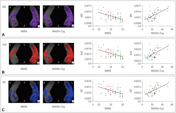
Fig. 5 Linear relationships between DTI metrics and clinical scores. Clusters showing linear relationships with DTI indices: MD (A), AxD (B), and RD (C). RD versus MMSE and MADAS-Cog scores (purple, MD; red, AxD; blue, RD). Scatter plots showing the correlations between result clusters and clinical scores (green, EMCI; blue, LMCI; red, AD). A: anterior, AD: Alzheimer's disease, AxD: axial diffusivity, DTI: diffusion-tensor imaging, EMCI: early mild cognitive impairment, LMCI: late mild cognitive impairment, MADAS-Cog: Modified Alzheimer's Disease Assessment Scale-Cognitive subscale, MD: mean diffusivity, MMSE: Mini Mental State Examination, P: posterior, RD: radial diffusivity, S: superior.
Cited by 1 articles
-
Medial-Vowel Writing Difficulty in Korean Syllabic Writing: A Characteristic Sign of Alzheimer's Disease
Ji Hye Yoon, Yong Jeong, Duk L. Na
J Clin Neurol. 2018;14(2):179-185. doi: 10.3988/jcn.2018.14.2.179.
Reference
-
1. Jack CR Jr, Knopman DS, Jagust WJ, Shaw LM, Aisen PS, Weiner MW, et al. Hypothetical model of dynamic biomarkers of the Alzheimer's pathological cascade. Lancet Neurol. 2010; 9:119–128. PMID: 20083042.
Article2. Shim YS, Morris JC. Biomarkers predicting Alzheimer's disease in cognitively normal aging. J Clin Neurol. 2011; 7:60–68. PMID: 21779293.
Article3. Aisen PS, Petersen RC, Donohue MC, Gamst A, Raman R, Thomas RG, et al. Clinical core of the Alzheimer's Disease neuroimaging initiative: progress and plans. Alzheimers Dement. 2010; 6:239–246. PMID: 20451872.
Article4. Weiner MW, Aisen PS, Jack CR Jr, Jagust WJ, Trojanowski JQ, Shaw L, et al. The Alzheimer's disease neuroimaging initiative: progress report and future plans. Alzheimers Dement. 2010; 6:202–211.e7. PMID: 20451868.
Article5. Qiu Y, Li L, Zhou TY, Lu W. Alzheimer's Disease Neuroimaging Initiative. Alzheimer's disease progression model based on integrated biomarkers and clinical measures. Acta Pharmacol Sin. 2014; 35:1111–1120. PMID: 25088003.
Article6. Wang L, Miller JP, Gado MH, McKeel DW, Rothermich M, Miller MI, et al. Abnormalities of hippocampal surface structure in very mild dementia of the Alzheimer type. Neuroimage. 2006; 30:52–60. PMID: 16243546.
Article7. Frisoni GB, Ganzola R, Canu E, Rüb U, Pizzini FB, Alessandrini F, et al. Mapping local hippocampal changes in Alzheimer's disease and normal ageing with MRI at 3 Tesla. Brain. 2008; 131(Pt 12):3266–3276. PMID: 18988639.
Article8. Costafreda SG, Dinov ID, Tu Z, Shi Y, Liu CY, Kloszewska I, et al. Automated hippocampal shape analysis predicts the onset of dementia in mild cognitive impairment. Neuroimage. 2011; 56:212–219. PMID: 21272654.
Article9. Hanseeuw BJ, Van Leemput K, Kavec M, Grandin C, Seron X, Ivanoiu A. Mild cognitive impairment: differential atrophy in the hippocampal subfields. AJNR Am J Neuroradiol. 2011; 32:1658–1661. PMID: 21835940.
Article10. Shen KK, Fripp J, Mériaudeau F, Chételat G, Salvado O, Bourgeat P. Alzheimer's Disease Neuroimaging Initiative. Detecting global and local hippocampal shape changes in Alzheimer's disease using statistical shape models. Neuroimage. 2012; 59:2155–2166. PMID: 22037419.
Article11. Ye BS, Seo SW, Kim CH, Jeon S, Kim GH, Noh Y, et al. Hippocampal and cortical atrophy in amyloid-negative mild cognitive impairments: comparison with amyloid-positive mild cognitive impairment. Neurobiol Aging. 2014; 35:291–300. PMID: 24080178.
Article12. Kim J, Valdes-Hernandez Mdel C, Royle NA, Park J. Hippocampal shape modeling based on a progressive template surface deformation and its verification. IEEE Trans Med Imaging. 2015; 34:1242–1261. PMID: 25532173.
Article13. Lee DY, Fletcher E, Carmichael OT, Singh B, Mungas D, Reed B, et al. Sub-regional hippocampal injury is associated with fornix degeneration in Alzheimer's disease. Front Aging Neurosci. 2012; 4:1. PMID: 22514534.
Article14. Thomann PA, Seidl U, Brinkmann J, Hirjak D, Traeger T, Wolf RC, et al. Hippocampal morphology and autobiographic memory in mild cognitive impairment and Alzheimer's disease. Curr Alzheimer Res. 2012; 9:507–515. PMID: 22372439.15. Bozzali M, Falini A, Franceschi M, Cercignani M, Zuffi M, Scotti G, et al. White matter damage in Alzheimer's disease assessed in vivo using diffusion tensor magnetic resonance imaging. J Neurol Neurosurg Psychiatry. 2002; 72:742–746. PMID: 12023417.
Article16. Takahashi S, Yonezawa H, Takahashi J, Kudo M, Inoue T, Tohgi H. Selective reduction of diffusion anisotropy in white matter of Alzheimer disease brains measured by 3.0 Tesla magnetic resonance imaging. Neurosci Lett. 2002; 332:45–48. PMID: 12377381.
Article17. Ringman JM, O'Neill J, Geschwind D, Medina L, Apostolova LG, Rodriguez Y, et al. Diffusion tensor imaging in preclinical and presymptomatic carriers of familial Alzheimer's disease mutations. Brain. 2007; 130(Pt 7):1767–1776. PMID: 17522104.
Article18. Stricker NH, Schweinsburg BC, Delano-Wood L, Wierenga CE, Bangen KJ, Haaland KY, et al. Decreased white matter integrity in late-myelinating fiber pathways in Alzheimer's disease supports retrogenesis. Neuroimage. 2009; 45:10–16. PMID: 19100839.
Article19. Sexton CE, Kalu UG, Filippini N, Mackay CE, Ebmeier KP. A meta-analysis of diffusion tensor imaging in mild cognitive impairment and Alzheimer's disease. Neurobiol Aging. 2011; 32:2322.e5–2322.e18.
Article20. Bosch B, Arenaza-Urquijo EM, Rami L, Sala-Llonch R, Junqué C, Solé-Padullés C, et al. Multiple DTI index analysis in normal aging, amnestic MCI and AD. Relationship with neuropsychological performance. Neurobiol Aging. 2012; 33:61–74. PMID: 20371138.
Article21. Nir TM, Jahanshad N, Villalon-Reina JE, Toga AW, Jack CR, Weiner MW, et al. Effectiveness of regional DTI measures in distinguishing Alzheimer's disease, MCI, and normal aging. Neuroimage Clin. 2013; 3:180–195. PMID: 24179862.
Article22. Zhuang L, Sachdev PS, Trollor JN, Reppermund S, Kochan NA, Brodaty H, et al. Microstructural white matter changes, not hippocampal atrophy, detect early amnestic mild cognitive impairment. PLoS One. 2013; 8:e58887. PMID: 23516569.
Article23. Patil RB, Ramakrishnan S. Analysis of sub-anatomic diffusion tensor imaging indices in white matter regions of Alzheimer with MMSE score. Comput Methods Programs Biomed. 2014; 117:13–19. PMID: 24986110.
Article24. Chen H, Wang K, Yao J, Dai J, Ma J, Li S, et al. White matter changes in Alzheimer's disease revealed by diffusion tensor imaging with TBSS. World J Neurosci. 2015; 5:58–65.25. Bozzali M, Cercignani M, Sormani MP, Comi G, Filippi M. Quantification of brain gray matter damage in different MS phenotypes by use of diffusion tensor MR imaging. AJNR Am J Neuroradiol. 2002; 23:985–988. PMID: 12063230.26. Coleman MP, Freeman MR. Wallerian degeneration, wld(s), and nmnat. Annu Rev Neurosci. 2010; 33:245–267. PMID: 20345246.
Article27. Walhovd KB, Fjell AM, Amlien I, Grambaite R, Stenset V, Bjørnerud A, et al. Multimodal imaging in mild cognitive impairment: metabolism, morphometry and diffusion of the temporal-parietal memory network. Neuroimage. 2009; 45:215–223. PMID: 19056499.
Article28. Kantarci K, Petersen RC, Boeve BF, Knopman DS, Weigand SD, O'Brien PC, et al. DWI predicts future progression to Alzheimer disease in amnestic mild cognitive impairment. Neurology. 2005; 64:902–904. PMID: 15753434.
Article29. Müller MJ, Greverus D, Dellani PR, Weibrich C, Wille PR, Scheurich A, et al. Functional implications of hippocampal volume and diffusivity in mild cognitive impairment. Neuroimage. 2005; 28:1033–1042. PMID: 16084115.
Article30. Fellgiebel A, Dellani PR, Greverus D, Scheurich A, Stoeter P, Müller MJ. Predicting conversion to dementia in mild cognitive impairment by volumetric and diffusivity measurements of the hippocampus. Psychiatry Res. 2006; 146:283–287. PMID: 16530394.
Article31. Fellgiebel A, Yakushev I. Diffusion tensor imaging of the hippocampus in MCI and early Alzheimer's disease. J Alzheimers Dis. 2011; 26(Suppl 3):257–262. PMID: 21971465.
Article32. Rose SE, Janke AL, Chalk JB. Gray and white matter changes in Alzheimer's disease: a diffusion tensor imaging study. J Magn Reson Imaging. 2008; 27:20–26. PMID: 18050329.
Article33. Douaud G, Menke RA, Gass A, Monsch AU, Rao A, Whitcher B, et al. Brain microstructure reveals early abnormalities more than two years prior to clinical progression from mild cognitive impairment to Alzheimer's disease. J Neurosci. 2013; 33:2147–2155. PMID: 23365250.
Article34. Jack CR Jr, Bernstein MA, Fox NC, Thompson P, Alexander G, Harvey D, et al. The Alzheimer's Disease Neuroimaging Initiative (ADNI): MRI methods. J Magn Reson Imaging. 2008; 27:685–691. PMID: 18302232.
Article35. Csernansky JG, Wang L, Swank J, Miller JP, Gado M, McKeel D, et al. Preclinical detection of Alzheimer's disease: hippocampal shape and volume predict dementia onset in the elderly. Neuroimage. 2005; 25:783–792. PMID: 15808979.
Article36. Smith SM, Jenkinson M, Johansen-Berg H, Rueckert D, Nichols TE, Mackay CE, et al. Tract-based spatial statistics: voxelwise analysis of multi-subject diffusion data. Neuroimage. 2006; 31:1487–1505. PMID: 16624579.
Article37. Smith SM, Nichols TE. Threshold-free cluster enhancement: addressing problems of smoothing, threshold dependence and localisation in cluster inference. Neuroimage. 2009; 44:83–98. PMID: 18501637.
Article38. Apostolova LG, Mosconi L, Thompson PM, Green AE, Hwang KS, Ramirez A, et al. Subregional hippocampal atrophy predicts Alzheimer's dementia in the cognitively normal. Neurobiol Aging. 2010; 31:1077–1088. PMID: 18814937.
Article39. Cho Y, Seong JK, Shin SY, Jeong Y, Kim JH, Qiu A, et al. A multi-resolution scheme for distortion-minimizing mapping between human subcortical structures based on geodesic construction on Riemannian manifolds. Neuroimage. 2011; 57:1376–1392. PMID: 21658456.
Article40. Schönheit B, Zarski R, Ohm TG. Spatial and temporal relationships between plaques and tangles in Alzheimer-pathology. Neurobiol Aging. 2004; 25:697–711. PMID: 15165691.
Article41. Bobinski M, Wegiel J, Wisniewski HM, Tarnawski M, Reisberg B, Mlodzik B, et al. Atrophy of hippocampal formation subdivisions correlates with stage and duration of Alzheimer disease. Dementia. 1995; 6:205–210. PMID: 7550600.
Article42. Bobinski M, Wegiel J, Tarnawski M, Bobinski M, Reisberg B, de Leon MJ, et al. Relationships between regional neuronal loss and neurofibrillary changes in the hippocampal formation and duration and severity of Alzheimer disease. J Neuropathol Exp Neurol. 1997; 56:414–420. PMID: 9100672.
Article43. Shi F, Liu B, Zhou Y, Yu C, Jiang T. Hippocampal volume and asymmetry in mild cognitive impairment and Alzheimer's disease: metaanalyses of MRI studies. Hippocampus. 2009; 19:1055–1064. PMID: 19309039.
Article44. Tondelli M, Wilcock GK, Nichelli P, De Jager CA, Jenkinson M, Zamboni G. Structural MRI changes detectable up to ten years before clinical Alzheimer's disease. Neurobiol Aging. 2012; 33:825.e25–825.e36.
Article45. Cappellani R, Bergsland N, Weinstock-Guttman B, Kennedy C, Carl E, Ramasamy DP, et al. Subcortical deep gray matter pathology in patients with multiple sclerosis is associated with white matter lesion burden and atrophy but not with cortical atrophy: a diffusion tensor MRI study. AJNR Am J Neuroradiol. 2014; 35:912–919. PMID: 24335548.
Article46. den Heijer T, der Lijn Fv, Vernooij MW, de Groot M, Koudstaal PJ, van der Lugt A, et al. Structural and diffusion MRI measures of the hippocampus and memory performance. Neuroimage. 2012; 63:1782–1789. PMID: 22960084.
Article47. Shipton OA, El-Gaby M, Apergis-Schoute J, Deisseroth K, Bannerman DM, Paulsen O, et al. Left-right dissociation of hippocampal memory processes in mice. Proc Natl Acad Sci U S A. 2014; 111:15238–15243. PMID: 25246561.
Article48. Basser PJ, Pierpaoli C. Microstructural and physiological features of tissues elucidated by quantitative-diffusion-tensor MRI. J Magn Reson B. 1996; 111:209–219. PMID: 8661285.
Article49. Kale RA, Gupta RK, Saraswat VA, Hasan KM, Trivedi R, Mishra AM, et al. Demonstration of interstitial cerebral edema with diffusion tensor MR imaging in type C hepatic encephalopathy. Hepatology. 2006; 43:698–706. PMID: 16557540.
Article50. Weston PS, Simpson IJ, Ryan NS, Ourselin S, Fox NC. Diffusion imaging changes in grey matter in Alzheimer's disease: a potential marker of early neurodegeneration. Alzheimers Res Ther. 2015; 7:47. PMID: 26136857.
Article51. Rulseh AM, Keller J, Tintěra J, Kozžísšek M, Vymazal J. Chasing shadows: what determines DTI metrics in gray matter regions? An in vitro and in vivo study. J Magn Reson Imaging. 2013; 38:1103–1110. PMID: 23440865.
Article52. Kamman RL, Go KG, Brouwer W, Berendsen HJ. Nuclear magnetic resonance relaxation in experimental brain edema: effects of water concentration, protein concentration, and temperature. Magn Reson Med. 1988; 6:265–274. PMID: 3362061.
Article53. Bartzokis G, Cummings JL, Sultzer D, Henderson VW, Nuechterlein KH, Mintz J. White matter structural integrity in healthy aging adults and patients with Alzheimer disease: a magnetic resonance imaging study. Arch Neurol. 2003; 60:393–398. PMID: 12633151.
- Full Text Links
-
- Actions
-
Cited
- CITED
-
- Close
- Share
-
- Similar articles
-
- Neuroimaging Markers for Alzheimer's Disease and Mild Cognitive Impairment in Alzheimer's Disease Neuroimaging Initiative (ADNI)
- Gene Interactions and Structural Brain Change in Early-Onset Alzheimer's Disease Subjects Using the Pipeline Environment
- Association of a History of Sleep Disorder With Risk of Mild Cognitive Impairment and Alzheimer’s Disease Dementia
- Changes in the Hippocampal Volume and Shape in Early-Onset Mild Cognitive Impairment
- Abnormal Integrity of Corticocortical Tracts in Mild Cognitive Impairment: A Diffusion Tensor Imaging Study
