Ovarian Clear Cell Carcinoma Sub-Typing by ARID1A Expression
- Affiliations
-
- 1Department of Pathology, Yonsei University College of Medicine, Seoul, Korea. cho1988@yuhs.ac
- 2Brain Korea 21 Plus Project for Medical Science, Yonsei University College of Medicine, Seoul, Korea.
- 3Department of Gynecology, Yonsei University College of Medicine, Seoul, Korea.
- 4Severance Biomedical Science Institute (SBSI), Yonsei University College of Medicine, Seoul, Korea.
- KMID: 2374189
- DOI: http://doi.org/10.3349/ymj.2017.58.1.59
Abstract
- PURPOSE
Loss of AT-rich DNA-interacting domain 1A (ARID1A) has been identified as a driving mutation of ovarian clear cell carcinoma (O-CCC), a triple-negative ovarian cancer that is intermediary between serous and endometrioid subtypes, in regards to molecular and clinical behaviors. However, about half of O-CCCs still express BAF250a, the protein encoded by ARID1A. Herein, we aimed to identify signatures of ARID1A-positive O-CCC in comparison with its ARID1A-negative counterpart.
MATERIALS AND METHODS
Seventy cases of O-CCC were included in this study. Histologic grades and patterns of primary tumor, molecular marker immunohistochemistry profiles, and clinical outcomes were analyzed.
RESULTS
Forty-eight (69%) O-CCCs did not express BAF250a, which were designated as "ARID1A-negative." The other 22 (31%) O-CCCs were designated as "ARID1A-positive." ARID1A-positive tumors were more likely to be histologically of high grades (41% vs. 10%, p=0.003), ERβ-positive (45% vs. 17%, p=0.011), and less likely to be HNF1β-positive (77% vs. 96%, p=0.016) and E-cadherin-positive (59% vs. 83%, p=0.028) than ARID1A-negative tumors. Patient age, parity, tumor stage were not significantly different in between the two groups. Cancer-specific survival was not significantly different either.
CONCLUSION
We classified O-CCCs according to ARID1A expression status. ARID1A-positive O-CCCs exhibited distinct immunohistochemical features from ARID1A-negative tumors, suggesting a different underlying molecular event during carcinogenesis.
Keyword
MeSH Terms
-
Adenocarcinoma, Clear Cell/*metabolism/mortality/pathology
Adult
Aged
Biomarkers, Tumor/metabolism
Cadherins/metabolism
Estrogen Receptor beta/metabolism
Female
Humans
Immunohistochemistry
Middle Aged
Mutation
Neoplasm Proteins/*metabolism
Nuclear Proteins/*metabolism
Ovarian Neoplasms/*metabolism/mortality/pathology
Transcription Factors/*metabolism
Biomarkers, Tumor
Cadherins
Estrogen Receptor beta
Neoplasm Proteins
Nuclear Proteins
Transcription Factors
Figure
-
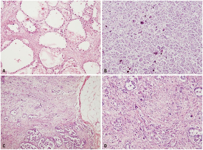
Fig. 1 Histologic grades of O-CCC. (A and B) Low grade O-CCC. (A) tubulocystic or alveolar patterns are the most common patterns seen in low grade tumor. Abortive small tubules are less differentiated (H-E, ×100). (B) Psammomatous calcifications (H-E, ×100). (C and D) High grade O-CCC. (C) Diffuse, solid areas are frequently seen in high grade tumor. Desmoplasia is frequently found in association with infiltrative cells (H-E, ×40). (D) highly cellular spindle cells are parallel with infiltrating tumor cells (H-E, ×200). O-CCC, clear cell carcinoma of ovary; H-E, hematoxylin-eosin.
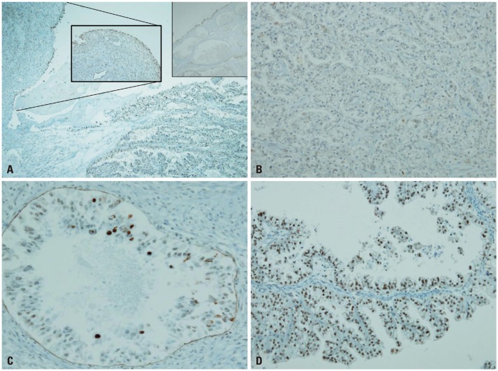
Fig. 2 ARID1A expressions in O-CCC. (A) In endometriosis, nuclear staining occurs along the epithelial lining (inlet), but CCC shows complete loss of ARID1A (DAB, ×40). (B) Total loss of ARID1A in a branched tubular pattern (DAB, ×100). (C) Focal expression (nearly total loss) in an alveolar pattern (DAB, ×200). (D) Moderate preservation of AR1D1A expression in a cystic papillary pattern (DAB, ×200). ARID1A, AT-rich DNA-interacting domain 1A; O-CCC, clear cell carcinoma of ovary; DAB, 3'-3' diaminobenzidine.
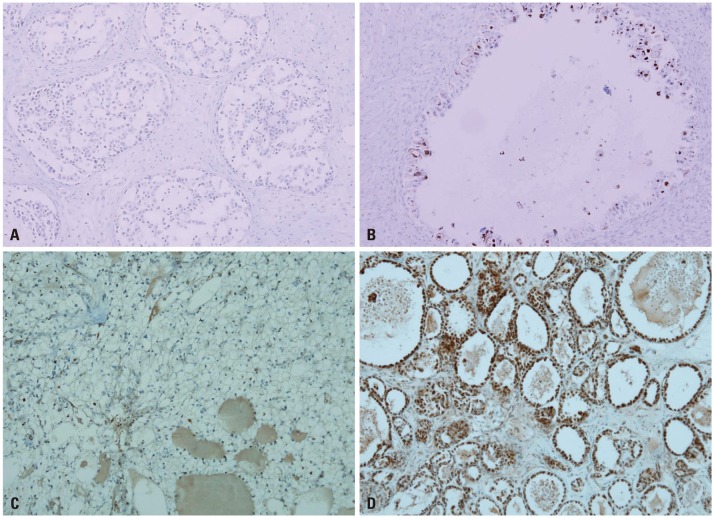
Fig. 3 HNF1β and ERβ expressions in O-CCC. (A and B) HNF1β expression. (A) Complete loss of HNF1-β is rarely found in CCC. However, negative expression of p53 or ER with loss of ARID1A is common (DAB, ×100). (B) Luminal hobnail cells express HNF1-β. More HNF1-β expression occurs in cells closer to the luminal side (DAB, ×200). (C and D) ERβ expression. (C) complete loss of ERβ is common (DAB, ×100). (D) Luminal protruding hobnail cells express ERβ (DAB, ×100). ERβ, estrogen receptor beta; O-CCC, clear cell carcinoma of ovary; ARID1A, AT-rich DNA-interacting domain 1A; DAB, 3'-3' diaminobenzidine.
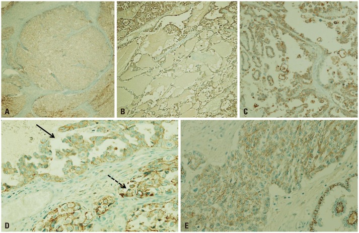
Fig. 4 E-cadherin expressions in O-CCC. Loss of CDH1, or E-cadherin was manifested variably. (A) Complete loss of CDH1 (DAB, ×10). (B) Partial and weak staining (DAB, ×100). (C) Aberrant CDH1 expression. Aberrant cytoplasmic expression with fragmented or broken linear forms instead of membrane staining is an indicator of E-cadherin disruption (DAB, ×100). (D) Within the same tumor, CDH1 loss is heterogeneous. The solid arrow points to a region of partial CDH1 loss as seen by weak staining along the membrane. The broken arrow points to disrupted fragments in the membrane (DAB, ×200). (E) Images taken at a higher magnification show aberrant staining, such as and non-continuous, fragmented membrane, or cytoplasmic staining (DAB, ×200). O-CCC, clear cell carcinoma of ovary; DAB, 3'-3' diaminobenzidine.
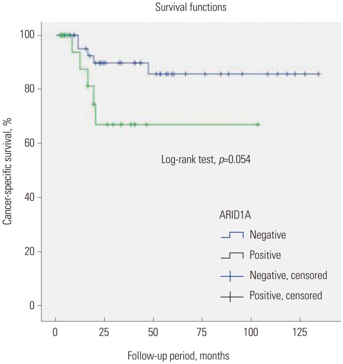
Fig. 5 Kaplan-Meier curve of cancer-specific survival for O-CCC, stratified by ARID1A expression status. O-CCC, clear cell carcinoma of ovary; ARID1A, AT-rich DNA-interacting domain 1A.
Reference
-
1. Ahmed Q, Alosh B, Bandyopadhyay S, Ali-Fehmi R. Gynecologic cancers: molecular updates. Clin Lab Med. 2013; 33:911–925. PMID: 24267195.2. Jones S, Wang TL, Shih IeM, Mao TL, Nakayama K, Roden R, et al. Frequent mutations of chromatin remodeling gene ARID1A in ovarian clear cell carcinoma. Science. 2010; 330:228–231. PMID: 20826764.3. Wiegand KC, Shah SP, Al-Agha OM, Zhao Y, Tse K, Zeng T, et al. ARID1A mutations in endometriosis-associated ovarian carcinomas. N Engl J Med. 2010; 363:1532–1543. PMID: 20942669.4. Yamamoto S, Tsuda H, Takano M, Tamai S, Matsubara O. Loss of ARID1A protein expression occurs as an early event in ovarian clear-cell carcinoma development and frequently coexists with PIK3CA mutations. Mod Pathol. 2012; 25:615–624. PMID: 22157930.
Article5. Chene G, Ouellet V, Rahimi K, Barres V, Provencher D, Mes-Masson AM. The ARID1A pathway in ovarian clear cell and endometrioid carcinoma, contiguous endometriosis, and benign endometriosis. Int J Gynaecol Obstet. 2015; 130:27–30. PMID: 25912412.6. Reisman D, Glaros S, Thompson EA. The SWI/SNF complex and cancer. Oncogene. 2009; 28:1653–1668. PMID: 19234488.
Article7. Nagl NG Jr, Zweitzig DR, Thimmapaya B, Beck GR Jr, Moran E. The c-myc gene is a direct target of mammalian SWI/SNF-related complexes during differentiation-associated cell cycle arrest. Cancer Res. 2006; 66:1289–1293. PMID: 16452181.
Article8. Samartzis EP, Noske A, Dedes KJ, Fink D, Imesch P. ARID1A mutations and PI3K/AKT pathway alterations in endometriosis and endometriosis-associated ovarian carcinomas. Int J Mol Sci. 2013; 14:18824–18849. PMID: 24036443.9. Tanase Y, Yamada Y, Shigetomi H, Kajihara H, Oonogi A, Yoshizawa Y, et al. Modulation of estrogenic action in clear cell carcinoma of the ovary (review). Exp Ther Med. 2012; 3:18–24. PMID: 22969838.
Article10. LaGrenade A, Silverberg SG. Ovarian tumors associated with atypical endometriosis. Hum Pathol. 1988; 19:1080–1084. PMID: 3417292.
Article11. Prefumo F, Todeschini F, Fulcheri E, Venturini PL. Epithelial abnormalities in cystic ovarian endometriosis. Gynecol Oncol. 2002; 84:280–284. PMID: 11812087.
Article12. Inoue H, Furukawa T, Giannakopoulos S, Zhou S, King DS, Tanese N. Largest subunits of the human SWI/SNF chromatin-remodeling complex promote transcriptional activation by steroid hormone receptors. J Biol Chem. 2002; 277:41674–41685. PMID: 12200431.
Article13. Lazennec G. Estrogen receptor beta, a possible tumor suppressor involved in ovarian carcinogenesis. Cancer Lett. 2006; 231:151–157. PMID: 16399219.
Article14. Banine F, Bartlett C, Gunawardena R, Muchardt C, Yaniv M, Knudsen ES, et al. SWI/SNF chromatin-remodeling factors induce changes in DNA methylation to promote transcriptional activation. Cancer Res. 2005; 65:3542–3547. PMID: 15867346.
Article15. Tsuchiya A, Sakamoto M, Yasuda J, Chuma M, Ohta T, Ohki M, et al. Expression profiling in ovarian clear cell carcinoma: identification of hepatocyte nuclear factor-1 beta as a molecular marker and a possible molecular target for therapy of ovarian clear cell carcinoma. Am J Pathol. 2003; 163:2503–2512. PMID: 14633622.16. Tomassetti A, De Santis G, Castellano G, Miotti S, Mazzi M, Tomasoni D, et al. Variant HNF1 modulates epithelial plasticity of normal and transformed ovary cells. Neoplasia. 2008; 10:1481–1492. PMID: 19048126.
Article17. Ye S, Yang J, You Y, Cao D, Huang H, Wu M, et al. Clinicopathologic significance of HNF-1β, AIRD1A, and PIK3CA expression in ovarian clear cell carcinoma: a tissue microarray study of 130 cases. Medicine (Baltimore). 2016; 95:e3003. PMID: 26945423.18. Okamoto A, Glasspool RM, Mabuchi S, Matsumura N, Nomura H, Itamochi H, et al. Gynecologic Cancer InterGroup (GCIG) consensus review for clear cell carcinoma of the ovary. Int J Gynecol Cancer. 2014; 24(9 Suppl 3):S20–S25. PMID: 25341576.
Article19. Lee YY, Kim TJ, Kim MJ, Kim HJ, Song T, Kim MK, et al. Prognosis of ovarian clear cell carcinoma compared to other histological subtypes: a meta-analysis. Gynecol Oncol. 2011; 122:541–547. PMID: 21640372.
Article20. Itamochi H, Oumi N, Oishi T, Shoji T, Fujiwara H, Sugiyama T, et al. Loss of ARID1A expression is associated with poor prognosis in patients with stage I/II clear cell carcinoma of the ovary. Int J Clin Oncol. 2015; 20:967–973. PMID: 25744580.
Article21. Zeppernick F, Meinhold-Heerlein I. The new FIGO staging system for ovarian, fallopian tube, and primary peritoneal cancer. Arch Gynecol Obstet. 2014; 290:839–842. PMID: 25082067.
Article22. Xiao W, Awadallah A, Xin W. Loss of ARID1A/BAF250a expression in ovarian endometriosis and clear cell carcinoma. Int J Clin Exp Pathol. 2012; 5:642–650. PMID: 22977660.23. Ayhan A, Mao TL, Seckin T, Wu CH, Guan B, Ogawa H, et al. Loss of ARID1A expression is an early molecular event in tumor progression from ovarian endometriotic cyst to clear cell and endometrioid carcinoma. Int J Gynecol Cancer. 2012; 22:1310–1315. PMID: 22976498.
Article24. Lin K, Zhan H, Ma J, Xu K, Wu R, Zhou C, et al. Increased steroid receptor RNA activator protein (SRAP) accompanied by decreased estrogen receptor-beta (ER-β) levels during the malignant transformation of endometriosis associated ovarian clear cell carcinoma. Acta Histochem. 2014; 116:878–882. PMID: 24704270.
Article25. Yan HB, Wang XF, Zhang Q, Tang ZQ, Jiang YH, Fan HZ, et al. Reduced expression of the chromatin remodeling gene ARID1A enhances gastric cancer cell migration and invasion via downregulation of E-cadherin transcription. Carcinogenesis. 2014; 35:867–876. PMID: 24293408.
Article26. Gounaris I, Brenton JD. Molecular pathogenesis of ovarian clear cell carcinoma. Future Oncol. 2015; 11:1389–1405. PMID: 25952785.
Article27. Faleiro-Rodrigues C, Macedo-Pinto I, Pereira D, Lopes CS. Prognostic value of E-cadherin immunoexpression in patients with primary ovarian carcinomas. Ann Oncol. 2004; 15:1535–1542. PMID: 15367415.
Article28. Rahman M, Nakayama K, Rahman MT, Katagiri H, Katagiri A, Ishibashi T, et al. Clinicopathologic analysis of loss of AT-rich interactive domain 1A expression in endometrial cancer. Hum Pathol. 2013; 44:103–109. PMID: 22939958.
Article29. Ryu SY, Park SI, Nam BH, Kim I, Yoo CW, Nam JH, et al. Prognostic significance of histological grade in clear-cell carcinoma of the ovary: a retrospective study of Korean Gynecologic Oncology Group. Ann Oncol. 2009; 20:1032–1036. PMID: 19193704.
Article30. Earp MA, Cunningham JM. DNA methylation changes in epithelial ovarian cancer histotypes. Genomics. 2015; 106:311–321. PMID: 26363302.
Article31. Yamaguchi K, Huang Z, Matsumura N, Mandai M, Okamoto T, Baba T, et al. Epigenetic determinants of ovarian clear cell carcinoma biology. Int J Cancer. 2014; 135:585–597. PMID: 24382740.
Article32. Shen H, Fridley BL, Song H, Lawrenson K, Cunningham JM, Ramus SJ, et al. Epigenetic analysis leads to identification of HNF1B as a subtype-specific susceptibility gene for ovarian cancer. Nat Commun. 2013; 4:1628. PMID: 23535649.33. Sung HY, Park AK, Ju W, Ahn JH. Overexpression of mucin 13 due to promoter methylation promotes aggressive behavior in ovarian cancer cells. Yonsei Med J. 2014; 55:1206–1213. PMID: 25048476.
Article34. Sung HY, Ju W, Ahn JH. DNA hypomethylation-mediated overexpression of carbonic anhydrase 9 induces an aggressive phenotype in ovarian cancer cells. Yonsei Med J. 2014; 55:1656–1663. PMID: 25323905.
Article35. Yamamoto S, Kasajima A, Takano M, Yaegashi N, Fujiwara H, Kuzuya K, et al. Validation of the histologic grading for ovarian clear cell adenocarcinoma: a retrospective multi-institutional study by the Japan Clear Cell Carcinoma Study Group. Int J Gynecol Pathol. 2011; 30:129–138. PMID: 21293288.
- Full Text Links
-
- Actions
-
Cited
- CITED
-
- Close
- Share
-
- Similar articles
-
- Decreased ARID1A expression is correlated with chemoresistance in epithelial ovarian cancer
- Suppression of ARID1A associated with decreased CD8 T cells improves cell survival of ovarian clear cell carcinoma
- Two Cases of Clear Cell Carcinoma of Ovary
- The roles of ARID1A in gynecologic cancer
- P21 and p53 expression in patients with epithelial ovarian carcinoma
