Radiographic features of plasma cell leukemia in the maxilla: A case report
- Affiliations
-
- 1Division of Oral and Maxillofacial Radiology, Oral and Maxillofacial Diagnostic Sciences/Radiology, Colleges of Dentistry/Medicine, University of Florida, Gainesville, FL, USA. nairomfr@gmail.com
- KMID: 2362746
- DOI: http://doi.org/10.5624/isd.2016.46.4.273
Abstract
- Plasma cell leukemia (PCL) is an aggressive form of multiple myeloma where there is hematogenous spread of abnormal plasma cells into the periphery. This is opposed to multiple myeloma, where the abnormal plasma cells stay in the bone marrow. PCL is more common in males than females, and is also more common in African-Americans than Caucasians. Signs and symptoms of PCL include, but are not limited to, renal insufficiency, hypercalcemia, anemia, lytic bone lesions, thrombocytopenia, hepatomegaly, and splenomegaly. Here, we discussed a case of a 71-year-old Caucasian female recently diagnosed with primary PCL with radiographic features of this disease throughout the body, with an emphasis on the maxillofacial skeleton and relevance from a dental standpoint.
Keyword
MeSH Terms
Figure
-
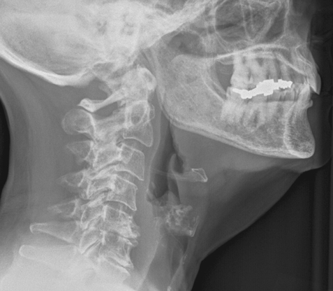
Fig. 1 Lateral skull radiograph (Aug. 2014) depicting osseous changes in the jaws, degenerative changes in the cervical spine, and a loss of intervertebral disc space between C5 and C6.

Fig. 2 Conventional plain film radiographs (Aug. 2014) of the left humerus (A) and left femur (B) depicting osseous changes associated with plasma cell leukemia.

Fig. 3 Lateral skull radiograph (Aug. 2014) depicting multiple punched-out radiolucent lesions consistent with a myeloproliferative disorder.

Fig. 4 Paraxial slice from a multidetector computed tomography study (Nov. 2014) depicts an intact right maxillary sinus free of gross disease.

Fig. 5 Dental panoramic radiograph acquired in April 2016 exhibits a relative radiolucency and loss of trabeculation apical to the maxillary right premolars.

Fig. 6 Periapical radiograph (Apr. 2016). Note the characteristic widening of the periodontal ligament spaces and sparse trabeculation in the periapical region surrounding the premolars.

Fig. 7 Paraxial slice from a cone beam computed tomography study (Apr. 2016) reveals the loss of the buccal and lingual cortical borders in the region of the premolars.

Fig. 8 Parasagittal slice from a cone beam computed tomography study (Apr. 2016) shows the altered trabeculation in the periapical region of the premolars, an oroantral communication, and the reactive mucositis of the right maxillary sinus.

Fig. 9 Photomicrographs exhibit a histopathological appearance of the known myeloproliferative disorder. Note the malignant neoplastic proliferation of atypical monomorphic lymphocytes forming cords, sheets, and clusters with normal nuclei and an eosinophilic cytoplasm typical of a myeloproliferative disorder (H&E stain, A. ×10, B. ×20).
Reference
-
1. Jimenez-Zepeda VH, Dominguez VJ. Plasma cell leukemia: a rare condition. Ann Hematol. 2006; 85:263–267.
Article2. Yamamoto JF, Goodman MT. Patterns of leukemia incidence in the United States by subtype and demographic characteristics, 1997-2002. Cancer Causes Control. 2008; 19:379–390.
Article3. Tiedemann RE, Gonzalez-Paz N, Kyle RA, Santana-Davila R, Price-Troska T, Van Wier SA, et al. Genetic aberrations and survival in plasma cell leukemia. Leukemia. 2008; 22:1044–1052.
Article4. Albarracin F, Fonseca R. Plasma cell leukemia. Blood Rev. 2011; 25:107–112.
Article5. Sher T, Miller KC, Deeb G, Lee K, Chanan-Khan A. Plasma cell leukaemia and other aggressive plasma cell malignancies. Br J Haematol. 2010; 150:418–427.
Article6. Han X, Kilfoy B, Zheng T, Holford TR, Zhu C, Zhu Y, et al. Lymphoma survival patterns by WHO subtype in the United States, 1973-2003. Cancer Causes Control. 2008; 19:841–858.
Article7. Campo E, Swerdlow SH, Harris NL, Pileri S, Stein H, Jaffe ES. The 2008 WHO classification of lymphoid neoplasms and beyond: evolving concepts and practical applications. Blood. 2011; 117:5019–5032.
Article8. Hayman SR, Fonseca R. Plasma cell leukemia. Curr Treat Options Oncol. 2001; 2:205–216.
Article9. Keraliya AR, Krajewski KM, Giardino AA, Tirumani SH, Shinagare AB, Ramaiya NH, et al. Imaging of nervous system involvement in hematologic malignancies: what radiologists need to know. AJR Am J Roentgenol. 2015; 205:604–617.
Article10. Fernandez de Larrea C, Kyle RA, Durie BG, Ludwig H, Usmani S, Vesole DH, et al. Plasma cell leukemia: consensus statement on diagnostic requirements, response criteria and treatment recommendations by the International Myeloma Working Group. Leukemia. 2013; 27:780–791.
Article11. Moulopoulos LA, Dimopoulos MA. Magnetic resonance imaging of the bone marrow in hematologic malignancies. Blood. 1997; 90:2127–2147.
Article12. Ali A, Paul Y, Nwabudike SM, Ogbonna O, Grantham M, Taddesse-Heath L. Plasma cell leukemia presenting as a chest wall mass: a case report. Case Rep Oncol. 2016; 9:338–343.
Article13. Epstein JB, Voss NJ, Stevenson-Moore P. Maxillofacial manifestations of multiple myeloma. An unusual case and review of the literature. Oral Surg Oral Med Oral Pathol. 1984; 57:267–271.14. Ruggiero SL. Diagnosis and staging of medication-related osteonecrosis of the jaw. Oral Maxillofac Surg Clin North Am. 2015; 27:479–487.
Article15. Kumar G, Manjunatha B. Metastatic tumors to the jaws and oral cavity. J Oral Maxillofac Pathol. 2013; 17:71–75.
Article
- Full Text Links
-
- Actions
-
Cited
- CITED
-
- Close
- Share
-
- Similar articles
-
- SOLITARY PLASMA CELL MYELOMA ON ANTERIOR MAXILLA: A CASE REPORT
- A case of primary plasma cell leukemia
- A case of primary plasma cell leukemia exhibiting hemophagocytic plasma cells relapsed with multiple cutaneous plasmacytoma
- Plasma cell leukemia with rouleaux formation involving neoplastic cells and RBC
- A Case of Metastatic Ovarian Cancer Arising in Plasma Cell Leukemia
