J Korean Ophthalmol Soc.
2016 Oct;57(10):1542-1548. 10.3341/jkos.2016.57.10.1542.
Utility of the Swept Source Optical Coherence Tomography for Measurements of Central Corneal Thickness
- Affiliations
-
- 1Cheil Eye Hospital, Daegu, Korea. vit.s0324@gmail.com
- 2Department of Ophthalmology, Keimyung University School of Medicine, Daegu, Korea.
- KMID: 2355408
- DOI: http://doi.org/10.3341/jkos.2016.57.10.1542
Abstract
- PURPOSE
To evaluate the efficacy of swept source optical coherence tomography (SS-OCT) by comparing the measurement of central corneal thickness (CCT) to the measurement obtained using Orbscan II, anterior segment optical coherence tomography (AS-OCT) and ultrasound pachymetry.
METHODS
One examiner measured the CCT in 65 eyes of 65 healthy subjects using Orbscan II, AS-OCT, SS-OCT and ultrasound pachymetry. The mean values and correlations were analyzed.
RESULTS
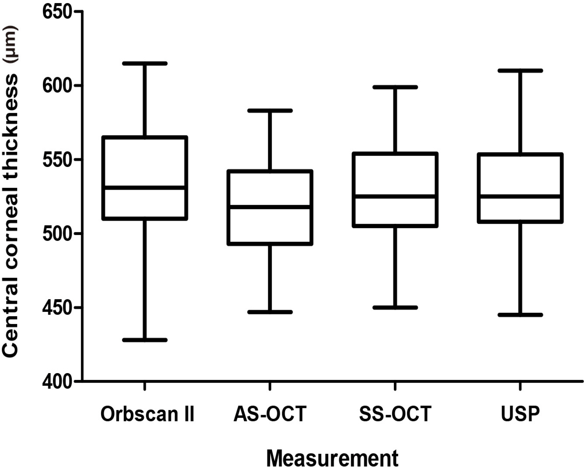
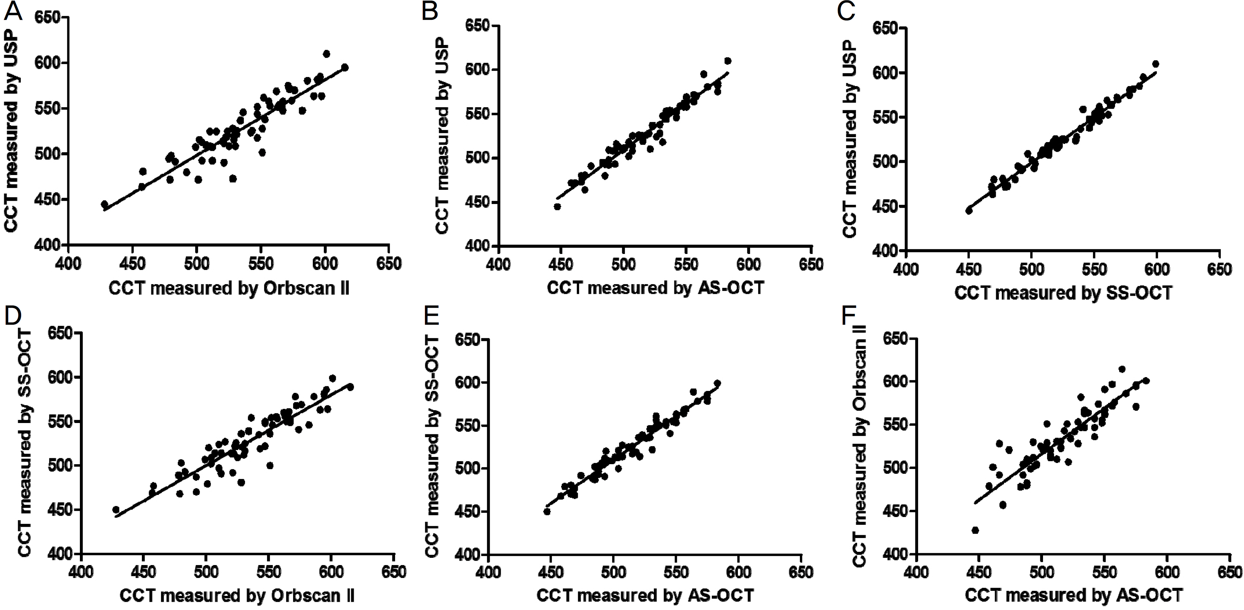
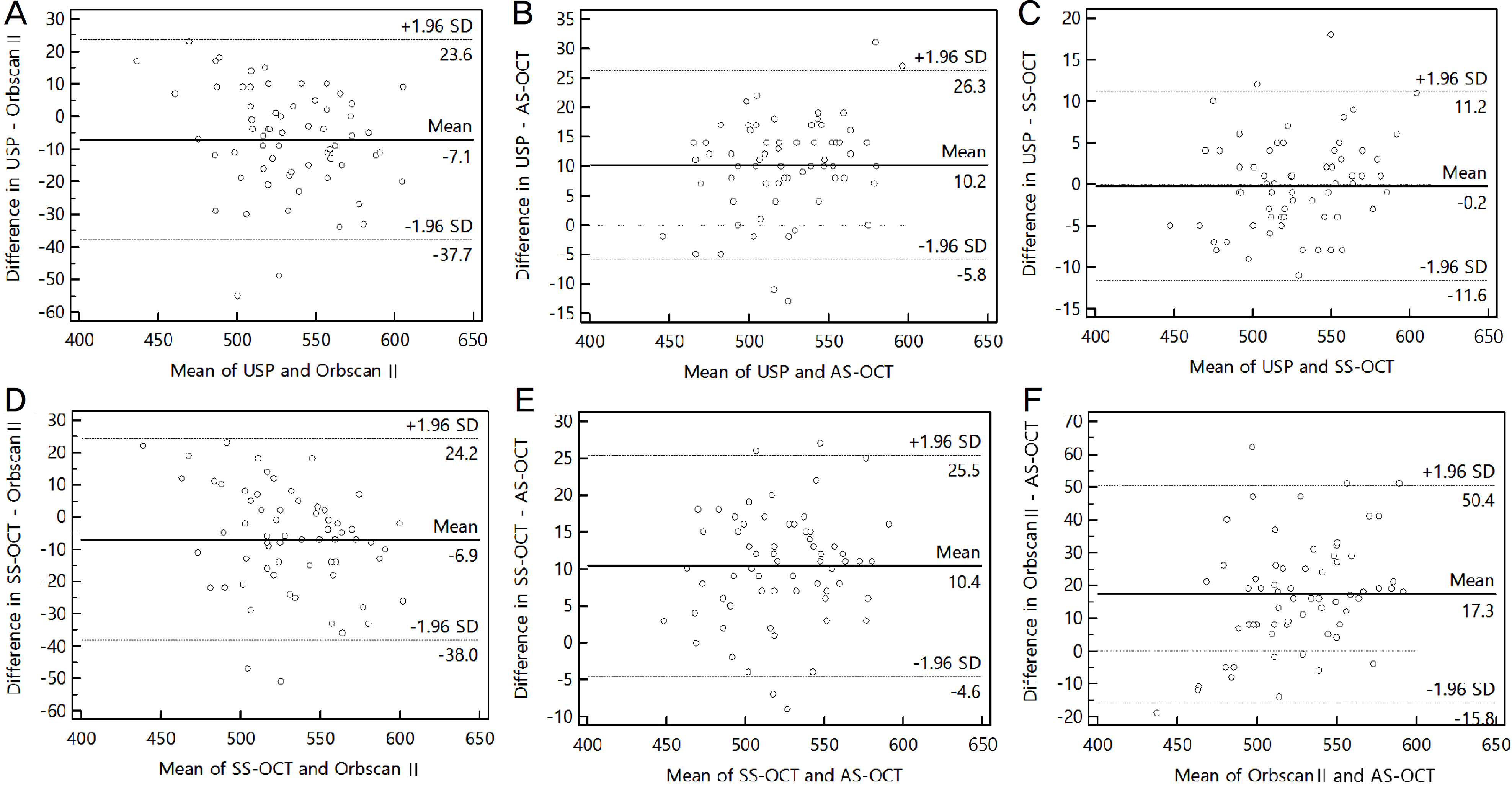
The average CCT measurements obtained using Orbscan II, AS-OCT, SS-OCT and ultrasound pachymetry were 534.83 ± 38.46, 517.80 ± 32.48, 528.22 ± 33.71 and 528.02 ± 34.90 µm, respectively. A significant linear correlation was observed among Orbscan II, AS-OCT, SS-OCT and ultrasound pachymetry (r > 0.894, p < 0.001). There was no significant difference between the SS-OCT and ultrasound pachymetry (p = 0.782).
CONCLUSIONS
The results of the 4 methods were significantly correlated and the SS-OCT reached a high level of agreement when CCT was determined using ultrasound pachymetry. The CCT measurements using SS-OCT is a better alternative for ultrasound pachymetry than Orbscan II and AS-OCT.
Keyword
Figure
Reference
-
References
1. Ou RJ, Shaw EL, Glasgow BJ. Keratectasia after laser in situ abdominal (LASIK): evaluation of the calculated residual stromal bed thickness. Am J Ophthalmol. 2002; 134:771–3.2. Wang Z, Chen J, Yang B. Posterior corneal surface topographic changes after laser in situ keratomileusis are related to residual abdominal bed thickness. Ophthalmology. 1999; 106:406–9. discussion 409–10.3. Grieshaber MC, Schoetzau A, Zawinka C, et al. Effect of central corneal thickness on dynamic contour tonometry and Goldmann applanation tonometry in primary open-angle glaucoma. Arch Ophthalmol. 2007; 125:740–4.
Article4. Jonas JB, Stroux A, Velten I, et al. Central corneal thickness corabdominal with glaucoma damage and rate of progression. Invest Ophthalmol Vis Sci. 2005; 46:1269–74.5. Kim DH, Kim MS, Kim JH. Early corneal-thickness changes after penetrating keratoplasty. J Korean Ophthalmol Soc. 1997; 38:1355–61.6. Solomon OD. Corneal indentation during ultrasonic pachometry. Cornea. 1999; 18:214–5.
Article7. Choi KS, Nam SM, Lee HK, et al. Comparison of central corneal thickness after the instillation of topical anesthetics: proparacaine versus oxybuprocaine. J Korean Ophthalmol Soc. 2005; 46:757–62.8. Li EY, Mohamed S, Leung CK, et al. Agreement among 3 methods to measure corneal thickness: ultrasound pachymetry, Orbscan II, and Visante anterior segment optical coherence tomography. Ophthalmology. 2007; 114:1842–7.
Article9. Akman A, Asena L, Güngör SG. Evaluation and comparison of the new swept source OCT-based IOLMaster 700 with the IOLMaster 500. Br J Ophthalmol. 2016; 100:1201–5.
Article10. Baum G, Greenwood I. The application of ultrasonic locating abdominals to ophthalmology. II. Ultrasonic slit lamp in the ultrasonic visualization of soft tissues. AMA Arch Ophthalmol. 1958; 60:263–79.11. Marsich MW, Bullimore MA. The repeatability of corneal abdominal measures. Cornea. 2000; 19:792–5.12. Miglior S, Albe E, Guareschi M, et al. Intraobserver and interobserver reproducibility in the evaluation of ultrasonic pachymetry measurements of central corneal thickness. Br J Ophthalmol. 2004; 88:174–7.
Article13. Ling T, Ho A, Holden BA. Method of evaluating ultrasonic pachometers. Am J Optom Physiol Opt. 1986; 63:462–6.
Article14. Copt RP, Thomas R, Mermoud A. Corneal thickness in ocular abdominal, primary open-angle glaucoma, and normal tension glaucoma. Arch Ophthalmol. 1999; 117:14–6.15. Al-Farhan HM, Al-Otaibi WM. Comparison of central corneal thickness measurements using ultrasound pachymetry, ultrasound biomicroscopy, and the Artemis-2 VHF scanner in normal eyes. Clin Ophthalmol. 2012; 6:1037–43.16. Khaja WA, Grover S, Kelmenson AT, et al. Comparison of central corneal thickness: ultrasound pachymetry versus abdominal optical coherence tomography, specular microscopy, and Orbscan. Clin Ophthalmol. 2015; 9:1065–70.17. Jonuscheit S, Doughty MJ. Regional repeatability measures of corneal thickness: Orbscan II and ultrasound. Optom Vis Sci. 2007; 84:52–8.
Article18. Choi SH, Kim JH, Han NS, et al. Comparison of corneal thickness measurements with optical low coherence reflectometry, orbscan system and ultrasound pachymeter. J Korean Ophthalmol Soc. 2006; 47:19–24.19. Thomas J, Wang J, Rollins AM, Sturm J. Comparison of corneal thickness measured with optical coherence tomography, abdominal pachymetry, and a scanning slit method. J Refract Surg. 2006; 22:671–8.20. Bao F, Wang Q, Cheng S, et al. Comparison and evaluation of abdominal corneal thickness using 2 new noncontact specular microscopes and conventional pachymetry devices. Cornea. 2014; 33:576–81.21. Nam SM, Im CY, Lee HK, et al. Accuracy of RTVue optical abdominal tomography, Pentacam, and ultrasonic pachymetry for the measurement of central corneal thickness. Ophthalmology. 2010; 117:2096–103.22. Uçakhan OO, Ozkan M, Kanpolat A. Corneal thickness abdominals in normal and keratoconic eyes: Pentacam comprehensive eye scanner versus noncontact specular microscopy and ultrasound pachymetry. J Cataract Refract Surg. 2006; 32:970–7.23. Christensen A, Nárvaez J, Zimmerman G. Comparison of central corneal thickness measurements by ultrasound pachymetry, konan noncontact optical pachymetry, and orbscan pachymetry. Cornea. 2008; 27:862–5.
Article24. Kunert KS, Peter M, Blum M, et al. Repeatability and agreement in optical biometry of a new swept-source optical coherence abdominal-based biometer versus partial coherence interferometry and optical low-coherence reflectometry. J Cataract Refract Surg. 2016; 42:76–83.25. Yaylali V, Kaufman SC, Thompson HW. Corneal thickness abdominals with the Orbscan Topography System and ultrasonic pachymetry. J Cataract Refract Surg. 1997; 23:1345–50.26. Kang PS, Yang YS, Kim JD. Comparison of corneal thickness measurements with the orbscan and ultrasonic pachymetry. J Korean Ophthalmol Soc. 2000; 41:1697–703.27. Liu Z, Huang AJ, Pflugfelder SC. Evaluation of corneal thickness and topography in normal eyes using the Orbscan corneal topography system. Br J Ophthalmol. 1999; 83:774–8.
Article28. Bechmann M, Thiel MJ, Neubauer AS, et al. Central corneal abdominal measurement with a retinal optical coherence tomography device versus standard ultrasonic pachymetry. Cornea. 2001; 20:50–4.29. Wong AC, Wong CC, Yuen NS, Hui SP. Correlational study of abdominal corneal thickness measurements on Hong Kong Chinese using optical coherence tomography, Orbscan and ultrasound pachymetry. Eye (Lond). 2002; 16:715–21.30. Shim HS, Choi CY, Lee HG, et al. Utility of the anterior segment optical coherence tomography for measurements of central corneal thickness. J Korean Ophthalmol Soc. 2007; 48:1643–8.
Article31. Zhao PS, Wong TY, Wong WL, et al. Comparison of central abdominal thickness measurements by visante anterior segment abdominal coherence tomography with ultrasound pachymetry. Am J Ophthalmol. 2007; 143:1047–9.32. Fukuda R, Usui T, Miyai T, et al. Corneal thickness and volume measurements by swept source anterior segment optical coherence tomography in normal subjects. Curr Eye Res. 2013; 38:531–6.
Article33. Prakash G, Agarwal A, Jacob S, et al. Comparison of four-ier-domain and time-domain optical coherence tomography for assessment of corneal thickness and intersession repeatability. Am J Ophthalmol. 2009; 148:282–90.e2.
Article34. Unterhuber A, Povazay B, Hermann B, et al. In vivo retinal optical coherence tomography at 1040 nm-enhanced penetration into the choroid. Opt Express. 2005; 13:3252–8.
- Full Text Links
- Actions
-
Cited
- CITED
-
- Close
- Share
- Similar articles
-
- Comparison of Anterior Segment Measurements between Scheimpflug-Placido Camera and New Swept-source Optical Coherence Tomography
- Comparison of Anterior Segment Measurements Between Swept-source Optical Coherence Tomography and Schiempflug Coherence Interferometer
- Comparison of Ocular Biometric Measurements Using New Swept-source Optical-coherence Tomography, Low-coherence Reflectometry, A-Scan Biometry, and Autokeratometry
- Diagnostic Ability of Macular Ganglion Cell Layer Measurements in Glaucoma Using Swept Source Optical Coherence Tomography
- Utility of the Noncontact Specular Microscopy for Measurements of Central Corneal Thickness


