Utility of the Noncontact Specular Microscopy for Measurements of Central Corneal Thickness
- Affiliations
-
- 1Department of Ophthalmology, Chosun University College of Medicine, Gwangju, Korea. ophkoh@hanmail.net
Abstract
- PURPOSE
To evaluate the efficacy of noncontact specular microscopy (NCSM) by comparing the measurement of central corneal thickness (CCT) to the measurement with anterior segment optical coherence tomography (AS-OCT) and ultrasound pachymetry (USP).
METHODS
One examiner measured the CCT of 50 eyes of 50 healthy young subjects using NCSM, AS-OCT, and USP. The mean values and correlations were analyzed.
RESULTS
The mean CCT value was 546.92 +/- 32.06 microm with NCSM, 535.24 +/- 30.54 microm with AS-OCT, and 546.38 +/- 30.70 microm with USP. The CCT measurements with NCSM and USP were significantly thicker than with AS-OCT (p < 0.001, p < 0.001, respectively). There were no significant differences between the measurements obtained with NCSM and USP (p = 0.505). The 3 instruments were significantly correlated (r > 0.900 in all groups, p < 0.001 in all groups).
CONCLUSIONS
CCT measurements of healthy eyes using NCSM are more correlated with USP than AS-OCT. The CCT measurement using NCSM is a better alternative for USP than AS-OCT.
Keyword
Figure
-
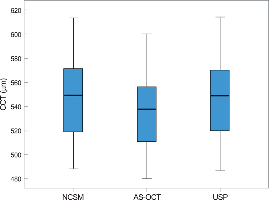
Figure 1. Mean value, 95% confidence interval (CI) and range of central corneal thickness (CCT) measured by noncontact specular microscopy (NCSM), anterior segment optical coherence tomography (AS-OCT) and ultrasound pachymetry (USP).

Figure 2. The correlation plot of the central corneal thickness (CCT) measured by noncontact specular microscopy (NCSM), anterior segment optical coherence tomography (AS-OCT) and ultrasound pachymetry (USP). (A) Correlation between NCSM and USP (r = 0.984, p < 0.001); The best-fit line (y = 30.8 +0.94x) is designated by solid line, and the equivalent line (y=x) by dashed line. (B) Correlation between NCSM and AS-OCT (r = 0.965, p < 0.001); The best-fit line (y = 32.7 +0.92x) is designated by solid line, and the equivalent line (y=x) by dashed line. (C) Correlation between AS-OCT and USP (r = 0.971, p < 0.001); The best-fit line (y = 24.1+ 0.98x) is designated by solid line, and the equivalent line (y = x) by dashed line.
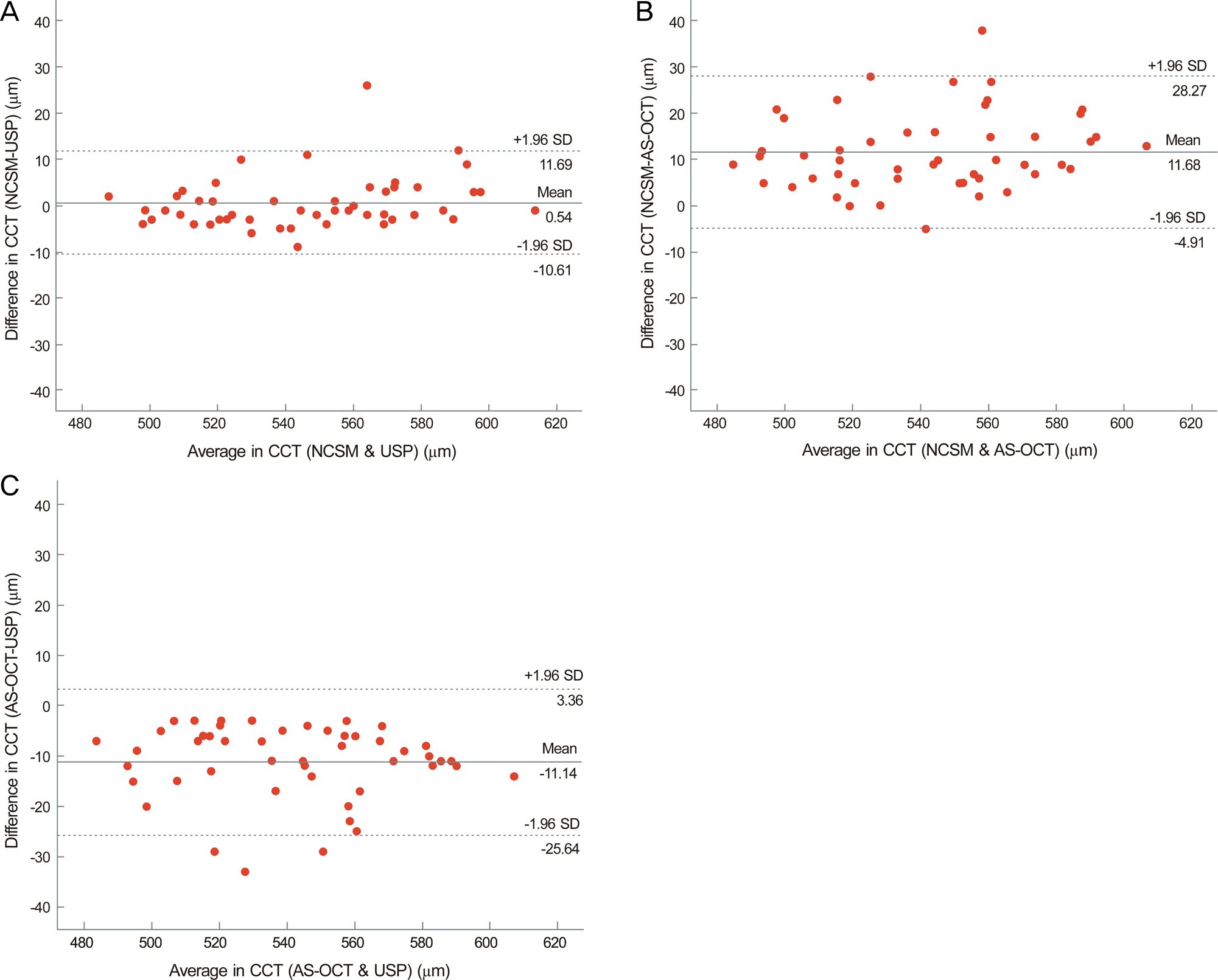
Figure 3. Bland-Altman plots between the 2 instruments. The middle solid line represents the mean difference in central corneal thickness (CCT) values and the upper and lower dashed lines represent the crude 95% limits of agreement (LoA). (A) Noncontact specular microscopy (NCSM) and ultrasound pachymetry (USP). (B) NCSM and anterior segment optical coherence tomography (AS-OCT). (C) AS-OCT and USP.
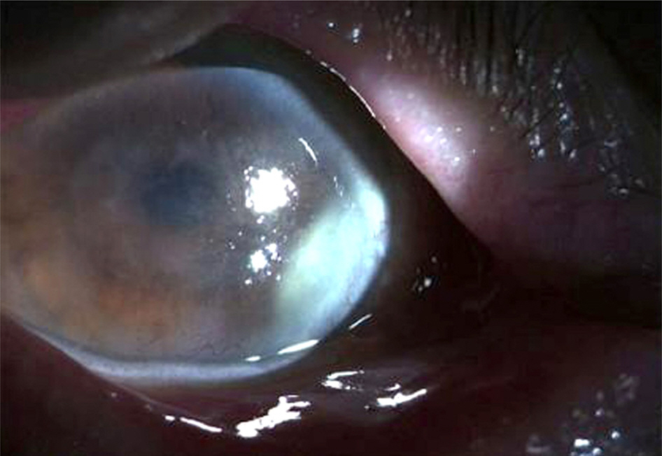
Figure 4. Anterior segment photo in a patient with keratouvei-tis, shows corneal edema with peripheral neovascularization. Central corneal thickness (CCT) measured by ultrasound pachymetry (USP) was 780 μm, measured by anterior segment optical coherence tomography (AS-OCT) was 757 μm, and measured by noncontact specular microscopy (NCSM) was 485 μm.
Reference
-
References
1. Reinstein DZ, Aslanides IM, Silverman RH. . High-frequency ultrasound corneal pachymetry in the assessment of corneal scars for therapeutic planning. CLAO J. 1994; 20:198–203.
Article2. Cheng H, Bates AK, Wood L, McPherson K.Positive correlation of corneal thickness and endothelial cell loss. Serial measurements after cataract surgery. Arch Ophthalmol. 1988; 106:920–2.3. Ou RJ, Shaw EL, Glasgow BJ.Keratectasia after laser in situ kera-tomileusis (LASIK): evaluation of the calculated residual stromal bed thickness. Am J Ophthalmol. 2002; 134:771–3.
Article4. Wang Z, Chen J, Yang B.Posterior corneal surface topographic changes after laser in situ keratomileusis are related to residual cor-neal bed thickness. Ophthalmology. 1999; 106:406–9. discussion 409-10.5. Jonas JB, Stroux A, Velten I. . Central corneal thickness corre-lated with glaucoma damage and rate of progression. Invest Ophthalmol Vis Sci. 2005; 46:1269–74.
Article6. Choi KS, Nam SM, Lee HK. . Comparison of central corneal thickness after the instillation of topical anesthetics: proparacaine versus oxybuprocaine. J Korean Ophthalmol Soc. 2005; 46:757–62.7. Giers U, Epple C.Comparison of A-scan device accuracy. J Cataract Refract Surg. 1990; 16:235–42.
Article8. Li EY, Mohamed S, Leung CK. . Agreement among 3 methods to measure corneal thickness: ultrasound pachymetry, Orbscan II, and Visante anterior segment optical coherence tomography. Ophthalmology. 2007; 114:1842–7.
Article9. Kim HS, Kim JH, Kim HM, Song JS.Comparison of corneal thick-ness measured by specular, US pachymetry, and orbscan in post-PKP eyes. J Korean Ophthalmol Soc. 2007; 48:245–50.10. Lee WH, Hwang YH, Kim SJ. . Comparison and repeatability of anterior segment parameters obtained by galilei and slit-lamp optical coherence tomography. J Korean Ophthalmol Soc. 2011; 52:53–9.
Article11. Shim HS, Choi CY, Lee HG. . Utility of the anterior segment optical coherence tomography for measurements of central corneal thickness. J Korean Ophthalmol Soc. 2007; 48:1643–8.
Article12. Jung YG, Song JS, Kim HM, Jung HR. Comparison of corneal thickness measurements with noncontact specular microscope and ultrasonic pachymeter. J Korean Ophthalmol Soc. 2004; 45:1060–5.13. Kim HY, Budenz DL, Lee PS. . Comparison of central corneal thickness using anterior segment optical coherence tomography vs ultrasound pachymetry. Am J Ophthalmol. 2008; 145:228–32.
Article14. Kim DW, Yi KY, Choi DG, Shin YJ.Corneal thickness measured by dual scheimpflug, anterior segment optical coherence tomog-raphy, and ultrasound pachymetry. J Korean Ophthalmol Soc. 2012; 53:1412–8.
Article15. Miglior S, Albe E, Guareschi M. . Intraobserver and interob-server reproducibility in the evaluation of ultrasonic pachymetry measurements of central corneal thickness. Br J Ophthalmol. 2004; 88:174–7.
Article16. Solomon OD.Corneal indentation during ultrasonic pachometry. Cornea. 1999; 18:214–5.
Article17. Ventura AC, Wälti R, Böhnke M.Corneal thickness and endothe-lial density before and after cataract surgery. Br J Ophthalmol. 2001; 85:18–20.
Article18. Almubrad TM, Osuagwu UL, Alabbadi I, Ogbuehi KC.Comparison of the precision of the Topcon SP-3000P specular microscope and an ultrasound pachymeter. Clin Ophthalmol. 2011; 5:871–6.
Article19. Thomas J, Wang J, Rollins AM, Sturm J.Comparison of corneal thickness measured with optical coherence tomography, ultrasonic pachymetry, and a scanning slit method. J Refract Surg. 2006; 22:671–8.
Article20. Bovelle R, Kaufman SC, Thompson HW, Hamano H.Corneal thickness measurements with the Topcon SP-2000P specular mi-croscope and an ultrasound pachymeter. Arch Ophthalmol. 1999; 117:868–70.
Article21. Módis L Jr, Langenbucher A, Seitz B.Corneal thickness measure-ments with contact and noncontact specular microscopic and ultra-sonic pachymetry. Am J Ophthalmol. 2001; 132:517–21.
Article22. Bechmann M, Thiel MJ, Neubauer AS. . Central corneal thick-ness measurement with a retinal optical coherence tomography de-vice versus standard ultrasonic pachymetry. Cornea. 2001; 20:50–4.
Article23. Prakash G, Agarwal A, Jacob S. . Comparison of fourier-do-main and time-domain optical coherence tomography for assess-ment of corneal thickness and intersession repeatability. Am J Ophthalmol. 2009; 148:282–90.e2.
Article24. Azen SP, Burg KA, Smith RE, Maguen E.A comparison of three methods for the measurement of corneal thickness. Invest Ophthalmol Vis Sci. 1979; 18:535–8.25. Zhao PS, Wong TY, Wong WL. . Comparison of central cor-neal thickness measurements by visante anterior segment optical coherence tomography with ultrasound pachymetry. Am J Ophthalmol. 2007; 143:1047–9.
Article26. Li H, Leung CK, Wong L. . Comparative study of central cor-neal thickness measurement with slit-lamp optical coherence tomog-raphy and visante optical coherence tomography. Ophthalmology. 2008; 115:796–801.e2.
Article
- Full Text Links
-
- Actions
-
Cited
- CITED
-
- Close
- Share
-
- Similar articles
-
- Comparison of Central Corneal Thickness Measurements between Noncontact Specular Microscopy and Ultrasound Pachymetry
- Comparison of Corneal Thickness Measured by Specular, US Pachymetry, and Orbscan in Post-PKP Eyes
- Comparison of Corneal Thickness Measurements with Noncontact Specular Microscope and Ultrasonic Pachymeter
- Central Corneal Thickness Measured by Noncontact Specular Microscopy, Dual Rotating Scheimpflug Camera and Ultrasound Pachymetry
- Comparision of central Corneal Endotherial Cell Density with Peripheral Areas According to Age by Specular Microscopy
