In vitro evaluation of a newly produced resin-based endodontic sealer
- Affiliations
-
- 1Department of Conservative Dentistry, School of Dentistry and Institute of Oral Bioscience, Chonbuk National University, Jeonju, Korea. endomin@gmail.com
- 2Department of Conservative Dentistry, Wonkwang University Dental Hospital, Iksan, Korea.
- 3Biomedical Research Institute of Chonbuk National University Hospital, Jeonju, Korea.
- 4Department of Conservative Dentistry, School of Dentistry, Dankook University, Cheonan, Korea.
- KMID: 2346767
- DOI: http://doi.org/10.5395/rde.2016.41.3.189
Abstract
OBJECTIVES
A variety of root canal sealers were recently launched to the market. This study evaluated physicochemical properties, biocompatibility, and sealing ability of a newly launched resin-based sealer (Dia-Proseal, Diadent) compared to the existing root canal sealers (AHplus, Dentsply DeTrey and ADseal, Metabiomed).
MATERIALS AND METHODS
The physicochemical properties of the tested sealers including pH, solubility, dimensional change, and radiopacity were evaluated. Biocompatibility was measured using the 3-(4,5-dimethylthiazol-2-yl)-2,5-diphenyltetrazolium bromide (MTT) assay. For microleakage test, single-rooted teeth were instrumented, and obturated with gutta-percha and one of the sealers (n = 10). After immersion in 1% methylene blue solution for 2 weeks, the specimens were split longitudinally. Then, the maximum length of staining was measured. Statistical analysis was performed by one-way analysis of variance followed by Tukey test (p = 0.05).
RESULTS
Dia-Proseal showed the highest pH value among the tested sealers (p < 0.05). ADseal showed higher dimensional change compared to AHplus and Dia-Proseal (p < 0.05). The solubility values of AHplus and Dia-Proseal were similar, whereas ADseal had the lowest solubility value (p < 0.05). The flow values of sealer in increasing order were AHplus, DiaProseal, and ADseal (p < 0.05). The radiopacity of AHplus was higher than those of ADseal and Dia-Proseal (p < 0.05). The cell viability of the tested materials was statistically similar throughout the experimental period. There were no significant differences in microleakage values among the tested samples.
CONCLUSIONS
The present study indicates that Dia-Proseal has acceptable physicochemical properties, biocompatibility, and sealing ability.
Keyword
MeSH Terms
Figure
-
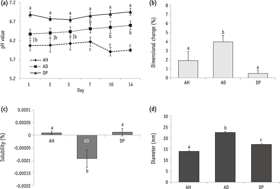
Figure 1 Changes in physicochemical properties of the tested sealers. (a) pH (n = 3); (b) dimensional change (n = 5); (c) solubility (n = 5); (d) flow (n = 3). Groups identified by the same symbols were not significantly different in the same gene group. AH, AHplus; AD, ADseal; DP, Dia-Proseal.
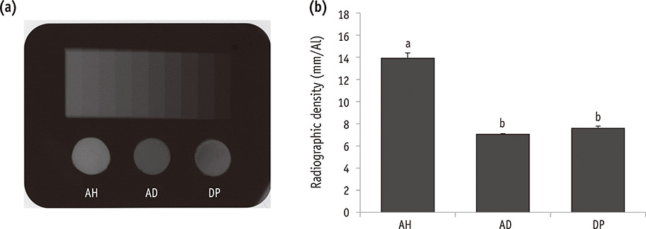
Figure 2 Radiopacity of the tested materials (n = 3). (a) Radiograph showing the radiopacity of each material and its equivalence to that of the aluminum step-wedge; (b) Relative radiographic density of each material in comparison with that of a 10-step aluminum step-wedge. Groups identified by the same symbols were not significantly different in the same gene group. AH, AHplus; AD, ADseal; DP, Dia-Proseal.
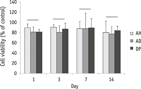
Figure 3 Biocompatibility of the tested materials (n = 4). Cell viability tested by the MTT assay. Groups connected with the bar were not significantly different in the same gene group. AH, AHplus; AD, ADseal; DP, Dia-Proseal.
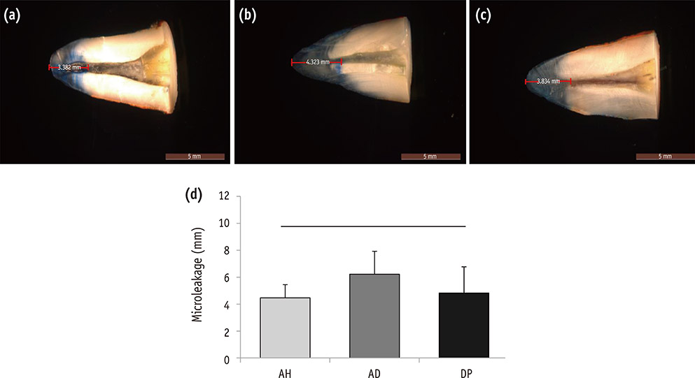
Figure 4 Representative photographs showing the measurement of the apical dye penetration of (a) AHplus, (b) ADseal, and (c) DiaProseal; (d) Average length of dye penetration. Values are shown as the mean ± standard deviation (n = 10). There was no significant difference in microleakage value among the tested groups. AH, AHplus; AD, ADseal; DP, Dia-Proseal.
Cited by 3 articles
-
Micro-computed tomographic evaluation of a new system for root canal filling using calcium silicate-based root canal sealers
Mario Tanomaru-Filho, Fernanda Ferrari Esteves Torres, Jader Camilo Pinto, Airton Oliveira Santos-Junior, Karina Ines Medina Carita Tavares, Juliane Maria Guerreiro-Tanomaru
Restor Dent Endod. 2020;45(3):e34. doi: 10.5395/rde.2020.45.e34.Flow characteristics and alkalinity of novel bioceramic root canal sealers
Anastasios Katakidis, Konstantinos Sidiropoulos, Elisabeth Koulaouzidou, Christos Gogos, Nikolaos Economides
Restor Dent Endod. 2020;45(4):e42. doi: 10.5395/rde.2020.45.e42.A micro-computed tomographic study using a novel test model to assess the filling ability and volumetric changes of bioceramic root repair materials
Fernanda Ferrari Esteves Torres, Jader Camilo Pinto, Gabriella Oliveira Figueira, Juliane Maria Guerreiro-Tanomaru, Mario Tanomaru-Filho
Restor Dent Endod. 2020;46(1):e2. doi: 10.5395/rde.2021.46.e2.
Reference
-
1. Lucena-Martín C, Ferrer-Luque CM, González-Rodríguez MP, Robles-Gijón V, Navajas-Rodríguez de. A comparative study of apical leakage of Endomethasone, Topseal and Roeko Seal cements. J Endod. 2002; 28:423–426.2. Grossman LI. Endodontic practice. 10th ed. Philadelphia: Henry Kimpton Publishers;1981. p. 297.3. Donnelly A, Sword J, Nishitani Y, Yoshiyama M, Agee K, Tay FR, Pashley DH. Water sorption and solubility of methacrylate resin-based root canal sealers. J Endod. 2007; 33:990–994.
Article4. Schröeder A. The impermeability of root canal filling material and first demonstrations of new root filling materials. SSO Schweiz Monatsschr Zahnheilkd. 1954; 64:921–931.5. Versiani MA, Carvalho-Junior JR, Padilha MI, Lacey S, Pascon EA, Sousa-Neto MD. A comparative study of physicochemical properties of AH Plus and Epiphany root canal sealants. Int Endod J. 2006; 39:464–471.
Article6. Bouillaguet S, Shaw L, Barthelemy J, Krejci I, Wataha JC. Long-term sealing ability of Pulp Canal Sealer, AH Plus, GuttaFlow and Epiphany. Int Endod J. 2008; 41:219–226.
Article7. Ørstavik D. Materials used for root canal obturation: technical, biological and clinical testing. Endod Topics. 2005; 12:25–38.
Article8. Lim ES, Park YB, Kwon YS, Shon WJ, Lee KW, Min KS. Physical properties and biocompatibility of an injectable calcium-silicate-based root canal sealer: in vitro and in vivo study. BMC Oral Health. 2015; 15:129.
Article9. Silva EJ, Rosa TP, Herrera DR, Jacinto RC, Gomes BP, Zaia AA. Evaluation of cytotoxicity and physicochemical properties of calcium silicate-based endodontic sealer MTA Fillapex. J Endod. 2013; 39:274–277.
Article10. Islam I, Chng HK, Yap AU. Comparison of the physical and mechanical properties of MTA and portland cement. J Endod. 2006; 32:193–197.
Article11. Marciano MA, Guimarães BM, Ordinola-Zapata R, Bramante CM, Cavenago BC, Garcia RB, Bernardineli N, Andrade FB, Moraes IG, Duarte MA. Physical properties and interfacial adaptation of three epoxy resin-based sealers. J Endod. 2011; 37:1417–1421.
Article12. Silva EJ, Perez R, Valentim RM, Belladonna FG, De-Deus GA, Lima IC, Neves AA. Dissolution, dislocation and dimensional changes of endodontic sealers after a solubility challenge: a micro-CT approach. Int Endod J. 2016; 03. 22. DOI: 10.1111/iej.12636. [Epub ahead of print].
Article13. Almeida JF, Gomes BP, Ferraz CC, Souza-Filho FJ, Zaia AA. Filling of artificial lateral canals and microleakage and flow of five endodontic sealers. Int Endod J. 2007; 40:692–699.
Article14. Chang SW, Lee YK, Zhu Q, Shon WJ, Lee WC, Kum KY, Baek SH, Lee IB, Lim BS, Bae KS. Comparison of the rheological properties of four root canal sealers. Int J Oral Sci. 2015; 23:56–61.
Article15. Kim YK, Grandini S, Ames JM, Gu LS, Kim SK, Pashley DH, Gutmann JL, Tay FR. Critical review on methacrylate resin-based root canal sealers. J Endod. 2010; 36:383–399.
Article16. Zmener O, Pameijer CH, Macri E. Evaluation of the apical seal in root canals prepared with a new rotary system and obturated with a methacrylate based endodontic sealer: an in vitro study. J Endod. 2005; 31:392–395.
Article17. Aptekar A, Ginnan K. Comparative analysis of microleakage and seal for 2 obturation materials: Resilon/Epiphany and gutta-percha. J Can Dent Assoc. 2006; 72:245.18. De-Deus G, Audi C, Murad C, Fidel S, Fidel RA. Sealing ability of oval-shaped canals filled using the System B heat source with either gutta-percha or Resilon: an ex vivo study using a polymicrobial leakage model. Oral Surg Oral Med Oral Pathol Oral Radiol Endod. 2007; 104:e114–e119.19. Baumgartner G, Zehnder M, Paqué F. Enterococcus faecalis type strain leakage through root canals filled with Gutta-Percha/AH plus or Resilon/Epiphany. J Endod. 2007; 33:45–47.
Article20. Tunga U, Bodrumlu E. Assessment of the sealing ability of a new root canal obturation material. J Endod. 2006; 32:876–878.
Article21. Sagsen B, Er O, Kahraman Y, Orucoglu H. Evaluation of microleakage of roots filled with different techniques with a computerized fluid filtration technique. J Endod. 2006; 32:1168–1170.
Article22. Kaya BU, Kececi AD, Belli S. Evaluation of the sealing ability of gutta-percha and thermoplastic synthetic polymer-based systems along the root canals through the glucose penetration model. Oral Surg Oral Med Oral Pathol Oral Radiol Endod. 2007; 104:e66–e73.
Article23. Wu MK, Wesselink PR. Endodontic leakage studies reconsidered. Part I. Methodology, application and relevance. Int Endod J. 1993; 26:37–43.
Article24. Oliver CM, Abbott PV. Correlation between clinical success and apical dye penetration. Int Endod J. 2001; 34:637–644.
Article25. Delivanis PD, Chapman KA. Comparison and reliability of techniques for measuring leakage and marginal penetration. Oral Surg Oral Med Oral Pathol. 1982; 53:410–416.
Article26. Martell B, Chandler NP. Electrical and dye leakage comparison of three root-end restorative materials. Quintessence Int. 2002; 33:30–34.
- Full Text Links
-
- Actions
-
Cited
- CITED
-
- Close
- Share
-
- Similar articles
-
- Cytotoxicity of resin-based root canal sealer, adseal
- Retention of fiber posts to the optimally and over-prepared dowel spaces
- Micro-computed tomographic evaluation of single-cone obturation with three sealers
- Biocompatibility and Bioactivity of Four Different Root Canal Sealers in Osteoblastic Cell Line MC3T3-El
- Physical properties of a new resin-based root canal sealer in comparison with AH Plus Jet
