Acquired Adult Flatfoot: Pathophysiology, Diagnosis, and Nonoperative Treatment
- Affiliations
-
- 1Department of Orthopaedic Surgery, Samsung Medical Center, Sungkyunkwan University School of Medicine, Seoul, Korea. kissung@gmail.com
- KMID: 2333470
- DOI: http://doi.org/10.14193/jkfas.2014.18.3.87
Abstract
- Acquired adult flatfoot is a deformity characterized by a decreased medial longitudinal arch and a hindfoot valgus with or without forefoot abduction. The etiologies of this deformity include posterior tibial tendon dysfunction, rheumatoid arthritis, trauma, Charcot's joint, neurologic deficit, and damage to the medial spring ligament complex or plantar fascia. Among these, posterior tibial tendon dysfunction is the most well-known cause. Although posterior tibial tendon dysfunction has been regarded as a synonym of acquired adult acquired flatfoot, failure of the ligaments supporting the arch can also result in progressive deformity even without a posterior tibial tendon problem. The authors describe the pathophysiology, diagnosis, and nonoperative treatment of acquired adult flatfoot, focusing on posterior tibial tendon dysfunction.
Keyword
MeSH Terms
Figure
-

Figure 1. Medial longitudinal arch is reestablished when an examiner extends the great toe while the patient is standing in the case of flexible flat foot.

Figure 2. This figure shows ‘too many toes sign’ in right side. When looking at the heel from the back of the patient, usually only the fifth toe and half of the fourth toe are seen. In a flatfoot deformity (right foot), more of the little toe can be seen.
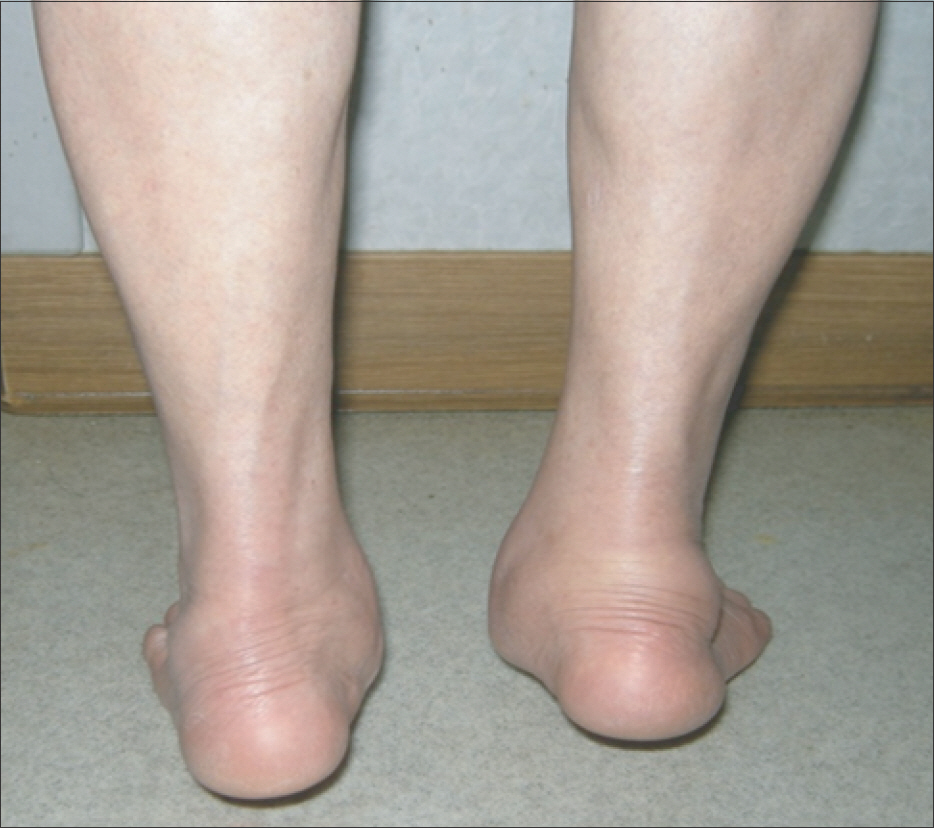
Figure 3. During the double-limb heel-rise test, the right heel shows a failed restoration of inversion.

Figure 4. On standing foot anteroposterior view, talo-calcaneal angle (A) and talo-navicular coverage angle (B) can be measured. α: talocalcaneal angle is the angle between the long axis of talus and the long axis of calcaneus. β: talo-navicular coverage angle is formed between the line through base of distal articular cap of talus and the line through proximal articular cap of navicula.

Figure 5. On standing lateral view, talo-1st metatarsal angle (α) and calcaneal pitch angle (β) can be measured.
Cited by 2 articles
-
Diagnosis of Flatfoot Deformity
Tae Hoon Lee, Suh Woo Chay, Hak Jun Kim
J Korean Foot Ankle Soc. 2016;20(1):1-5. doi: 10.14193/jkfas.2016.20.1.1.Treatment of Flatfoot Deformity
Dong-Oh Lee, Hong-Geun Jung
J Korean Foot Ankle Soc. 2016;20(1):6-11. doi: 10.14193/jkfas.2016.20.1.6.
Reference
-
1.Johnson KA. Tibialis posterior tendon rupture1 Clin Orthop Relat Res1. 1983. 177:140–71.2.Mann RA. Rupture of the tibialis posterior tendon1 Instr Course Lect1. 1984. 33:302–91.3.Myerson MS. Adult acquired flatfoot deformity: treatment of dysfunction of the posterior tibial tendon1 Instr Course Lect1. 1997. 46:393–4051.4.Williams G., Widnall J., Evans P., Platt S. Could failure of the spring ligament complex be the driving force behind the development of the adult flatfoot deformity? J Foot Ankle Surg1. 2014. 53:152–51.
Article5.Niki H., Ching RP., Kiser P., Sangeorzan BJ. The effect of posterior tibial tendon dysfunction on hindfoot kinematics1 Foot Ankle Int1. 2001. 22:292–3001.6.Jennings MM., Christensen JC. The effects of sectioning the spring ligament on rearfoot stability and posterior tibial tendon efficiency1 J Foot Ankle Surg1. 2008. 47:219–241.7.Yeap JS., Birch R., Singh D. Long-term results of tibialis posterior tendon transfer for drop-foot1 Int Orthop1. 2001. 25:114–81.8.Tao K., Ji WT., Wang DM., Wang CT., Wang X. Relative contributions of plantar fascia and ligaments on the arch static stability: a finite element study1 Biomed Tech (Berl)1. 2010. 55:265–711.9.Iaquinto JM., Wayne JS. Computational model of the lower leg and foot/ankle complex: application to arch stability1 J Biomech Eng1. 2010. 132:0210091.10.Shuen V., Prem H. Acquired unilateral pes planus in a child caused by a ruptured plantar calcaneonavicular (spring) liga-ment1 J Pediatr Orthop B1. 2009. 18:129–301.11.Tryfonidis M., Jackson W., Mansour R., Cooke PH., Teh J., Ostlere S, et al. Acquired adult flat foot due to isolated plantar calcaneonavicular (spring) ligament insufficiency with a normal tibialis posterior tendon1 Foot Ankle Surg1. 2008. 14:89–951.12.Orr JD., Nunley JA 2nd. Isolated spring ligament failure as a cause of adult-acquired flatfoot deformity1 Foot Ankle Int1. 2013. 34:818–231.13.Blake RL., Anderson K., Ferguson H. Posterior tibial tendinitis1 A literature review with case reports1 J Am Podiatr Med Assoc1. 1994. 84:141–91.14.Helal B. Tibialis posterior tendon synovitis and rupture1 Acta Or-thop Belg1. 1989. 55:457–601.15.Supple KM., Hanft JR., Murphy BJ., Janecki CJ., Kogler GF. Posterior tibial tendon dysfunction1 Semin Arthritis Rheum1. 1992. 22:106–131.16.Basmajian JV., Stecko G. The role of muscles in arch support of the foot1 J Bone Joint Surg Am1. 1963. 45:1184–901.17.Chhabra A., Soldatos T., Chalian M., Faridian-Aragh N., Fritz J., Fayad LM, et al. 3-Tesla magnetic resonance imaging evaluation of posterior tibial tendon dysfunction with relevance to clinical staging1 J Foot Ankle Surg1. 2011. 50:320–81.18.Trnka HJ. Dysfunction of the tendon of tibialis posterior1 J Bone Joint Surg Br1. 2004. 86:939–461.19.Deland JT. Adult-acquired flatfoot deformity1 J Am Acad Orthop Surg1. 2008. 16:399–4061.20.Khoury NJ., el-Khoury GY., Saltzman CL., Brandser EA. MR imaging of posterior tibial tendon dysfunction1 AJR Am J Roentgenol1. 1996. 167:675–821.21.Weinraub GM., Saraiya MJ. Adult flatfoot/posterior tibial tendon dysfunction: classification and treatment1 Clin Podiatr Med Surg1. 2002. 19:345–701.22.Frey C., Shereff M., Greenidge N. Vascularity of the posterior tibial tendon1 J Bone Joint Surg Am1. 1990. 72:884–81.23.Petersen W., Hohmann G., Stein V., Tillmann B. The blood supply of the posterior tibial tendon1 J Bone Joint Surg Br1. 2002. 84:141–41.24.Rosenberg ZS. Chronic rupture of the posterior tibial tendon1 Magn Reson Imaging Clin N Am1. 1994. 2:79–871.25.Mosier SM., Pomeroy G., Manoli A 2nd. Pathoanatomy and etiology of posterior tibial tendon dysfunction1 Clin Orthop Relat Res1. 1999. 365:12–221.26.Holmes GB Jr., Mann RA. Possible epidemiological factors associated with rupture of the posterior tibial tendon1 Foot Ankle1. 1992. 13:70–91.27.Smith CF. Anatomy, function, and pathophysiology of the posterior tibial tendon1 Clin Podiatr Med Surg1. 1999. 16:399–4061.28.DeOrio JK., Shapiro SA., McNeil RB., Stansel J. Validity of the posterior tibial edema sign in posterior tibial tendon dysfunction1 Foot Ankle Int1. 2011. 32:189–921.29.Sangeorzan BJ., Mosca V., Hansen ST Jr. Effect of calcaneal lengthening on relationships among the hindfoot, midfoot, and forefoot1 Foot Ankle1. 1993. 14:136–411.30.Johnson KA., Strom DE. Tibialis posterior tendon dysfunction1 Clin Orthop Relat Res1. 1989. 239:196–2061.31.Kulig K., Burnfield JM., Requejo SM., Sperry M., Terk M. Selective activation of tibialis posterior: evaluation by magnetic resonance imaging1 Med Sci Sports Exerc1. 2004. 36:862–71.32.Neville C., Lemley FR. Effect of ankle-foot orthotic devices on foot kinematics in Stage II posterior tibial tendon dysfunction1 Foot Ankle Int1. 2012. 33:406–141.33.Chao W., Wapner KL., Lee TH., Adams J., Hecht PJ. Nonoperative management of posterior tibial tendon dysfunction1 Foot Ankle Int1. 1996. 17:736–411.34.Augustin JF., Lin SS., Berberian WS., Johnson JE. Nonoperative treatment of adult acquired flat foot with the Arizona brace1 Foot Ankle Clin1. 2003. 8:491–5021.35.Alvarez RG., Marini A., Schmitt C., Saltzman CL. Stage I and II posterior tibial tendon dysfunction treated by a structured nonoperative management protocol: an orthosis and exercise pro-gram1 Foot Ankle Int1. 2006. 27:2–81.36.Jahss MH. Spontaneous rupture of the tibialis posterior tendon: clinical findings, tenographic studies, and a new technique of repair1 Foot Ankle1. 1982. 3:158–661.37.Ross JA. Posterior tibial tendon dysfunction in the athlete1 Clin Podiatr Med Surg1. 1997. 14:479–881.
- Full Text Links
-
- Actions
-
Cited
- CITED
-
- Close
- Share
-
- Similar articles
-
- Adult flatfoot
- Diagnosis of Flatfoot Deformity
- Operative Treatment of Acquired Adult Flatfoot
- Adult Idiopathic Flexible Flat Foot Treated with Medial Sliding Calcaneal Osteotomy and Subtalar Arthroereisis: Report of 1 Case
- Operative Treatment of Adult Flexible Flatfoot with Young's Tenosuspension: Case Report
