Adult flatfoot
- Affiliations
-
- 1Department of Orthopaedic Surgery, Hanyang University College of Medicine, Seoul, Korea. lbgwej@hanmail.net
- KMID: 2064881
- DOI: http://doi.org/10.5124/jkma.2014.57.3.243
Abstract
- Flatfoot deformity in adults is a type of postural deformity of the foot in which the arch collapses. This condition includes a wide spectrum of clinical situations, ranging from asymptomatic to progressive and disabling pathology. The common causes of adult-acquired flatfoot deformity are sustained flexible flatfoot from childhood, posterior tibial tendon insufficiency, tarsal coalition, generalized inflammatory diseases, neuropathic arthropathy, and posttraumatic deformities. The treatment of adult acquired flatfoot deformity should be individualized in each case, depending on the causes, symptoms, severity of deformity, and flexibility of the deformity. Therefore, it is mandatory for physicians to be acquainted with the basic pathomechanics of flatfoot deformity as well as the diagnostic procedures and treatments for each condition. The treatment usually begins with conservative methods and variable surgical procedures could be selectively performed. This article reviews the basic pathoanatomy, the diagnostic procedures for various causes and the treatment of flatfoot deformity in adult.
Keyword
MeSH Terms
Figure
-

Figure 1 (A) Posteroanterior view of both feet, hindfoot valgus deformity is prominent on left side. (B) Medial view of left foot, medial longitudinal arch is markedly collapsed.
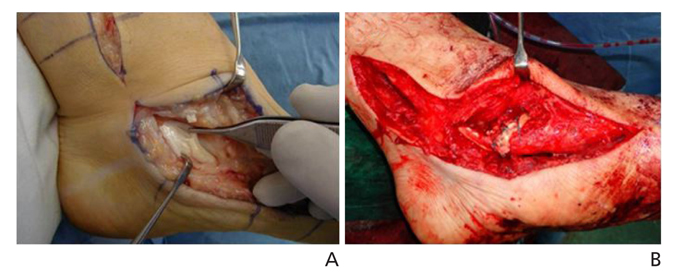
Figure 2 Photographs, taken at the surgical field, markedly degenerated posterior tibial tendon was shown (A). Flexor digitorum longus tendon was transferred to the navicular bone, adjacent to the insertion of degenerated posterior tibial tendon (B).
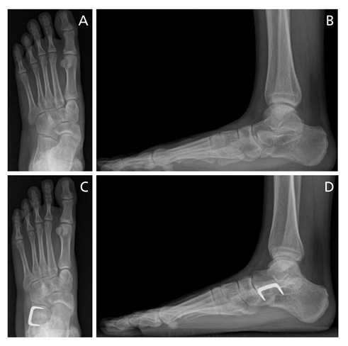
Figure 3 Example of anterior calcaneal distraction osteotomy (Evan's procedure). Preoperative anteroposterior radiographs reveals accessory navicular bone, abducted forefoot, pronation of entire foot in weight bearing position (A). Lateral radiographs showing talocalcaneal coalition, negative talar first metatarsal angle, decreased calcaneal pitch angle (B). Excision of bone bridge and anterior calcaneal distraction osteotomy were done (C,D). Note that the osteotomy plane was made just posterior to the calcaneocuboidal joint.
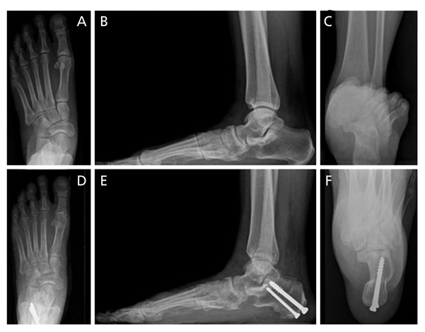
Figure 4 Preoperative anteroposterior radiograph (A). Lateral radiographs shows negative talar-first metatarsal angle (B). Hindfoot alignment view reveals marked laterally translated hindfoot (C). Postoperative radiographs. Medial calcaneal displacement osteotomy and the flexor digitorum longus tendon transfer were done (D,E,F).
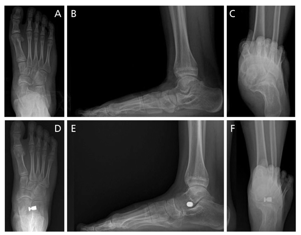
Figure 5 Preoperative radiographs reveals markedly pronated entire foot position and abducted forefoot, note that the uncoverage of medial talar head due to lateral subluxation of navicular bone (A). Markedly negative talar first metatarsal angle and calcaneal pitch angle were seen (B), and hindfoot alignment radiographs reveals severe valgus angulation of hindfoot (C). Arthroereisis procedure was done. Postoperatively, forefoot abduction and pronation of foot were corrected (D). Talar first metatarsal angle and calcaneal pitch angle were restored (E), and valgus angulation of hindfoot was greatly improved (F).
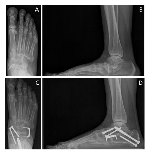
Figure 6 Severe inflammatory arthritis on triple joint accompanying collapse of medial longitudinal arch were observed in patient with rheumatoid arthritis (A,B). Medial calcaneal displacement osteotomy and triple arthrodesis with correction of joint alignment were done, postoperatively, medial longitudinal arch was greatly restored (C,D).
Cited by 1 articles
-
Diagnosis of Flatfoot Deformity
Tae Hoon Lee, Suh Woo Chay, Hak Jun Kim
J Korean Foot Ankle Soc. 2016;20(1):1-5. doi: 10.14193/jkfas.2016.20.1.1.
Reference
-
1. Lee KT, Gwak GD, Kim DY, Kim ES, Kim CR, Kim JY, Song JY, Song HH, Ahn JH, Yang KW. Foot and ankle surgery. Seoul: Koonja Publishing;2004.2. Sarrafian SK. Sarrafian's anatomy of the foot and ankle: descriptive, topographic, functional. 2nd ed. Philadelphia: Lippincott;1993.3. Hayda R, Tremaine MD, Tremaine K, Banco S, Teed K. Effect of metatarsal pads and their positioning: a quantitative assessment. Foot Ankle Int. 1994; 15:561–566.
Article4. Cavanagh PR, Rodgers MM, Liboshi A. Pressure distribution under symptom-free feet during barefoot standing. Foot Ankle. 1987; 7:262–276.
Article5. Scott SH, Winter DA. Biomechanical model of the human foot: kinematics and kinetics during the stance phase of walking. J Biomech. 1993; 26:1091–1104.
Article6. Perry J. Gait analysis: normal and pathological function. Thorofare: SLACK;1992.7. Anderson M, Blais MM, Green WT. Lengths of the growing foot. J Bone Joint Surg Am. 1956; 38:998–1000.
Article8. Staheli LT, Chew DE, Corbett M. The longitudinal arch: a survey of eight hundred and eighty-two feet in normal children and adults. J Bone Joint Surg Am. 1987; 69:426–428.
Article9. Ferciot CF. The etiology of developmental flatfoot. Clin Orthop Relat Res. 1972; 85:7–10.
Article10. Chang TJ, Lee J. Subtalar joint arthroereisis in adult-acquired flatfoot and posterior tibial tendon dysfunction. Clin Podiatr Med Surg. 2007; 24:687–697.
Article11. Harris RI, Beath T. Hypermobile flat-foot with short tendo achillis. J Bone Joint Surg Am. 1948; 30A:116–140.
Article12. Deland JT. Adult-acquired flatfoot deformity. J Am Acad Orthop Surg. 2008; 16:399–406.
Article13. Coughlin MJ, Mann RA, Saltzman CL. Surgery of the foot an ankle. 8th ed. Philadelphia: Mosby;2007.14. Murphy GA. Disorders of tendons and fascia and adolescent and adult pes planus. In : Campbell WC, Canale ST, Beaty JH, editors. Campbell's operative orthopaedics. 11th ed. Philadelphia: Elsevier;2013. p. 3907–3978.15. Deland JT, de Asla RJ, Sung IH, Ernberg LA, Potter HG. Posterior tibial tendon insufficiency: which ligaments are involved? Foot Ankle Int. 2005; 26:427–435.
Article16. Deland JT, Page A, Sung IH, O'Malley MJ, Inda D, Choung S. Posterior tibial tendon insufficiency results at different stages. HSS J. 2006; 2:157–160.
Article17. Alvarez RG, Marini A, Schmitt C, Saltzman CL. Stage I and II posterior tibial tendon dysfunction treated by a structured non-operative management protocol: an orthosis and exercise program. Foot Ankle Int. 2006; 27:2–8.
Article18. Augustin JF, Lin SS, Berberian WS, Johnson JE. Nonoperative treatment of adult acquired flat foot with the Arizona brace. Foot Ankle Clin. 2003; 8:491–502.
Article19. Chao W, Wapner KL, Lee TH, Adams J, Hecht PJ. Nonoperative management of posterior tibial tendon dysfunction. Foot Ankle Int. 1996; 17:736–741.
Article20. Myerson MS. Adult acquired flatfoot deformity: treatment of dysfunction of the posterior tibial tendon. Instr Course Lect. 1997; 46:393–405.21. Lin JS, Myerson MS. The management of complications following the treatment of flatfoot deformity. Instr Course Lect. 2011; 60:321–334.22. Kim BC, Choi SJ, Yoo CI, Eun IS, Kim JK. Operative treatment of adult flexible flatfoot with young's tenosuspension: case report. J Korean Foot Ankle Soc. 2005; 9:110–112.23. Choi HJ, Cho JH, Wang BG. Calcaneal lengthening osteotomy for the symptomatic flexible flatfoot in adults. J Korean Foot Ankle Soc. 2013; 17:115–120.24. Tien TR, Parks BG, Guyton GP. Plantar pressures in the forefoot after lateral column lengthening: a cadaver study comparing the Evans osteotomy and calcaneocuboid fusion. Foot Ankle Int. 2005; 26:520–525.
Article25. Park JH, Moon JS, Lee WC, Bae WH, Seo JG. Short-term results of medial displacement calcaneal osteotomy for flexible flatfoot. J Korean Foot Ankle Soc. 2009; 13:113–117.26. Otis JC, Deland JT, Kenneally S, Chang V. Medial arch strain after medial displacement calcaneal osteotomy: an in vitro study. Foot Ankle Int. 1999; 20:222–226.
Article27. Michelson JD, Mizel M, Jay P, Schmidt G. Effect of medial displacement calcaneal osteotomy on ankle kinematics in a cadaver model. Foot Ankle Int. 1998; 19:132–136.
Article28. Hirose CB, Johnson JE. Plantarflexion opening wedge medial cuneiform osteotomy for correction of fixed forefoot varus associated with flatfoot deformity. Foot Ankle Int. 2004; 25:568–574.
Article29. Moon JS, Bae WH, Seo JG, Lee WC. Clinical results of the subtalar arthroereisis for the flat foot. J Korean Foot Ankle Soc. 2008; 12:117–121.30. Lee KT, Bae JW. The strain of the spring ligament complex at different arthrodesis of the hindfoot for treatment of the flatfoot. J Korean Soc Foot Surg. 1997; 1:38–42.
- Full Text Links
-
- Actions
-
Cited
- CITED
-
- Close
- Share
-
- Similar articles
-
- Diagnosis of Flatfoot Deformity
- Operative Treatment of Adult Flexible Flatfoot with Young's Tenosuspension: Case Report
- Acquired Adult Flatfoot: Pathophysiology, Diagnosis, and Nonoperative Treatment
- Prevalence of the Flatfoot and Its Relation with the Practice of Wearing Footwear of Primary School Children in Korea
- Adult Idiopathic Flexible Flat Foot Treated with Medial Sliding Calcaneal Osteotomy and Subtalar Arthroereisis: Report of 1 Case
