Operative Treatment of Acquired Adult Flatfoot
- Affiliations
-
- 1Department of Orthopaedic Surgery, Seoul St. Mary's Hospital, College of Medicine, The Catholic University of Korea, Seoul, Korea. jahn@catholic.ac.kr
- KMID: 2333471
- DOI: http://doi.org/10.14193/jkfas.2014.18.3.93
Abstract
- Acquired adult flatfoot deformity is characterized by flattening of the medial longitudinal arch and dysfunction of the posteromedial soft tissues, including the posterior tibial tendon. When the non-operative treatment fails to result in improvement of symptoms, surgery should be considered. Operative techniques include flexor digitorum longus tendon transfer, calcaneal medial slide osteotomy, lateral column lengthening, and arthrodesis of the hindfoot. The principle of correcting the deformity while avoiding overcorrection and excessive stiffness is important in achievement of good outcomes in these patients.
Keyword
Figure
-
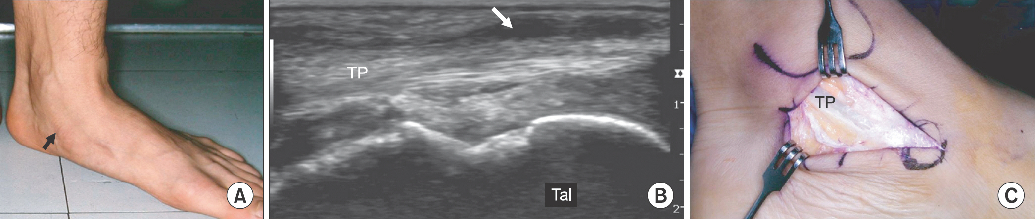
Figure 1. (A) The photograph of left foot showed swelling (black arrow) around the tibialis posterior tendon. (B) The ultrasonograph of left foot demonstrated mild thickening of the tendon with surrounding effusion (white arrow). (C) Under the diagnosis of stage 1 acquired adult flatfoot deformity or tibialis posterior tenosynovitis, a tenosynovectomy was performed. TP: tibialis posterior tendon, Tal: talus.

Figure 2. (A) The photograph of both heel showed a valgus deformity and ‘too many toes’ sign in the left side. (B) The standing anteroposterior radiograph of left foot demonstrated mild abduction in the midfoot. The talonavicular uncoverage was measured to be 25%. (C) Under the diagnosis of stage 2a acquired adult flatfoot deformity, a flexor digitorum longus tendon transfer was performed. (D) The tendon transfer was combined with a calcaneal medial slide osteotomy.
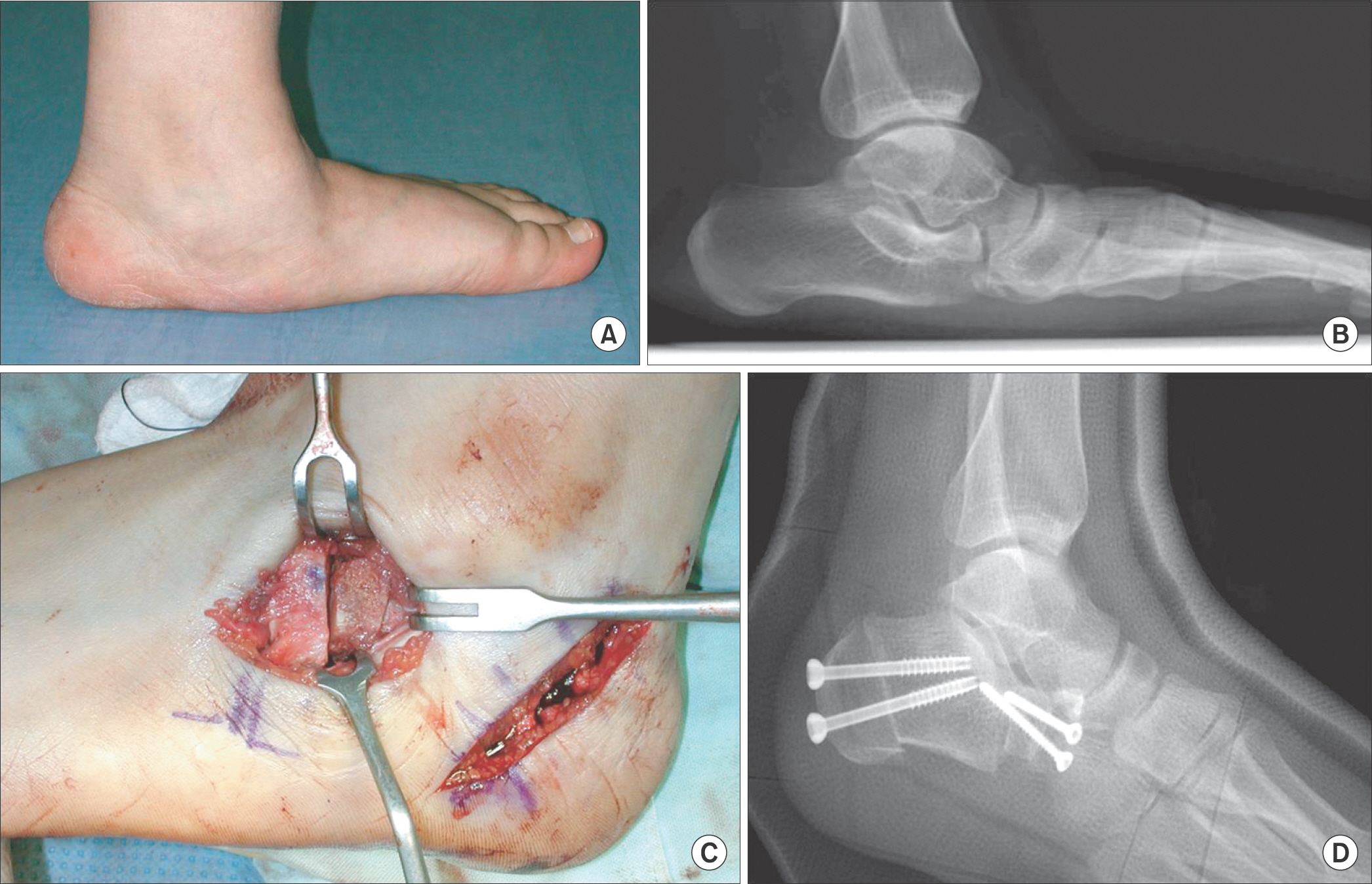
Figure 3. The photograph (A) and standing lateral radiograph (B) of left foot showed a severe flatfoot deformity, which was diagnosed as stage 2b acquired adult flatfoot deformity. (C) A calcaneal medial slide osteotomy and a lateral column lengthening were performed at the same time to correct the severe deformity. (D) Postoperative lateral radiograph showed the well-fixed double osteotomies.
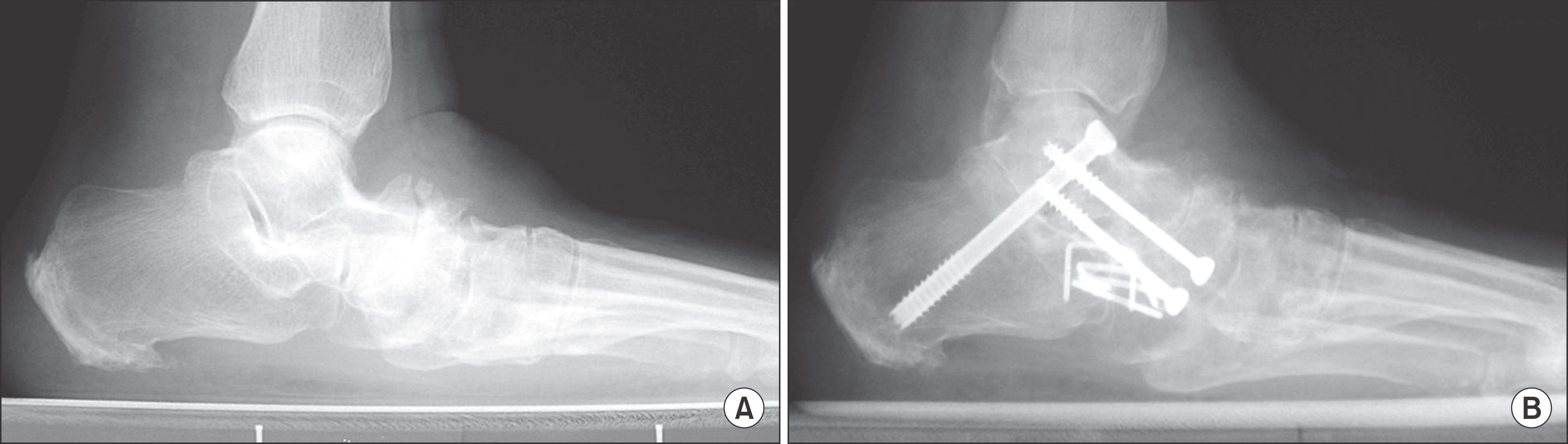
Figure 4. (A) Stage 3 acquired adult flatfoot deformity was seen at preoperative standing lateral radiograph of 47-year-old male patient. (B) Triple arthrodesis was performed for the rigid flatfoot deformity.

Figure 5. Preoperative standing anteroposterior (A) and lateral (B) radiographs of a 62-year-old female patient showed a valgus deformity of ankle joint with a severe flatfoot deformity. Postoperative anteroposterior (C) and lateral (D) radiographs showed the triple arthrodesis and extended medial column fusion combined with a deltoid ligament reconstruction, which were performed under the diagnosis of stage 4 acquired adult flatfoot deformity.
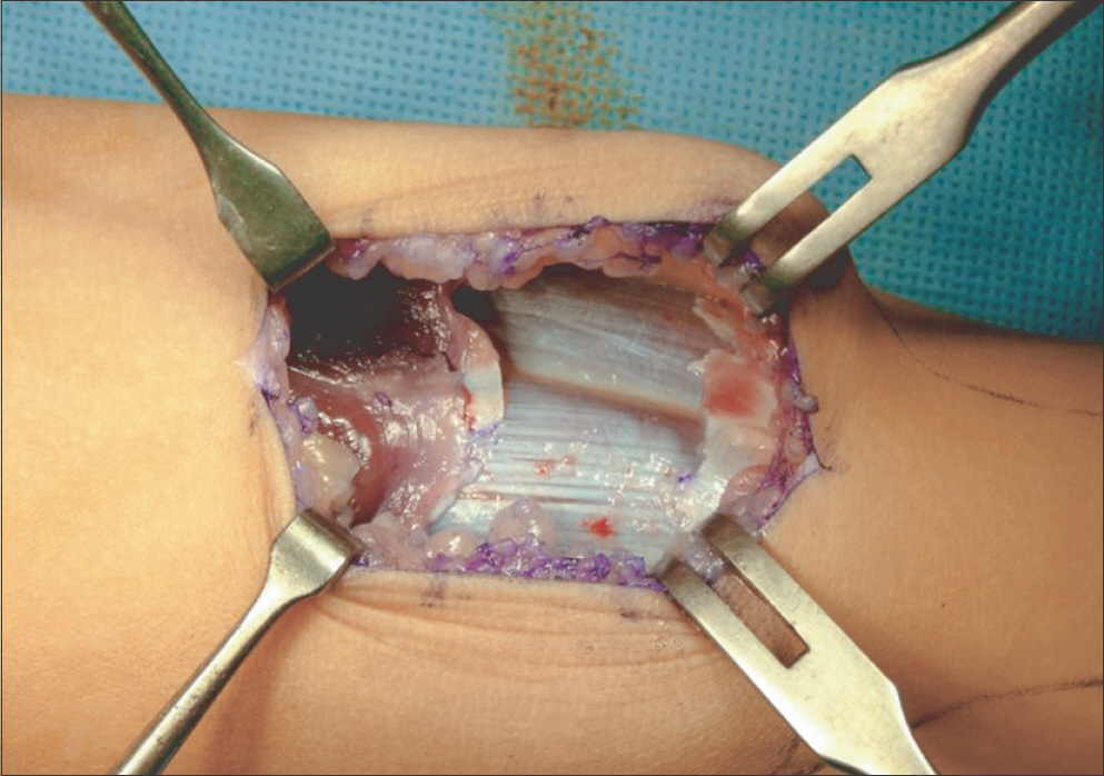
Figure 6. This photograph demonstrates the technique of gastrocnemius recession.
Cited by 1 articles
-
Diagnosis of Flatfoot Deformity
Tae Hoon Lee, Suh Woo Chay, Hak Jun Kim
J Korean Foot Ankle Soc. 2016;20(1):1-5. doi: 10.14193/jkfas.2016.20.1.1.
Reference
-
1.Johnson KA. Tibialis posterior tendon rupture1 Clin Orthop Relat Res1. 1983. 177:140–71.2.Mann RA., Thompson FM. Rupture of the posterior tibial tendon causing flat foot1 Surgical treatment1 J Bone Joint Surg Am1. 1985. 67:556–611.3.Myerson MS. Adult acquired flatfoot deformity: treatment of dysfunction of the posterior tibial tendon1 Instr Course Lect1. 1997. 46:393–4051.4.Johnson KA., Strom DE. Tibialis posterior tendon dysfunction1 Clin Orthop Relat Res1. 1989. 239:196–2061.5.Pinney SJ., Lin SS. Current concept review: acquired adult flatfoot deformity1 Foot Ankle Int1. 2006. 27:66–751.6.Deland JT., de Asla RJ., Sung IH., Ernberg LA., Potter HG. Posterior tibial tendon insufficiency: which ligaments are involved? Foot Ankle Int1. 2005. 26:427–351.
Article7.Holmes GB Jr., Mann RA. Possible epidemiological factors associated with rupture of the posterior tibial tendon1 Foot Ankle1. 1992. 13:70–91.8.Deland JT. Adult-acquired flatfoot deformity1 J Am Acad Orthop Surg1. 2008. 16:399–4061.9.Deland JT., Page A., Sung IH., O’Malley MJ., Inda D., Choung S. Posterior tibial tendon insufficiency results at different stages1 HSS J1. 2006. 2:157–601.10.Vora AM., Tien TR., Parks BG., Schon LC. Correction of moderate and severe acquired flexible flatfoot with medializing calcaneal osteotomy and flexor digitorum longus transfer1 J Bone Joint Surg Am1. 2006. 88:1726–341.11.Deland JT., de Asla RJ., Segal A. Reconstruction of the chronically failed deltoid ligament: a new technique1 Foot Ankle Int1. 2004. 25:795–91.12.Teasdall RD., Johnson KA. Surgical treatment of stage I posterior tibial tendon dysfunction1 Foot Ankle Int1. 1994. 15:646–81.13.Myerson MS., Badekas A., Schon LC. Treatment of stage II posterior tibial tendon deficiency with flexor digitorum longus tendon transfer and calcaneal osteotomy1 Foot Ankle Int1. 2004. 25:445–501.14.Otis JC., Deland JT., Kenneally S., Chang V. Medial arch strain after medial displacement calcaneal osteotomy: an in vitro study1 Foot Ankle Int1. 1999. 20:222–61.15.Hiller L., Pinney SJ. Surgical treatment of acquired flatfoot deformity: what is the state of practice among academic foot and ankle surgeons in 2002? Foot Ankle Int1. 2003. 24:701–51.
Article16.Needleman RL. A surgical approach for flexible flatfeet in adults including a subtalar arthroereisis with the MBA sinus tarsi im-plant1 Foot Ankle Int1. 2006. 27:9–181.17.Hirose CB., Johnson JE. Plantarflexion opening wedge medial cuneiform osteotomy for correction of fixed forefoot varus associated with flatfoot deformity1 Foot Ankle Int1. 2004. 25:568–741.18.Evans D. Calcaneo-valgus deformity1 J Bone Joint Surg Br1. 1975. 57:270–81.19.Pomeroy GC., Pike RH., Beals TC., Manoli A 2nd. Acquired flatfoot in adults due to dysfunction of the posterior tibial tendon1 J Bone Joint Surg Am1. 1999. 81:1173–821.20.Tien TR., Parks BG., Guyton GP. Plantar pressures in the forefoot after lateral column lengthening: a cadaver study comparing the Evans osteotomy and calcaneocuboid fusion1 Foot Ankle Int1. 2005. 26:520–51.21.Deland JT., Otis JC., Lee KT., Kenneally SM. Lateral column lengthening with calcaneocuboid fusion: range of motion in the triple joint complex1 Foot Ankle Int1. 1995. 16:729–331.22.Thomas RL., Wells BC., Garrison RL., Prada SA. Preliminary results comparing two methods of lateral column lengthening1 Foot Ankle Int1. 2001. 22:107–191.23.Dolan CM., Henning JA., Anderson JG., Bohay DR., Kornmesser MJ., Endres TJ. Randomized prospective study comparing tri-cortical iliac crest autograft to allograft in the lateral column lengthening component for operative correction of adult acquired flatfoot deformity1 Foot Ankle Int1. 2007. 28:8–121.24.Cohen BE., Johnson JE. Subtalar arthrodesis for treatment of posterior tibial tendon insufficiency1 Foot Ankle Clin1. 2001. 6:121–81.25.Deland JT., Page AE., Kenneally SM. Posterior calcaneal osteotomy with wedge: cadaver testing of a new procedure for insufficiency of the posterior tibial tendon1 Foot Ankle Int1. 1999. 20:290–51.26.Coetzee JC., Hansen ST. Surgical management of severe deformity resulting from posterior tibial tendon dysfunction1 Foot Ankle Int1. 2001. 22:944–91.
- Full Text Links
-
- Actions
-
Cited
- CITED
-
- Close
- Share
-
- Similar articles
-
- Acquired Adult Flatfoot: Pathophysiology, Diagnosis, and Nonoperative Treatment
- Adult flatfoot
- Diagnosis of Flatfoot Deformity
- Operative Treatment of Adult Flexible Flatfoot with Young's Tenosuspension: Case Report
- Adult Idiopathic Flexible Flat Foot Treated with Medial Sliding Calcaneal Osteotomy and Subtalar Arthroereisis: Report of 1 Case
