Tuberc Respir Dis.
2008 Sep;65(3):230-234.
A Case of Extensive Stage Small Cell Lung Cancer Presenting as an Acute Appendicitis with Perforation
- Affiliations
-
- 1Department of Internal Medicine, Bucheon Hospital, Soonchunhyang University School of Medicine, Bucheon, Korea. kdj@schbc.ac.kr
- 2Department of Surgery, Bucheon Hospital, Soonchunhyang University School of Medicine, Bucheon, Korea.
- 3Department of Pathology, Bucheon Hospital, Soonchunhyang University School of Medicine, Bucheon, Korea.
- 4Department of Radiology, Bucheon Hospital, Soonchunhyang University School of Medicine, Bucheon, Korea.
Abstract
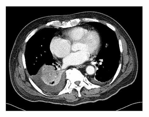
- The incidence of appendiceal metastatic cancer is quite low. In particular, in small cell lung cancer, there is a very low incidence of a metastasis to the appendix. A 75-years old man with right lower quadrant pain, cough and sputum was transferred to our hospital. Abdominal CT revealed acute appendicitis with a perforation. The patient underwent surgery. The frozen sections of the tissue obtained during surgery, indicated a malignancy, but a right hemicolectomy was not performed due to the patient's poor general condition. The histology findings of the appendix were identified as a small cell carcinoma. The abdominal CT scan and chest x-ray at admission day showed a mass in the right lower lobe, and a further evaluation of the lesion was performed including positron emission tomography and flexible bronchoscopy with a biopsy. The pathology findings of the lung mass were also small cell lung cancer. The specimens from both sites stained positive for cytokeratin, cluster designation 56, synaptophysin, chromogranin-A and thyroid transcription factor 1. It was concluded that the appendiceal small cell cancer originated from the lung.
Keyword
MeSH Terms
-
Appendicitis
Appendix
Biopsy
Bronchoscopy
Carcinoma, Small Cell
Cough
Frozen Sections
Humans
Incidence
Keratins
Lung
Neoplasm Metastasis
Nuclear Proteins
Positron-Emission Tomography
Small Cell Lung Carcinoma
Sputum
Synaptophysin
Thorax
Thyroid Gland
Transcription Factors
Keratins
Nuclear Proteins
Synaptophysin
Transcription Factors
Figure
Reference
-
1. Cohen MH, Matthews MJ. Small cell bronchogenic carcinoma: a distinct clinicopathologic entity. Semin Oncol. 1978. 5:234–243.2. Sridhar KS, Hussein AM, Thurer RJ. Evolving role of surgical treatment in limited-disease small cell lung carcinoma. J Surg Oncol. 1989. 40:155–161.3. Johnson BE. Management of small cell lung cancer. Clin Chest Med. 2002. 23:225–239.4. Pang LC. Metastasis-induced acute appendicitis in small cell bronchogenic carcinoma. South Med J. 1988. 81:1461–1462.5. Goldstein EB, Savel RH, Walter KL, Rankin LF, Satheesan R, Lehman HE, et al. Extensive stage small cell lung cancer presenting as an acute perforated appendix: case report and review of the literature. Am Surg. 2004. 70:706–709.6. Levchenko AM, Vasechko VN, Erusalimskiĭ EL. Metastasis of small-cell lung cancer to the appendix. Klin Khir. 1985. 5:56–57.7. Park IJ, Yu CS, Kim HC, Kim JC. Clinical features and prognostic factors in primary adenocarcinoma of the appendix. Korean J Gastroenterol. 2004. 43:29–34.8. Cortina R, McCormick J, Kolm P, Perry RR. Management and prognosis of adenocarcinoma of the appendix. Dis Colon Rectum. 1995. 38:848–852.9. Ryo H, Sakai H, Ikeda T, Hibino S, Goto I, Yoneda S, et al. Gastrointestinal metastasis from lung cancer. Nihon Kyobu Shikkan Gakkai Zasshi. 1996. 34:968–972.10. Lobins R, Floyd J. Small cell carcinoma of unknown primary. Semin Oncol. 2007. 34:39–42.11. Lau SK, Luthringer DJ, Eisen RN. Thyroid transcription factor-1: a review. Appl Immunohistochem Mol Morphol. 2002. 10:97–102.12. Ordonez NG. Value of thyroid transcription factor-1 immunostaining in distinguishing small cell lung carcinomas from other small cell carcinomas. Am J Surg Pathol. 2000. 24:1217–1223.13. Fletcher MS. Gastric perforation secondary to metastatic carcinoma of the lung: a case report. Cancer. 1980. 46:1879–1882.14. Morgan MW, Sigel B, Wolcott MW. Perforation of a metastatic carcinoma of the jejunum after cancer chemotherapy. Surgery. 1961. 49:687–689.15. Shen YY, Shiau YC, Wang JJ, Ho ST, Kao CH. Whole-body 18F-2-deoxyglucose positron emission tomography in primary staging small cell lung cancer. Anticancer Res. 2002. 22:1257–1264.
- Full Text Links
- Actions
-
Cited
- CITED
-
- Close
- Share
- Similar articles
-
- Treatment for Extensive stage Small cell Lung Cancer
- A Case of Left Ureteral Obstruction due to Acute Appendicitis
- Acute Appendicitis Presenting with Escherichia coli Bacteremia without Perforation in a Healthy Male
- Etoposide and cisplatin combination chemotherapy in extensive stage small cell lung cancer
- A Clinical Therapeutic Results on Small Cell Lung Cancer


