Taxol and Taurine Protect the Renal Tissue of Rats after Unilateral Ureteral Obstruction: A Stereological Survey
- Affiliations
-
- 1Histomorphometry and Stereology Research Center, Shiraz University of Medical Sciences, Shiraz, Iran. noora@sums.ac.ir
Abstract
- PURPOSE
Blockage of the urinary tract induces changes in renal structure including tubular dilatation or atrophy, tubular cell death, inflammatory processes, and progressive interstitial fibrosis with the loss of renal parenchyma. The present study was conducted to survey the protective effects of Taxol and taurine on the renal structure after unilateral ureteral obstruction (UUO).
MATERIALS AND METHODS
UUO was induced in three groups of rats (n=6) who then received distilled water, Taxol (0.3 mg/kg/d), or taurine (7.5 mg/kg/d). Stereological methods were used to gather quantitative as well as comparative data.
RESULTS
Less than -8% of the volume of the glomeruli, proximal convoluted tubules (PCT), distal convoluted tubules (DCT), Henle's loop, and collecting ducts were preserved after UUO. After treatment of the UUO rats with Taxol, between -32% and 88% of the parameters mentioned above remained intact, and after treatment of the UUO rats with taurine, between -16% and 46% of the parameters remained intact (p<0.01). Compared with the untreated UUO animals, the volume of necrotic and fibrotic tissues decreased -53% and -63% in the UUO rats treated with Taxol and taurine, respectively (p<0.01). Less than -3% of the lengths of the renal tubules (PCT, DCT, Henle's loop, and collecting) were preserved in the UUO rats. After treatment with Taxol and taurine, -61% to 70% and -43% to 53% of the length of the renal tubules were preserved, respectively (p<0.01).
CONCLUSIONS
Taurine and Taxol are effective in preventing some structural renal damage in a direct ureteral obstruction model. Taxol was more effective in renal protection.
Keyword
MeSH Terms
Figure
-
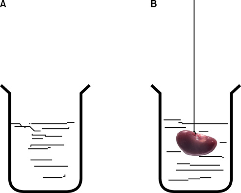
FIG. 1 Estimation of kidney volume by use of the immersion method. (A) A laboratory jar was filled with distilled water, was placed on the scale, and weighed. (B) The kidney was suspended by a thin thread in the laboratory jar. The kidney did not have to touch the bottom or sides of the jar. The new weight in grams minus the weight of the laboratory jar and water divided by the specific gravity of the distilled water (-1.0) was considered the primary volume of the kidney in cubic centimeters.
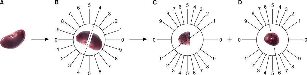
FIG. 2 Isotropic uniform random sectioning by use of the orientator method. (A) The kidney was cleaned. (B) It was placed on the circle such that each half of it was divided into 10 equal parts. A random number between 0 and 9 was selected. The kidney was sectioned into two parts at the direction of the selected number (here 4). (C) The cut surface of one part of the kidney was, then, placed on the 0-0 direction of the second circle with 10 unequal divisions. The circle division was done according to the cosine of the angels. Then, another random number was selected and the second cut was done (here 1). The parts were sectioned into parallel slabs (D) The cut surface of the other part of the kidney was placed vertically on the second circle. Again, a new number and direction (here 6) was selected and cut. This part was also sectioned into parallel slabs.
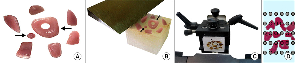
FIG. 3 Estimation of shrinkage and slide preparation. (A) The entire kidney was sectioned into slabs with a blade and was placed in the direction of the isotropic uniform random cut with an interval of -0.5 mm. Then, the slabs (8 to 12 slabs) were collected. A circle (left arrow) was punched from a kidney slab (right arrow) by a trocar. The diameter of the circular piece and the area of the circle were estimated using the usual formula for calculating the area of a circle. (B) The cut surface of the slabs and the circle were embedded in the paraffin block. (C) Sectioning of the block using the microtome blade (5 µm thicknesses) is demonstrated. After staining with Heidenhain's Azan trichrome, the area of the circular piece was measured again and the volume shrinkage was calculated by using the following formula: volume shrinkage:=1-(AA/AB)1.5 where AA and AB, respectively,represent the area of the circular piece after and before the processing, the sectioning, and the staining. After estimating the shrinkage, the final volume of the kidney (the reference space) was computed by using the following formula: Vfinal:=Vprimary×(1-volume shrinkage). (D) The microscopic fields were sampled in a systematic random design. The fields were sampled and analyzed at equal intervals along the X- and Y-axis by using a stage micrometer. This procedure was continued until all the sections had been studied.
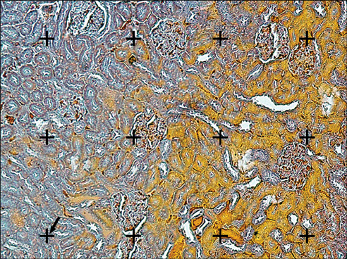
FIG. 4 The volume density estimation using the point-counting method. A gird of points was superimposed upon the images of the renal sections were viewed on the monitor. The volume density (fractional volume) (Vv) of the renal cortex, the medulla, the glomeruli, the proximal convoluted tubules, the distal convoluted tubule, the collecting ducts, the Henle's loop, the vessels, and the connective tissue was obtained by using the point-counting method and the following formula: Vv=P(structure)/P(total) where P(structure) and P(total) represent the sum of the number of the points hitting the structure and all sampled fields of the kidney, respectively. The point is the right upper corner of the cross (the arrow) (Heidenhain's AZAN trichrome stain, ×200).
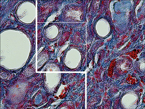
FIG. 5 The length density estimation in a kidney with an obstructed ureter. The counting frame with exclusion lines (bold lines of the left and lower borders) and inclusion lines (thin lines of the right and upper borders) was superimposed on the images. The tubules that were either completely or partly inside the counting frame and did not touch the exclusion lines were counted. The length density (Lv) of each tubule was calculated by using the following formula: Lv:=2×ΣQ/ΣAT where ΣQ denotes the total number of each tubule profiles counted per kidney and AT was the test area that was calculated by multiplying the area of the frame (here was 2,700 µm2) and the total number of the counted frames (Heidenhain's AZAN trichrome stain, ×400).

FIG. 6 A comparison of the renal tissue in different groups. (A) The normal tissue. (B) The renal tissue after the unilateral ureteral obstruction (UUO). (C) The renal tissue after the UUO+taxol. (D) The renal tissue after the UUO+taurine (Heidenhain's AZAN trichrome stain, ×200).
Reference
-
1. Sato S, Yamate J, Saito T, Hosokawa T, Saito S, Kurasaki M. Protective effect of taurine against renal interstitial fibrosis of rats induced by cisplatin. Naunyn Schmiedebergs Arch Pharmacol. 2002. 365:277–283.2. Chesney RW, Han X, Patters AB. Taurine and the renal system. J Biomed Sci. 2010. 17:Suppl 1. S4.3. Trachtman H, Futterweit S, Maesaka J, Ma C, Valderrama E, Fuchs A, et al. Taurine ameliorates chronic streptozocin-induced diabetic nephropathy in rats. Am J Physiol. 1995. 269(3 Pt 2):F429–F438.4. Cruz CI, Ruiz-Torres P, del Moral RG, Rodriguez-Puyol M, Rodríguez-Puyol D. Age-related progressive renal fibrosis in rats and its prevention with ACE inhibitors and taurine. Am J Physiol Renal Physiol. 2000. 278:F122–F129.5. Marcinkiewicz J, Kurnyta M, Biedron R, Bobek M, Kontny E, Maślinski W. Anti-inflammatory effects of taurine derivatives (taurine chloramine, taurine bromamine, and taurolidine) are mediated by different mechanisms. Adv Exp Med Biol. 2006. 583:481–492.6. Odobasic D, Kitching AR, Semple TJ, Holdsworth SR. Endogenous myeloperoxidase promotes neutrophil-mediated renal injury, but attenuates T cell immunity inducing crescentic glomerulonephritis. J Am Soc Nephrol. 2007. 18:760–770.7. Suliman ME, Barany P, Filho JC, Lindholm B, Bergstrom J. Accumulation of taurine in patients with renal failure. Nephrol Dial Transplant. 2002. 17:528–529.8. Erdem A, Gundogan NU, Usubutun A, Kilinç K, Erdem SR, Kara A, et al. The protective effect of taurine against gentamicin-induced acute tubular necrosis in rats. Nephrol Dial Transplant. 2000. 15:1175–1182.9. Sun L, Zhang D, Liu F, Xiang X, Ling G, Xiao L, et al. Low-dose paclitaxel ameliorates fibrosis in the remnant kidney model by down-regulating miR-192. J Pathol. 2011. 225:364–377.10. Zhang D, Sun L, Xian W, Liu F, Ling G, Xiao L, et al. Low-dose paclitaxel ameliorates renal fibrosis in rat UUO model by inhibition of TGF-beta/Smad activity. Lab Invest. 2010. 90:436–447.11. Sommardahl CS, Woychik RP, Sweeney WE, Avner ED, Wilkinson JE. Efficacy of taxol in the orpk mouse model of polycystic kidney disease. Pediatr Nephrol. 1997. 11:728–733.12. Zhou J, Zhong DW, Wang QW, Miao XY, Xu XD. Paclitaxel ameliorates fibrosis in hepatic stellate cells via inhibition of TGF-beta/Smad activity. World J Gastroenterol. 2010. 16:3330–3334.13. Hyde DM, Tyler NK, Plopper CG. Morphometry of the respiratory tract: avoiding the sampling, size, orientation, and reference traps. Toxicol Pathol. 2007. 35:41–48.14. Scherle W. A simple method for volumetry of organs in quantitative stereology. Mikroskopie. 1970. 26:57–60.15. Nyengaard JR. Stereologic methods and their application in kidney research. J Am Soc Nephrol. 1999. 10:1100–1123.16. Gundersen HJ, Bendtsen TF, Korbo L, Marcussen N, Moller A, Nielsen K, et al. Some new, simple and efficient stereological methods and their use in pathological research and diagnosis. APMIS. 1988. 96:379–394.17. Gundersen HJ, Bagger P, Bendtsen TF, Evans SM, Korbo L, Marcussen N, et al. The new stereological tools: disector, fractionator, nucleator and point sampled intercepts and their use in pathological research and diagnosis. APMIS. 1988. 96:857–881.18. Yoon HY, Kang NI, Lee HK, Jang KY, Park JW, Park BH. Sulforaphane protects kidneys against ischemia-reperfusion injury through induction of the Nrf2-dependent phase 2 enzyme. Biochem Pharmacol. 2008. 75:2214–2223.19. Guerrero-Beltran CE, Calderon-Oliver M, Tapia E, Medina-Campos ON, Sanchez-Gonzalez DJ, Martínez-Martinez CM, et al. Sulforaphane protects against cisplatin-induced nephrotoxicity. Toxicol Lett. 2010. 192:278–285.20. Mostafavi-Pour Z, Zal F, Monabati A, Vessal M. Protective effects of a combination of quercetin and vitamin E against cyclosporine A-induced oxidative stress and hepatotoxicity in rats. Hepatol Res. 2008. 38:385–392.21. Sanchez-Gonzalez PD, Lopez-Hernandez FJ, Perez-Barriocanal F, Morales AI, Lopez-Novoa JM. Quercetin reduces cisplatin nephrotoxicity in rats without compromising its anti-tumour activity. Nephrol Dial Transplant. 2011. 26:3484–3495.22. Yousef MI, Omar SA, El-Guendi MI, Abdelmegid LA. Potential protective effects of quercetin and curcumin on paracetamol-induced histological changes, oxidative stress, impaired liver and kidney functions and haematotoxicity in rat. Food Chem Toxicol. 2010. 48:3246–3261.23. Guerrero-Beltran CE, Mukhopadhyay P, Horvath B, Rajesh M, Tapia E, Garcia-Torres I, et al. Sulforaphane, a natural constituent of broccoli, prevents cell death and inflammation in nephropathy. J Nutr Biochem. 2012. 23:494–500.24. Koz OG, Ozhuy S, Tezel GG, Karaman N, Unlu N, Yarangumeli A, et al. The effect of paclitaxel on conjunctival wound healing: a pilot study. J Glaucoma. 2007. 16:610–615.25. Devi SL, Viswanathan P, Anuradha CV. Regression of liver fibrosis by taurine in rats fed alcohol: effects on collagen accumulation, selected cytokines and stellate cell activation. Eur J Pharmacol. 2010. 647:161–170.26. Devi SL, Anuradha CV. Oxidative and nitrosative stress in experimental rat liver fibrosis: Protective effect of taurine. Environ Toxicol Pharmacol. 2010. 29:104–110.
- Full Text Links
-
- Actions
-
Cited
- CITED
-
- Close
- Share
-
- Similar articles
-
- New Application of the Partial Unilateral Ureteral Obstruction in Neonatal Rat Model
- Expression of Heat Shock Protein 70 in Ipsilateral Rat Kidney with Unilateral Partial Ureteral Obstruction
- Significance of Renal Resistive Index in Children with Unilateral Ureteral Obstruction
- Renal Expression of an Ammonia Transporter in Rats with a Unilateral Ureteral Obstruction
- A radiological study of recovery from hydronephrosis by ureteral ligation
