Lab Anim Res.
2013 Mar;29(1):55-62. 10.5625/lar.2013.29.1.55.
Establishment of a murine model for radiation-induced bone loss using micro-computed tomography in adult C3H/HeN mice
- Affiliations
-
- 1General Toxicity Team, Korea Testing & Research Institute, Seoul, Korea.
- 2Radiological Effect Research Department, Korea Institute of Radiological & Medical Science, Seoul, Korea.
- 3College of Veterinary Medicine, Chonnam National University, Gwangju, Korea. shokim@chonnam.ac.kr
- 4Division of Radiation Biotechnology, Advanced Radiation Technology Institute, Jeongeup, Korea.
- 5Faculty of Animal Science & Biotechnology, Kyungpook National University, Sangju, Korea.
- KMID: 2312104
- DOI: http://doi.org/10.5625/lar.2013.29.1.55
Abstract
- Bone changes are common sequela of radiation therapy for cancer. The purpose of this study was to establish an experimental model of radiation-induced bone loss in adult mice using micro-computed tomography (microCT). The extent of changes following 2 Gy gamma irradiation (2 Gy/min) was studied at 4, 8, 12 or 16 weeks after exposure. Adult mice that received 1, 2, 4 or 6 Gy of gamma-rays were examined 12 weeks after irradiation. Tibiae were analyzed using microCT. Serum markers and biomechanical properties were measured and the osteoclast surface was examined. A significant loss of trabecular bone in tibiae was evident 12 weeks after exposure. Measurements performed after irradiation showed a dose-related decrease in trabecular bone volume fraction (BV/TV) and bone mineral density (BMD), respectively. The best-fitting dose-response curves were linear-quadratic. Taking the controls into accounts, the lines of best fit were as follows: BV/TV (%)= -0.071D2-1.799D+18.835 (r2=0.968, D=dose in Gy) and BMD (mg/cm3) = -3.547D2-14.8D+359.07 (r2=0.986, D=dose in Gy). Grip strength and body weight did not differ among the groups. No dose-dependent differences were apparent among the groups with regard to mechanical and anatomical properties of tibia, serum biochemical markers and osteoclast activity. The findings provide the basis required for better understanding of the results that will be obtained in any further studies of radiation-induced bone responses.
Keyword
MeSH Terms
Figure
-
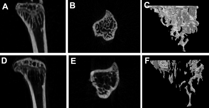
Figure 1 Micro-CT images of the tibia for control (A, B, C) and irradiated (D, E, F) C3H/HeN mice. Cross sectional view (A, D), vertical view (B, E) and reconstructed three-dimensional image (C, F).
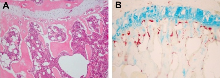
Figure 2 Hematoxylin & eosin (A) and tartrate-resistant acid phosphatase (TRAP) staining (B) of trabecular bone in tibia (× 40).
Reference
-
1. Aziz NM, Rowland JH. Trends and advances in cancer survivorship research: challenge and opportunity. Semin Radiat Oncol. 2003; 13(3):248–266. PMID: 12903014.
Article2. Oeffinger KC, Mertens AC, Sklar CA, Kawashima T, Hudson MM, Meadows AT, Friedman DL, Marina N, Hobbie W, Kadan-Lottick NS, Schwartz CL, Leisenring W, Robison LL. Chronic health conditions in adult survivors of childhood cancer. N Engl J Med. 2006; 355(15):1572–1582. PMID: 17035650.
Article3. Zhao W, Diz DI, Robbins ME. Oxidative damage pathways in relation to normal tissue injury. Br J Radiol. 2007; 80(Spec No 1):S23–S31. PMID: 17704323.
Article4. Zhao W, Robbins ME. Inflammation and chronic oxidative stress in radiation-induced late normal tissue injury: therapeutic implications. Curr Med Chem. 2009; 16(2):130–143. PMID: 19149566.
Article5. Howland WJ, Loeffler RK, Starchman DE, Johnson RG. Postirradiation atrophic changes of bone and related complications. Radiology. 1975; 117(3 Pt 1):677–685. PMID: 1188119.
Article6. Ergün H, Howland WJ. Postradiation atrophy of mature bone. CRC Crit Rev Diagn Imaging. 1980; 12(3):225–243. PMID: 6985580.7. Heuch F, Lauritzen C. Veränderungen von mineralgehalt und struktur des femur nach gyakologischer stralentherapie. Stralentherapie. 1967; 66:87–92.8. Chen HH, Lee BF, Guo HR, Su WR, Chiu NT. Changes in bone mineral density of lumbar spine after pelvic radiotherapy. Radiother Oncol. 2002; 62(2):239–242. PMID: 11937252.
Article9. Glatt V, Canalis E, Stadmeyer L, Bouxsein ML. Age-related changes in trabecular architecture differ in female and male C57BL/6J mice. J Bone Miner Res. 2007; 22(8):1197–1207. PMID: 17488199.
Article10. Szymczyk KH, Shapiro IM, Adams CS. Ionizing radiation sensitizes bone cells to apoptosis. Bone. 2004; 34(1):148–156. PMID: 14751572.
Article11. Bandstra ER, Pecaut MJ, Anderson ER, Willey JS, De Carlo F, Stock SR, Gridley DS, Nelson GA, Levine HG, Bateman TA. Long-term dose response of trabecular bone in mice to proton radiation. Radiat Res. 2008; 169(6):607–614. PMID: 18494551.
Article12. Willey JS, Lloyd SA, Robbins ME, Bourland JD, Smith-Sielicki H, Bowman LC, Norrdin RW, Bateman TA. Early increase in osteoclast number in mice after whole-body irradiation with 2 Gy X rays. Radiat Res. 2008; 170(3):388–392. PMID: 18763868.
Article13. Parfitt AM, Drezner MK, Glorieux FH, Kanis JA, Malluche H, Meunier PJ, Ott SM, Recker RR. Bone histomorphometry: standardization of nomenclature, symbols, and units. Report of the ASBMR Histomorphometry Nomenclature Committee. J Bone Miner Res. 1987; 2(6):595–610. PMID: 3455637.
Article14. Chappard D, Retailleau-Gaborit N, Legrand E, Baslé MF, Audran M. Comparison insight bone measurements by histomorphometry and microCT. J Bone Miner Res. 2005; 20(7):1177–1184. PMID: 15940370.15. Bouxsein ML, Boyd SK, Christiansen BA, Guldberg RE, Jepsen KJ, Müller R. Guidelines for assessment of bone microstructure in rodents using micro-computed tomography. J Bone Miner Res. 2010; 25(7):1468–1486. PMID: 20533309.
Article16. Beamer WG, Donahue LR, Rosen CJ, Baylink DJ. Genetic variability in adult bone density among inbred strains of mice. Bone. 1996; 18(5):397–403. PMID: 8739896.
Article17. Bower AL, Lang DH, Vogler GP, Vandenbergh DJ, Blizard DA, Stout JT, McClearn GE, Sharkey NA. QTL analysis of trabecular bone in BXD F2 and RI mice. J Bone Miner Res. 2006; 21(8):1267–1275. PMID: 16869725.
Article18. Willey JS, Lloyd SA, Nelson GA, Bateman TA. Ionizing Radiation and Bone Loss: Space Exploration and Clinical Therapy Applications. Clin Rev Bone Miner Metab. 2011; 9(1):54–62. PMID: 22826690.
Article
- Full Text Links
-
- Actions
-
Cited
- CITED
-
- Close
- Share
-
- Similar articles
-
- The effect of pentoxifylline on radiation-induced bone loss in C3H/HeN mice
- Morphometric Analysis of Tibial Bone in Three Strains of Mice Using Micro-computed Tomography
- Evaluation of effect of red ginseng on ovariectomy-induced bone loss in C3H/HeN mice
- The effect of Puerariae Radix on ovariectomy-induced bone loss in C3H/HeN mice
- Quantification of Microstructures in Mice Alveolar Bone using Micro-computed tomography (microCT)
