Vascular Endothelial Growth Factor (VEGF) and Advanced Glycation End Products (AGEs) Overexpression in the Retina and Serum and Lens Opacities of Streptozotocin-induced Diabetic Rats
- Affiliations
-
- 1Department of Herbal Pharmaceutical Development, Korea Institute of Oriental Medicine, Korea.
- 2Department of Pathology, Inje University, Sanggye Paik Hospital, Korea.
- KMID: 2298109
- DOI: http://doi.org/10.4093/kdj.2008.32.1.44
Abstract
-
BACKGROUND: Vascular Endothelial Growth Factor (VEGF) and Advanced Glycation End products (AGEs) have been implicated in the development of diabetic retinopathy. In this study, we examined the expression of VEGF and AGEs in the retina and serum, apoptosis in the retina, and lens opacities in streptozotocin (STZ)-induced diabetic rats.
METHODS
The localization of VEGF and AGEs in the retina of STZ-induced diabetic rats was determined by immunohistochemical analysis, and apoptotic cell death was assessed using the TUNEL assay. In the serum, STZ-induced diabetic rats were assayed for VEGF and AGEs by ELISA. Lenses were also isolated to detect the opacity.
RESULTS
Expression of VEGF and accumulation of AGEs were significantly increased in the retinal ganglion cell layers (GCL) and nuclear cell layers (NCL) of STZ-induced diabetic rats compared to normal control rats. In addition to cellular expression, serum VEGF and AGEs levels were also increased significantly in STZ-diabetic rats compared to normal rats (both P < 0.001) and there was a significant correlation between the serum VEGF and AGEs levels (r = 0.504). The lens opaque density of STZ-induced diabetic rats were significantly higher than in normal rats (P < 0.001).
CONCLUSIONS
AGEs could be involved in the development of diabetic retinopathy through the induction of VEGF. One could possibly correlate this lens opaque formation with elevation of AGE induced VEGF level. Thus, this study should be considered as a basic research for studying pathology of the retina and lens in diabetic experimental models.
Keyword
MeSH Terms
-
Animals
Apoptosis
Cataract
Cell Death
Diabetic Retinopathy
Enzyme-Linked Immunosorbent Assay
Glycosylation End Products, Advanced
In Situ Nick-End Labeling
Models, Theoretical
Rats
Retina
Retinal Ganglion Cells
Streptozocin
Vascular Endothelial Growth Factor A
Glycosylation End Products, Advanced
Streptozocin
Vascular Endothelial Growth Factor A
Figure
-
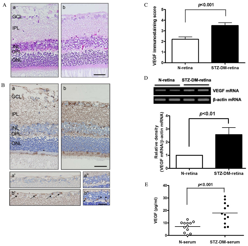
Fig. 1 Expression of VEGF in the retina and the expression of VEGF in the serum of STZ-induced diabetic rats. A. Hematoxylin and eosin staining (a, normal rats; b, diabetic rats). B. VEGF staining: The brown product of the peroxidase reaction is present in the GCL (a' and b') and in the INL (a'' and b'') after VEGF staining of the retinal sections. (n = 4 retinas per group). Scale bar = 50 µm. C. Distribution of VEGF immunostaining score in the retina: Each sample was scored from 0 to 5 based on the intensity of the positive area with VEGF immunostaining. Results are given as mean ± S.E.M. D. Expression of VEGF mRNA in the retina was quantified by a RT-PCR method. Results are given as mean ± S.E.M. (n = 6). E. Serum VEGF levels: Each dot represents the serum VEGF levels determined by ELISA for each animal. N, normal (n = 12); STZ-DM, STZ-induced diabetic rats (n = 12).

Fig. 2 Localization of AGEs and RAGE in the retina and expression of AGEs in the serum of STZ-induced diabetic rats. A. AGEs and RAGE double-immunofluorescence staining of STZ-induced diabetic rat retina. The sections were immunolabeled with anti-AGE antibody (a and b) and then RAGE antibody (a' and b'). Fluorescein (a and b) and Texas red (a' and b') show identical fields, respectively. Cell bodies showing AGEs immunoreactivity are present in the GCL and INL; The retinas of normal rats (a, a', and a'') and of STZ-induced diabetic rats (b, b', and b''). Scale bar = 50 µm. B. Serum AGEs levels. Each dot represents the serum AGEs levels determined by ELISA for each animal. The serum AGEs levels of STZ-induced diabetic rats are significantly higher than those of normal control rats. N, normal (n = 12); STZ-DM, STZ-induced diabetic rats (n = 12).
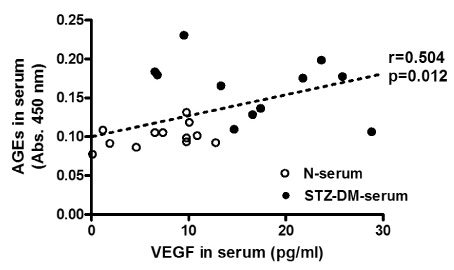
Fig. 3 Correlation between serum VEGF and AGEs levels. A strong correlation (r = 0.504) between serum VEGF levels measured by ELISA and serum AGEs levels was found in both normal rats (n = 12) and STZ-induced diabetic rats (n = 12).
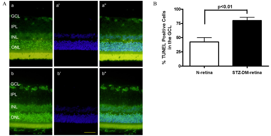
Fig. 4 Retinal cell apoptosis detected by TUNEL and DAPI staining. (A) The retinas of normal control rats (a, a', and a'') and STZ-induced diabetic rats (b, b', and b''): apoptotic retinal ganglion cell death was detected by TUNEL (green), and the nuclei (blue) were stained with DAPI. Merged images (a'' and b'') of TUNEL and DAPI. Scale bar = 50 µm. The number of TUNEL-positive cells was increased compared to that of normal control retinas (B) as indicated in the graph.
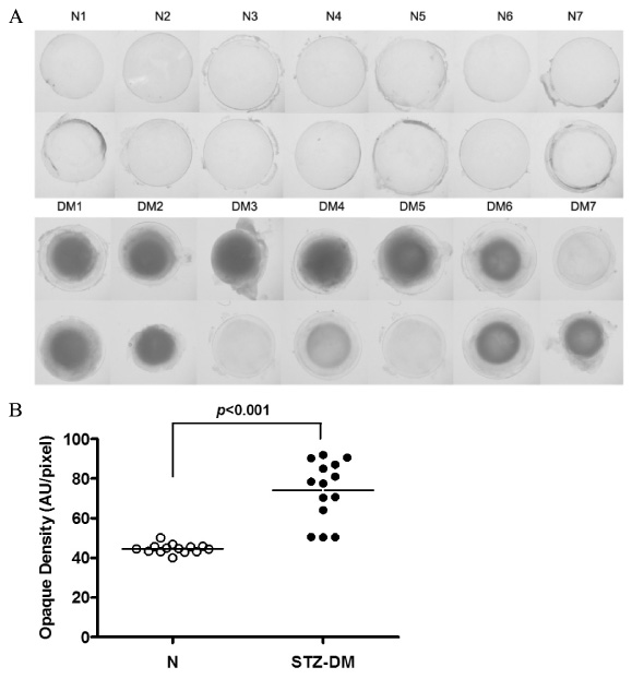
Fig. 5 Cataractogenesis in the lenses of STZ-induced diabetic rats. A. Images of rat lenses in normal and STZ-induced diabetic rats. B. Lens opacities. All lens opacities were analyzed. N, normal rats; DM, STZ-induced diabetic rats. Since the opacity changes associated with diabetes are usually asymmetrical, the eyes from each animal were always analyzed independently. All data are expressed as means ± S.E.M. (n = 14).
Reference
-
1. Gardiner TA, Archer DB, Curtis TM, Stitt AW. Arteriolar involvement in the microvascular lesions of diabetic retinopathy: implications for pathogenesis. Microcirculation. 2007. 14:25–38.2. Stitt AW. The role of advanced glycation in the pathogenesis of diabetic retinopathy. Exp Mol Path. 2003. 75:95–108.3. Yamagishi S-I, Nakamura K, Matsui T. Advanced glycation end products (AGEs) and their receptor (RAGE) system in diabetic retinopathy. Curr Drug Discov Technol. 2006. 3:83–88.4. Masuzawa K, Jesmin S, Maeda S, Zaedi S, Shimojo N, Miyauchi T, Goto K. Effect of endothelin dual receptor antagonist on VEGF levels in streptozotocin-induced diabetic rat retina. Exp Biol Med (Maywood). 2006. 231:1090–1094.5. Witmer AN, Vrensen GFJM, Van Noorden CJF, Schlingemann RO. Vascular endothelial growth factors and angiogenesis in eye disease. Prog Retin Eye Res. 2003. 22:1–29.6. Aiello LP, Pierce EA, Foley ED, Takagi H, Chen H, Riddle L, Ferrara N, King GL, Smith LE. Suppression of retinal neovascularization in vivo by inhibition of vascular endothelial growth factor (VEGF) using soluble VEGF-receptor chimeric proteins. Proc Natl Acad Sci USA. 1995. 92:10457–10461.7. Stitt AW, Curtis TM. Advanced glycation and retinal pathology during diabetes. Pharmacol Rep. 2005. 57:156–168.8. Yamagishi S, Amano S, Inagaki Y, Okamoto T, Koga K, Sasaki N, Yamamoto H, Takeuchi M, Makita Z. Advanced glycation end products-induced apoptosis and overexpression of vascular endothelial growth factor in bovine retinal pericytes. Biochem Biophys Res Commun. 2002. 290:973–978.9. Lecleire-Collet A, Tessier LH, Massin P, Forster V, Brasseur G, Sahel JA, Picaud S. Advanced glycation end products can induce glial reaction and neuronal degeneration in retinal explants. Br J Ophthalmol. 2005. 89:1631–1633.10. Stitt AW, Li YM, Gardiner TA, Bucala R, Archer DB, Vlassara H. Advanced glycation end products (AGEs) co-localize with AGE receptors in the retinal vasculature of diabetic and of AGE-infused rats. Am J Path. 1997. 150:523–531.11. Gavrieli Y, Gavrieli Y, Ben-Sasson SA. Identification of programmed cell death in situ via specific labeling of nuclear DNA fragmentation. J Cell Biol. 1992. 119:493–501.12. Westwood A, Bullock DG, Whitehead TP. An examination of the hexokinase method for serum glucose assay using external quality assessment data. Ann Clin Biochem. 1986. 23:92–96.13. Obrosova IG, Drel VR, Kumagai AK, Szabo C, Pacher P, Stevens MJ. Early diabetes-induced biochemical changes in the retina: comparison of rat and mouse models. Diabetologia. 2006. 49:2525–2533.14. Kim YS, Jung DH, Kim NH, Lee YM, Jang DS, Song G-Y, Kim JS. KIOM-79 inhibits high glucose or AGEs-induced VEGF expression in human retinal pigment epithelial cells. J Ethnopharmacol. 2007. 112:166–172.15. Yamagishi S, Nakamura K, Matsui T. Advanced glycation end products (AGEs) and their receptor (RAGE) system in diabetic retinopathy. Curr Drug Discov Technol. 2006. 3:83–88.16. Barile GR, Pachydaki SI, Tari SR, Lee SE, Donmoyer CM, Ma W, Rong LL, Buciarelli LG, Wendt T, Horig H, Hudson BI, Qu W, Weinberg AD, Yan SF, Schmidt AM. The RAGE axis in early diabetic retinopathy. Invest Ophthalmol Vis Sci. 2005. 46:2916–2924.17. Chew EY, Klein ML, Ferris FL III, Remaley NA, Murphy RP, Chantry K, Hoogwerf BJD, Miller D. Association of elevated serum lipid levels with retinal hard exudate in diabetic retinopathy. Early Treatment Diabetic Retinopathy Study (ETDRS) Report 22. Arch Ophthalmol. 1996. 114:1079–1084.18. Kaji Y, Usui T, Ishida S, Yamashiro K, Moore TC, Moore J, Yamamoto Y, Yamamoto H, Adamis AP. Inhibition of diabetic leukostasis and blood-retinal barrier breakdown with a soluble form of a receptor for advanced glycation end products. Invest Ophthalmol Vis Sci. 2007. 48:858–865.19. Yokoi M, Yamagishi SI, Takeuchi M, Ohgami K, Okamoto T, Saito W, Muramatsu M, Imaizumi T, Ohno S. Elevations of AGE and vascular endothelial growth factor with decreased total antioxidant status in the vitreous fluid of diabetic patients with retinopathy. Br J Ophthalmol. 2005. 89:673–685.20. Yokoi M, Yamagishi S, Takeuchi M, Matsui T, Yoshida Y, Ohgami K, Amano-Okamoto T, Ohno S. Positive correlation between vitreous levels of advanced glycation end products and vascular endothelial growth factor in patients with diabetic retinopathy sufficiently treated with photocoagulation. Br J Ophthalmol. 2007. 91:397–398.21. Meleth AD, Agrn E, Chan CC, Reed GF, Arora K, Byrnes G, Csaky KG, Ferris FL 3rd, Chew EY. Serum inflammatory markers in diabetic retinopathy. Invest Ophthalmol Vis Sci. 2005. 46:4295–4301.22. Zarina S, Zhao HR, Abraham EC. Advanced glycation end products in human senile and diabetic cataractous lenses. Mol Cell Biochem. 2000. 210:29–34.23. Franke S, Dawczynski J, Strobel J, Niwa T, Stahl P, Stein G. Increased levels of advanced glycation end products in human cataractous lenses. J Cataract Refract Surg. 2003. 29:998–1004.24. Padival S, Nagaraj RH. Pyridoxamine Inhibits Maillard Reactions in Diabetic Rat Lenses. Ophthalmic Res. 2006. 38:294–302.
- Full Text Links
-
- Actions
-
Cited
- CITED
-
- Close
- Share
-
- Similar articles
-
- The Role of Advanced Glycation End Products in Diabetic Vascular Complications
- Preventive effect of polydatin on diabetesrelated hypofunction of salivary gland in streptozotocin-induced diabetic rats
- Immunohistochemical Study of Transforming Growth Factor-beta2(TGF-beta2) and Vascular Endothelial Growth Factor(VEGF) after Laser Photocoagulation in the Ocular Tissues of White Rats
- Suppression of VEGF and STAT3 by Lipoic acid in Experimental Diabetic Rat Retina
- Formation of the Advanced Glycosylated End-products in the Peritoneum of Streptozotocin Diabetic Rats
