Qualitative and quantitative evaluation of root injury risk potentially burdening insertion of miniscrew implants
- Affiliations
-
- 1Department of Dentofacial Orthopedics and Orthodontics, Wroclaw Medical University, Poland.
- 2Department of Dentofacial Orthopedics and Orthodontics, Wroclaw Medical University, Poland.
- 3Department of Orthodontics, University of Homburg/Saar, Germany.
- 4Department of Orthodontics, School of Dentistry, Kyungpook National University, Deagu, Korea. parkhs@knu.ac.kr
- KMID: 2274247
- DOI: http://doi.org/10.4041/kjod.2011.41.2.112
Abstract
OBJECTIVE
Microscrew implants (MSIs) offer many advantages, but some complications are known to occur during their insertion. One of the most commonly reported complications is root injury. Our aim was to identify factors associated with root injury and to evaluate their qualitative and quantitative values.
METHODS
Thirty-five orthodontists placed MSIs (AbsoAnchor(R), Dentos Co. Ltd, Daegu, Korea) in the upper jaw of typodonts, labially between the second premolar and the first molar, in low and high vertical positions. Root contacts were counted, and distances between MSI apices and roots were measured. Fear level of the orthodontists was surveyed before and after the experiment. Wilcoxon's test, chi-square test, and Mann-Whitney test were used for statistical analysis.
RESULTS
Overall root contact rate of MSI insertion was 23.57%. The root contact rate was significantly higher in MSIs inserted at 90degrees (45.71%) than at 30degrees (1.43%). The distance between the dental root and MSI also increased significantly in MSIs inserted at 30degrees. Mean fear level before MSI insertion (4.6) significantly decreased after insertion (3.2); the causative factors were risk of injury to dental root and maxillary sinus or mandibular canal.
CONCLUSIONS
Root injury is relatively rare, and oblique angulation reduces the risk of root and MSI contact.
Keyword
Figure
-

Fig. 1 Typodonts (A, B) made of transparent silicone material; the roots are covered with opaque red tape (B).
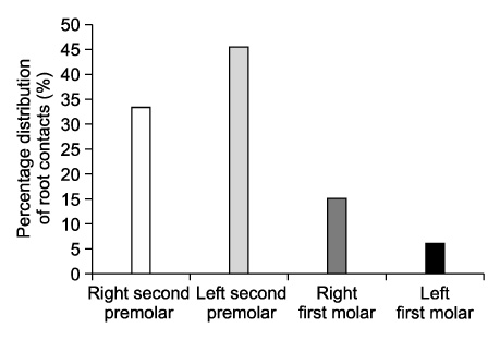
Fig. 2 Percentage distribution of root contacts on the upper premolars and molars.
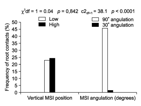
Fig. 3 Frequency of root contacts according to the vertical position and angulation of microscrew implant (MSI).
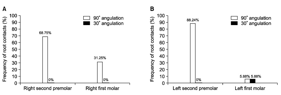
Fig. 4 Percentage distribution of root contacts according to either microscrew implant (MSI) angulation to the long axes of the teeth and jaw quadrant. A, Right side, low position group; B, left side, high position group.

Fig. 5 Statistical analysis; distance between each microscrew implant apex and the dental root of the premolar (DRMSI) versus microscrew implant (MSI) vertical position.

Fig. 6 Statistical analysis; distance between each microscrew implant apex and the dental root of the premolar (DRMSI) versus microscrew implant (MSI) angulation in (A) low position, (B) high position, and DRMSI versus position of MSI angulated at (C) 90° and (D) 30°.

Fig. 7 Comparison of fear level before and after microscrew implant (MSI) insertion.

Fig. 8 Statistical analysis; fear level before versus after microscrew implant (MSI) insertion.

Fig. 9 Percentage distribution of factors responsible for fear level before and after microscrew implant (MSI) insertion. RI, Root injury; MSI, maxillary sinus injury; MCI, maxillary canal injury; S, uncontrolled bur sliding while drilling; Br, breakage of either the drill or MSI; EBl, excessive bleeding; STI, soft tissue impaction into the drilled hole; BoN, bone necrosis; PC, postoperative complications; PA, patient's reluctance towards MSI insertion; LE, personal lack of experience.
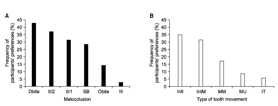
Fig. 10 Clinical applicability of microscrew implant (MSI) stated by participants in A, treatment of various malocclusions and B, tooth-movement. Dbite, Deep bite; II/2, class II/2; II/1, class II/1; SB, scissor bite; Obite, open bite; III, class III; IntI, intrusion of incisors; IntM, intrusion of molars; MM, mesialisation of molars; MU, molar uprighting; IT, impacted teeth.
Reference
-
1. Kanomi R. Mini-implant for orthodontic anchorage. J Clin Orthod. 1997. 31:763–767.2. Park HS. The skeletal cortical anchorage using titanium microscrew implants. Korean J Orthod. 1999. 29:699–706.3. Melsen B, Verna C. Miniscrew implants: The Aarhus anchorage system. Semin Orthod. 2005. 11:24–31.
Article4. Papadopoulos MA, Tarawneh F. The use of miniscrew implants for temporary skeletal anchorage in orthodontics: a comprehensive review. Oral Surg Oral Med Oral Pathol Oral Radiol Endod. 2007. 103:e6–e15.
Article5. Park HS, Kwon OW, Sung JH. Nonextraction treatment of an open bite with microscrew implant anchorage. Am J Orthod Dentofacial Orthop. 2006. 130:391–402.
Article6. Park HS, Kwon OW, Sung JH. Microscrew implant anchorage sliding mechanics. World J Orthod. 2005. 6:265–274.7. Kravitz ND, Kusnoto B. Risks and complications of orthodontic miniscrews. Am J Orthod Dentofacial Orthop. 2007. 131:4 Suppl. S43–S51.
Article8. Cho UH, Yu W, Kyung HM. Root contact during drilling for microimplant placement. Affect of surgery site and operator expertise. Angle Orthod. 2010. 80:130–136.9. Asscherickx K, Vannet BV, Wehrbein H, Sabzevar MM. Root repair after injury from mini-screw. Clin Oral Implants Res. 2005. 16:575–578.
Article10. Lee YK, Kim JW, Baek SH, Kim TW, Chang YI. Root and bone response to the proximity of a mini-implant under orthodontic loading. Angle Orthod. 2010. 80:452–458.
Article11. Schnelle MA, Beck FM, Jaynes RM, Huja SS. A radiographic evaluation of the availability of bone for placement of miniscrews. Angle Orthod. 2004. 74:832–837.12. Kuroda S, Yamada K, Deguchi T, Hashimoto T, Kyung HM, Takano-Yamamoto T. Root proximity is a major factor for screw failure in orthodontic anchorage. Am J Orthod Dentofacial Orthop. 2007. 131:4 Suppl. S68–S73.
Article13. Heránndez LC, Montoto G, Puente Rodríguez M, Galbán L, Marítnez V. 'Bone map' for a safe placement of miniscrews generated by computed tomography. Clin Oral Implants Res. 2008. 19:576–581.
Article14. Hu KS, Kang MK, Kim TW, Kim KH, Kim HJ. Relationships between dental roots and surrounding tissues for orthodontic miniscrew installation. Angle Orthod. 2009. 79:37–45.
Article15. Hembree M, Buschang PH, Carrillo R, Spears R, Rossouw PE. Effects of intentional damage of the roots and surrounding structures with miniscrew implants. Am J Orthod Dentofacial Orthop. 2009. 135:280.e1–280.e9.
Article16. Brisceno CE, Rossouw PE, Carrillo R, Spears R, Buschang PH. Healing of the roots and surrounding structures after intentional damage with miniscrew implants. Am J Orthod Dentofacial Orthop. 2009. 135:292–301.
Article17. Renjen R, Maganzini AL, Rohrer MD, Prasad HS, Kraut RA. Root and pulp response after intentional injury from miniscrew placement. Am J Orthod Dentofacial Orthop. 2009. 136:708–714.
Article18. Andreasen JO, Kristerson L. The effect of limited drying or removal of the periodontal ligament. Periodontal healing after replantation of mature permanent incisors in monkeys. Acta Odontol Scand. 1981. 39:1–13.19. Kang YG, Kim JY, Lee YJ, Chung KR, Park YG. Stability of mini-screws invading the dental roots and their impact on the paradental tissues in beagles. Angle Orthod. 2009. 79:248–255.
Article20. Poggio PM, Incorvati C, Velo S, Carano A. "Safe zones": a guide for miniscrew positioning in the maxillary and mandibular arch. Angle Orthod. 2006. 76:191–197.21. Park HS, Bae SM, Kyung HM, Sung JH. Micro-implant ananchorage for treatment of skeletal Class I bialveolar protrusion. J Clin Orthod. 2001. 35:417–422.22. Park HS, Jeong SH, Kwon OW. Factors affecting the clinical success of screw implants used as orthodontic anchorage. Am J Orthod Dentofacial Orthop. 2006. 130:18–25.
Article23. Miyawaki S, Koyama I, Inoue M, Mishima K, Sugahara T, Takano-Yamamoto T. Factors associated with the stability of titanium screws placed in the posterior region for orthodontic anchorage. Am J Orthod Dentofacial Orthop. 2003. 124:373–378.
Article24. Antoszewska J, Papadopoulos MA, Park HS, Ludwig B. Five-year experience with orthodontic miniscrew implants: a retrospective investigation of factors influencing success rates. Am J Orthod Dentofacial Orthop. 2009. 136:158.e1–158.e10.
Article
- Full Text Links
-
- Actions
-
Cited
- CITED
-
- Close
- Share
-
- Similar articles
-
- Removal torque and bone formation of orthodontic miniscrew implant
- Complications reported with the use of orthodontic miniscrews: A systematic review
- Three-dimensional evaluation of maxillary anterior alveolar bone for optimal placement of miniscrew implants
- A clinical study on skeletal anchorage system using miniscrew
- Histomorphometric evaluation of the bone surrounding orthodontic miniscrews according to their adjacent root proximity
