Histomorphometric evaluation of the bone surrounding orthodontic miniscrews according to their adjacent root proximity
- Affiliations
-
- 1Private Practice, Gimpo, Korea.
- 2Department of Orthodontics, The Institute of Cranio-Facial Deformity, College of Dentistry, Yonsei University, Seoul, Korea. hwang@yuhs.ac
- KMID: 2421313
- DOI: http://doi.org/10.4041/kjod.2018.48.5.283
Abstract
OBJECTIVE
This study was conducted to perform histomorphometric evaluations of the bone surrounding orthodontic miniscrews according to their proximity to the adjacent tooth roots in the posterior mandible of beagle dogs.
METHODS
Four male beagle dogs were used for this study. Six orthodontic miniscrews were placed in the interradicular spaces in the posterior mandible of each dog (n = 24). The implanted miniscrews were classified into no loading, immediate loading, and delayed loading groups according to the loading time. At 6 weeks after screw placement, the animals were sacrificed, and tissue blocks including the miniscrews were harvested for histological examinations. After analysis of the histological sections, the miniscrews were categorized into three additional groups according to the root proximity: high root proximity, low root proximity, and safe distance groups. Differences in the bone-implant contact (BIC, %) among the root proximity groups and loading time groups were determined using statistical analyses.
RESULTS
No BIC was observed within the bundle bone invaded by the miniscrew threads. Narrowing of the periodontal ligament space was observed in cases where the miniscrew threads touched the bundle bone. BIC (%) was significantly lower in the high root proximity group than in the low root proximity and safe distance groups. However, BIC (%) showed no significant differences among the loading time groups.
CONCLUSIONS
Regardless of the loading time, the stability of an orthodontic miniscrew is decreased if it is in contact with the bundle bone as well as the adjacent tooth root.
Keyword
Figure
-

Figure 1 Experimental protocol used in the present study. Immediately after implantation, the miniscrews on the left side were loaded. At 3 weeks after implantation, the miniscrews on the right side were loaded and the force on the left side was reactivated. At 4 weeks after implantation, the animals received intravenous tetracycline for fluorescence microscopy. At 6 weeks, the animals were sacrificed. TC, Tetracycline; IV, intravenous.
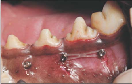
Figure 2 Miniscrews placed in interradicular spaces in the posterior mandible of beagle dogs. An orthodontic force is applied using an elastomeric chain. U, No loading group (screw placed between the second and third premolars); L, loading groups (one screw is placed between the third and fourth premolars, while another is placed between the fourth premolar and first molar).
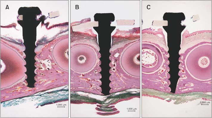
Figure 3 Classification of orthodontic miniscrews according to the proximity to the adjacent tooth roots using hematoxylin and eosin-stained sections (12.5×). A, High root proximity group. The miniscrew is in contact with the adjacent tooth root. B, Low root proximity group. The miniscrew is touching the bundle bone or the periodontal ligament but not contacting the adjacent root. C, Safe distance group. The miniscrew is placed in the interradicular septum, without any contact with the bundle bone.
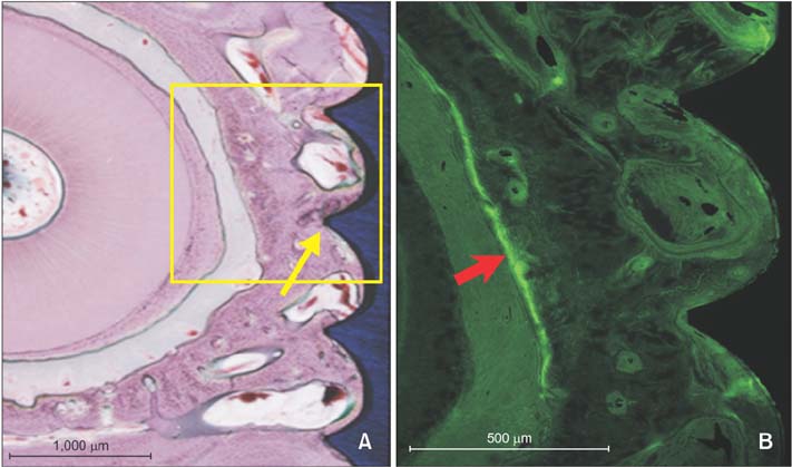
Figure 4 Root proximity of an orthodontic miniscrew without orthodontic loading and low root proximity, with atrophic changes in the adjacent periodontal ligament. A, A hematoxylin and eosin-stained section (50×). The arrow indicates a miniscrew thread touching the bundle bone. The distance from the miniscrew to the root surface is 0.494 mm. Narrowing of the periodontal ligament space can be observed (area in the box, shown in B). B, A fluorescence microscopy image with high magnification (100×). Active bone deposition can be observed as a bright green line (arrow) at the alveolar bone surface facing the periodontal ligament space.
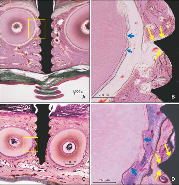
Figure 5 Effects of the proximity of an orthodontic miniscrew body to the bundle bone on the surrounding bone. The images show hematoxylin and eosin-stained sections. A and C, These are miniscrews with low root proximity. The miniscrew threads are invading the bundle bone (boxes, 12.5×). The miniscrew in A was loaded immediately after insertion, whereas the miniscrew in C was loaded at 3 weeks after insertion. The area in the box in A is shown in B, while the area in the box in C is shown in D. B and D, There is no direct bone–implant contact around the threads within the bundle bone (yellow arrows, 100×). Resorption of the adjacent tooth root can be observed (blue arrows).
Cited by 1 articles
-
Reader's Forum
Sun Mi Kwon
Korean J Orthod. 2019;49(1):1-2. doi: 10.4041/kjod.2019.49.1.1.
Reference
-
1. Liou EJ, Pai BC, Lin JC. Do miniscrews remain stationary under orthodontic forces? Am J Orthod Dentofacial Orthop. 2004; 126:42–47.
Article2. Kravitz ND, Kusnoto B. Risks and complications of orthodontic miniscrews. Am J Orthod Dentofacial Orthop. 2007; 131:4 Suppl. S43–S51.
Article3. Wang YC, Liou EJ. Comparison of the loading behavior of self-drilling and predrilled miniscrews throughout orthodontic loading. Am J Orthod Dentofacial Orthop. 2008; 133:38–43.
Article4. Papageorgiou SN, Zogakis IP, Papadopoulos MA. Failure rates and associated risk factors of orthodontic miniscrew implants: a meta-analysis. Am J Orthod Dentofacial Orthop. 2012; 142:577–595.
Article5. Kang YG, Kim JY, Lee YJ, Chung KR, Park YG. Stability of mini-screws invading the dental roots and their impact on the paradental tissues in beagles. Angle Orthod. 2009; 79:248–255.
Article6. Asscherickx K, Vande Vannet B, Wehrbein H, Sabzevar MM. Success rate of miniscrews relative to their position to adjacent roots. Eur J Orthod. 2008; 30:330–335.
Article7. Kuroda S, Yamada K, Deguchi T, Hashimoto T, Kyung HM, Takano-Yamamoto T. Root proximity is a major factor for screw failure in orthodontic anchorage. Am J Orthod Dentofacial Orthop. 2007; 131:4 Suppl. S68–S73.
Article8. Kim SH, Kang SM, Choi YS, Kook YA, Chung KR, Huang JC. Cone-beam computed tomography evaluation of mini-implants after placement: Is root proximity a major risk factor for failure? Am J Orthod Dentofacial Orthop. 2010; 138:264–276.
Article9. Janson G, Gigliotti MP, Estelita S, Chiqueto K. Influence of miniscrew dental root proximity on its degree of late stability. Int J Oral Maxillofac Surg. 2013; 42:527–534.
Article10. Wang Z, Li Y, Deng F, Song J, Zhao Z. A quantitative anatomical study on posterior mandibular interradicular safe zones for miniscrew implantation in the beagle. Ann Anat. 2008; 190:252–257.
Article11. Deguchi T, Yabuuchi T, Hasegawa M, Garetto LP, Roberts WE, Takano-Yamamoto T. Histomorphometric evaluation of cortical bone thickness surrounding miniscrew for orthodontic anchorage. Clin Implant Dent Relat Res. 2011; 13:197–205.
Article12. Cha JY, Kil JK, Yoon TM, Hwang CJ. Miniscrew stability evaluated with computerized tomography scanning. Am J Orthod Dentofacial Orthop. 2010; 137:73–79.
Article13. Marquezan M, Lima I, Lopes RT, Sant'Anna EF, de Souza MM. Is trabecular bone related to primary stability of miniscrews. Angle Orthod. 2014; 84:500–507.
Article14. Lee SY, Cha JY, Yoon TM, Park YC. The effect of loading time on the stability of mini-implant. Korean J Orthod. 2008; 38:149–158.
Article15. Freire JN, Silva NR, Gil JN, Magini RS, Coelho PG. Histomorphologic and histomophometric evaluation of immediately and early loaded mini-implants for orthodontic anchorage. Am J Orthod Dentofacial Orthop. 2007; 131:704.e1–704.e9.
Article16. Ure DS, Oliver DR, Kim KB, Melo AC, Buschang PH. Stability changes of miniscrew implants over time. Angle Orthod. 2011; 81:994–1000.
Article17. Motoyoshi M. Clinical indices for orthodontic mini implants. J Oral Sci. 2011; 53:407–412.18. Woods PW, Buschang PH, Owens SE, Rossouw PE, Opperman LA. The effect of force, timing, and location on bone-to-implant contact of miniscrew implants. Eur J Orthod. 2009; 31:232–240.
Article19. Youn JW, Cha JY, Yu H, Hwang CJ. Biologic evaluation of a hollow-type miniscrew implant: an experimental study in beagles. Am J Orthod Dentofacial Orthop. 2014; 145:626–637.
Article20. Cho YM, Cha JY, Hwang CJ. The effect of rotation moment on the stability of immediately loaded orthodontic miniscrews: a pilot study. Eur J Orthod. 2010; 32:614–619.
Article21. Watanabe H, Deguchi T, Hasegawa M, Ito M, Kim S, Takano-Yamamoto T. Orthodontic miniscrew failure rate and root proximity, insertion angle, bone contact length, and bone density. Orthod Craniofac Res. 2013; 16:44–55.
Article22. Nanci A. Dentin-pulp complex. In : Nanci A, editor. Ten cate's oral histology: development and function. 8th ed. St. Louis: Elsevier Mosby;2012. p. 220.23. Araújo M, Lindhe J. The edentulous alveolar ridge. In : Lindhe J, Lang NP, Karring T, editors. Clinical periodontology and implant dentistry. 5th ed. Oxford: Blackwell Munksgaard;2003. p. 53–63.24. Hubar JS. Quantification of the lamina dura. J Can Dent Assoc. 1993; 59:997–1000.25. Büchter A, Wiechmann D, Gaertner C, Hendrik M, Vogeler M, Wiesmann HP, et al. Load-related bone modelling at the interface of orthodontic microimplants. Clin Oral Implants Res. 2006; 17:714–722.
Article26. Kim H, Kim TW. Histologic evaluation of root-surface healing after root contact or approximation during placement of mini-implants. Am J Orthod Dentofacial Orthop. 2011; 139:752–760.
Article27. Lee YK, Kim JW, Baek SH, Kim TW, Chang YI. Root and bone response to the proximity of a mini-implant under orthodontic loading. Angle Orthod. 2010; 80:452–458.
Article28. Brisceno CE, Rossouw PE, Carrillo R, Spears R, Buschang PH. Healing of the roots and surrounding structures after intentional damage with miniscrew implants. Am J Orthod Dentofacial Orthop. 2009; 135:292–301.
Article29. Park HS, Yen S, Jeoung SH. Histologic and biomechanical characteristics of orthodontic self-drilling and self-tapping miniscrew implants. Korean J Orthod. 2006; 36:295–397.30. Cha JY, Hwang CJ, Kwon SH, Jung HS, Kim KM, Yu HS. Strain of bone-implant interface and insertion torque regarding different miniscrew thread designs using an artificial bone model. Eur J Orthod. 2015; 37:268–274.
Article31. Choi HW, Park YS, Chung SH, Jung MH, Moon W, Rhee SH. Comparison of mechanical and biological properties of zirconia and titanium alloy orthodontic micro-implants. Korean J Orthod. 2017; 47:229–237.
Article
- Full Text Links
-
- Actions
-
Cited
- CITED
-
- Close
- Share
-
- Similar articles
-
- Influence of immediate loading on the removal torque value of mini-screws
- A study on titanium miniscrew as orthodontic anchorage: An experimental investigation in dogs
- Root proximity of the anchoring miniscrews of orthodontic miniplates in the mandibular incisal area: Cone-beam computed tomographic analysis
- Consideration of maxillary sinus bone thickness when installing miniscrews
- Cortical bone thickness and root proximity at mandibular interradicular sites: implications for orthodontic mini-implant placement
