Korean J Obstet Gynecol.
2012 Dec;55(12):1015-1019. 10.5468/KJOG.2012.55.12.1015.
Small cell carcinoma, hypercalcemic type of the ovary
- Affiliations
-
- 1Department of Obstetrics and Gynecology, Busan Paik Hospital, Inje University College of Medicine, Busan, Korea. obgynjeong@hanmail.net
- 2Paik Institute for Clinical Research, Inje University, Busan, Korea.
- KMID: 2274217
- DOI: http://doi.org/10.5468/KJOG.2012.55.12.1015
Abstract
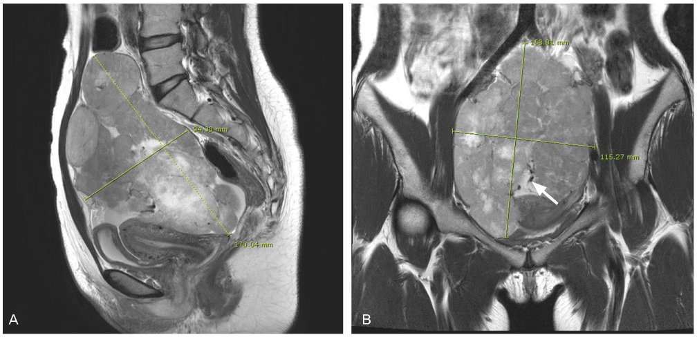
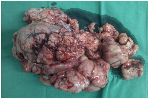
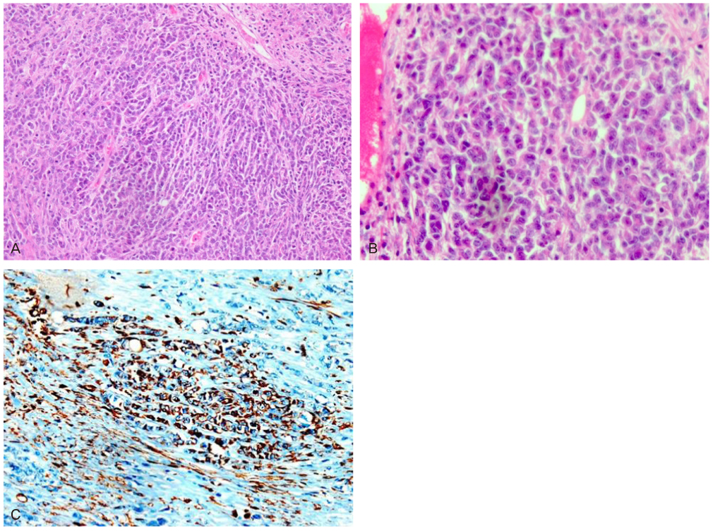
- The patient was a 22-year-old woman, who presented with abdominal pain and palpable huge mass. The initial investigation by ultrasound examination showed a huge size heterogenous soild mass and then magnetic resonance imaging presented multilobulated, huge solid mass (16x13 cm) with heterogenous enhancement in left ovary. Serum calcium level was slightly elevated (10.6 mg/dL, normal less than 10.4 mg/dL) and cancer antigen 125 level was normal. She underwent a laparotomy and left salpingo-ophorectomy. Grossly, ovary consists of yellowish solid portion but not cystic portion and outer capsule was ruptured focally. The pathologic finding, including immunohistochemical finding, confirmed small cell carcinoma, hypercalcemic type from ovary. Now, She has been treated with adjuvant chemotherapy (cisplatin, adriamycin, etoposide, cyclophophamide). We report this case with brief review of literature.
MeSH Terms
Figure
Reference
-
1. Siegel R, Naishadham D, Jemal A. Cancer statistics, 2012. CA Cancer J Clin. 2012. 62:10–29.2. National Cancer Information Center. Cancer incidence. c2011. cited 2012 Jan 2. Goyang (KR): National Cancer Information Center;Available from: http://www.cancer.go.kr/ncic/cics_f/01/012/index.html.3. Young RH, Oliva E, Scully RE. Small cell carcinoma of the ovary, hypercalcemic type. A clinicopathological analysis of 150 cases. Am J Surg Pathol. 1994. 18:1102–1116.4. Harrison ML, Hoskins P, du Bois A, Quinn M, Rustin GJ, Ledermann JA, et al. Small cell of the ovary, hypercalcemic type: analysis of combined experience and recommendation for management. A GCIG study. Gynecol Oncol. 2006. 100:233–238.5. Wynn D, Everett GD, Boothby RA. Small cell carcinoma of the ovary with hypercalcemia causes severe pancreatitis and altered mental status. Gynecol Oncol. 2004. 95:716–718.6. McCluggage WG, Oliva E, Connolly LE, McBride HA, Young RH. An immunohistochemical analysis of ovarian small cell carcinoma of hypercalcemic type. Int J Gynecol Pathol. 2004. 23:330–336.7. Ulbright TM, Roth LM, Stehman FB, Talerman A, Senekjian EK. Poorly differentiated (small cell) carcinoma of the ovary in young women: evidence supporting a germ cell origin. Hum Pathol. 1987. 18:175–184.8. Aguirre P, Thor AD, Scully RE. Ovarian small cell carcinoma. Histogenetic considerations based on immunohistochemical and other findings. Am J Clin Pathol. 1989. 92:140–149.9. Patsner B, Piver MS, Lele SB, Tsukada Y, Bielat K, Castillo NB. Small cell carcinoma of the ovary: a rapidly lethal tumor occurring in the young. Gynecol Oncol. 1985. 22:233–239.10. Dykgraaf RH, de Jong D, van Veen M, Ewing-Graham PC, Helmerhorst TJ, van der Burg ME. Clinical management of ovarian small-cell carcinoma of the hypercalcemic type: a proposal for conservative surgery in an advanced stage of disease. Int J Gynecol Cancer. 2009. 19:348–353.11. Rana S, Warren BK, Yamada SD. Stage IIIC small cell carcinoma of the ovary: survival with conservative surgery and chemotherapy. Obstet Gynecol. 2004. 103:1120–1123.12. Pautier P, Ribrag V, Duvillard P, Rey A, Elghissassi I, Sillet-Bach I, et al. Results of a prospective dose-intensive regimen in 27 patients with small cell carcinoma of the ovary of the hypercalcemic type. Ann Oncol. 2007. 18:1985–1989.13. Estel R, Hackethal A, Kalder M, Munstedt K. Small cell car cinoma of the ovary of the hypercalcaemic type: an analysis of clinical and prognostic aspects of a rare disease on the basis of cases published in the literature. Arch Gynecol Obstet. 2011. 284:1277–1282.
- Full Text Links
- Actions
-
Cited
- CITED
-
- Close
- Share
- Similar articles
-
- Small Cell Carcinoma of the Ovary Hypercalcemic Type: A Case Report
- Small Cell Carcinoma of the Ovary, Hypercalcemic Type, Large Cell Variant
- A case of small cell carcinoma of pulmonary type of ovary associated with huge mucinous cystadenocarcinoma
- Unusual malignant neoplasms of ovary in children: two cases report
- Primary small cell carcinoma of the ovary and the long-term survival after systemic chemotherapy


