Endovascular Management of Spontaneous Superficial Femoral Artery Pseudoaneurysm in a Renal Allograft Patient
- Affiliations
-
- 1Division of Cardiology, Cardiovascular Center, Internal Medicine, Myongji Hospital, Goyang, Korea. princette@hanmail.net
- KMID: 2198361
- DOI: http://doi.org/10.12997/jla.2014.3.1.49
Abstract
- We report a case of a superficial femoral artery pseudoaneurysm in 52-year old patient with a history of having renal allograft. The pseudoaneurysm spontaneously developed while standing up from squatting position after defecation, and it was successfully managed by an endovascular repair with an endograft. This case suggests that an atherosclerotic superficial femoral artery is vulnerable to torsion and tension movement during changing position from squatting to standing, which is repeatedly practiced by the people using the Korean traditional toilet. The endovascular therapy is also recommended for elderly patients with poor clinical conditions such as having a renal allograft and diffuse atherosclerosis of peripheral arteries.
Figure
-
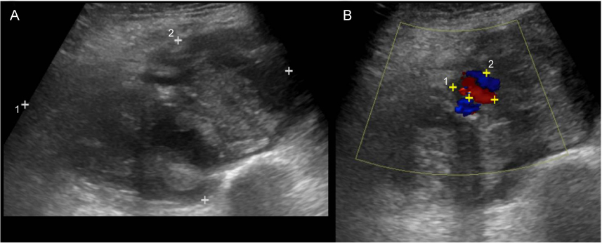
Fig. 1 Vascular ultrasound of the left thigh before endovascular treatment is shown. (A) The initial vascular ultrasound of the left thigh sonography showed a large (11.2×6.8×12 cm) hematoma including a 1.8×1.3 cm-sized pseudoaneurysm, (B) The pseudoaneurysm was communicated with the left superficial femoral artery, suggesting a spontaneous rupturing of left superficial femoral artery.
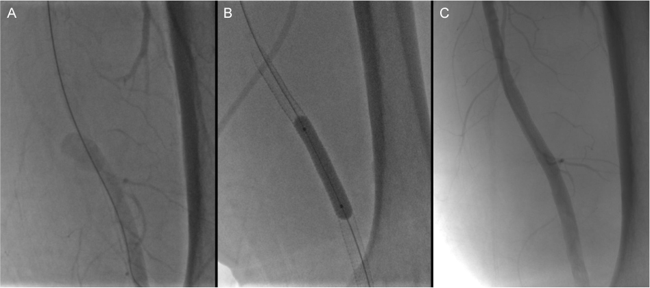
Fig. 2 Endovascular treatment of the pseudonaeurysm of left superficial femoral artery. (A) The peripheral angiography showed a pseudoaneurysm at the distal part of the left superficial femoral artery, (B) A self-expanding stent-graft (Viabahn® self-expanding stent-graft (8-mm diameter × 150-mm length)) was deployed to cover the pseudoaneurysm, and it was dilated and modeled by a 8 mm balloon, (C) The final angiography revealed a complete exclusion of the pseudoaneurysm.
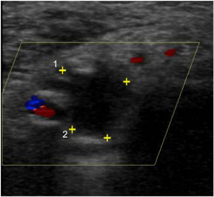
Fig. 3 Vascular ultrasound of the left thigh after endovascular treatment. The vascular ultrasound after endovascular treatment demonstrated a complete thrombosis of the pseudoaneurysm.
Reference
-
1. Applegate RJ, Sacrinty MT, Kutcher MA, Kahl FR, Gandhi SK, Santos RM, et al. Trends in vascular complications after diagnostic cardiac catheterization and percutaneous coronary intervention via the femoral artery, 1998 to 2007. JACC Cardiovasc Interv. 2008; 1:317–326.
Article2. Kanko M, Ciftci E. Spontaneous pseudoaneurysm of the superficial femoral artery in Behcet's disease endovascular stent-graft treatment combined with percutaneous drainage: a case report. Heart Surg Forum. 2007; 10:E84–E86.3. Merkus JW, Nieuwenhuijzen GA, Jacobs PP, van Roye SF, Koopman R, Pruszczynski MS, et al. Traumatic pseudoaneurysm of the superficial temporal artery. Injury. 1994; 25:468–471.
Article4. Blazick E, Keeling WB, Armstrong P, Letson D, Back M. Pseudoaneurysm of the superficial femoral artery associated with osteochondroma--a case report. Vasc Endovascular Surg. 2005; 39:355–358.
Article5. Yang KH, Park HW, Park SJ. Pseudoaneurysm of the superficial femoral artery after closed hip nailing with a Gamma nail: report of a case. J Orthop Trauma. 2002; 16:124–127.
Article6. Katzenschlager R, Ugurluoglu A, Ahmadi A, Hülsmann M, Koppensteiner R, Larch E, et al. Incidence of pseudoaneurysm after diagnostic and therapeutic angiography. Radiology. 1995; 195:463–466.
Article7. Goh BK, Chen CY, Hoe MN. Bilateral spontaneous rupture of the muscular branch of the superficial femoral artery with pseudoaneurysm formation. Ann Vasc Surg. 2004; 18:736–739.
Article8. Cadier MA, Watkin G, Pope FM, Marston A. Spontaneous rupture of the femoral arteries. J R Soc Med. 1993; 86:54.9. Calligaro KD, Savarese RP, Goldberg D, Doerr KJ, Dougherty MJ, DeLaurentis DA. Deep femoral artery pseudoaneurysm caused by acute trunk and hip torsion. Cardiovasc Surg. 1993; 1:392–394.10. Lossef SV, Gomes MN, Barth KH. Hemorrhage from spontaneous rupture of muscular branches of the superficial femoral artery. J Vasc Interv Radiol. 1994; 5:147–148.
Article11. Origuchi N, Shigematsu H, Nunokawa M, Yasuhura H, Muto T. Spontaneous perforation of a non-aneurysmal atherosclerotic abdominal aorta or femoral artery. Cardiovasc Surg. 1996; 4:351–355.
Article12. King JN, Kaupp HA. Spontaneous rupture of the superficial femoral artery with formation of a false aneurysm. J Cardiovasc Surg (Torino). 1970; 11:398–400.13. Helvie MA, Rubin JM, Silver TM, Kresowik TF. The distinction between femoral artery pseudoaneurysms and other causes of groin masses: value of duplex Doppler sonography. AJR Am J Roentgenol. 1988; 150:1177–1180.
Article14. Corriere MA, Guzman RJ. True and false aneurysms of the femoral artery. Semin Vasc Surg. 2005; 18:216–223.
Article15. Siani A, Flaishman I, Siani LM, Mounayergi F, Zaccaria A, Schioppa A, et al. Spontaneous rupture of the superficial femoral artery treated via an endovascular approach. Tex Heart Inst J. 2008; 35:66–68.
- Full Text Links
-
- Actions
-
Cited
- CITED
-
- Close
- Share
-
- Similar articles
-
- Pseudoaneurysm of the Superficial Femoral Artery Following Gamma nail Fixation for Trochanteric Fracture: A Case Report
- Successful Treatment of a Superficial Femoral Artery Pseudoaneurysm with Balloon Tamponade
- Pseudoaneurysm of Superficial Femoral Artery Following Proximal Femoral Nail Fixation
- Superficial Temporal Artery Pseudoaneurysm Treated with Manual Compression Alone
- Diagnosis and Treatment for Delayed Pseudoaneurysm of Deep Femoral Artery: A Case Report
