The Effects of Difumarate Salt S-15176 after Spinal Cord Injury in Rats
- Affiliations
-
- 1Department of Neurosurgery, Faculty of Medicine, Maltepe University, Istanbul, Turkey. drhakanerdogan@gmail.com
- 2Medical Biology Department, Cerrahpasa Faculty of Medicine, Istanbul University, Istanbul, Turkey.
- 3Department of Neurosurgery Taksim Education and Research Hospital, Istanbul, Turkey.
- 4Istanbul ME-DI ENT Surgery Center, Istanbul, Turkey.
- KMID: 2191253
- DOI: http://doi.org/10.3340/jkns.2015.57.6.445
Abstract
OBJECTIVE
In the present study we analyzed neuroprotective and antiapoptotic effect of the difumarate salt S-15176, as an anti-ischemic, an antioxidant and a stabilizer of mitochondrial membrane in secondary damage following spinal cord injury (SCI) in a rat model.
METHODS
Three groups were performed with 30 Wistar rats; control (1), trauma (2), and a trauma+S-15176 (10 mg/kg i.p., dimethyl sulfoxide) treatment (3). SCI was performed at the thoracic level using the weight-drop technique. Spinal cord tissues were collected following intracardiac perfusion in 3rd and 7th days of posttrauma. Hematoxylin and eosin staining for histopatology, terminal deoxynucleotidyl transferase dUTP nick end labeling assay for apoptotic cells and immunohistochemistry for proapoptotic cytochrome-c, Bax and caspase 9 were performed to all groups. Functional recovery test were applied to each group in 3rd and 7th days following SCI.
RESULTS
In trauma group, edematous regions, diffuse hemorrhage, necrosis, leukocyte infiltration and severe degeneration in motor neurons were observed prominently in gray matter. The number of apoptotic cells was significantly higher (p<0.05) than control group. In the S-15176-treated groups, apoptotic cell number in 3rd and 7th days (p<0.001), also cytochrome-c (p<0.001), Bax (p<0.001) and caspase 9 immunoreactive cells (p<0.001) were significantly decreased in number compared to trauma groups. Hemorrhage and edema in the focal areas were also noticed in gray matter of treatment groups. Results of the locomotor test were significantly increased in treatment group (p<0.05) when compared to trauma groups.
CONCLUSION
We suggest that difumarate salt S-15176 prevents mitochondrial pathways of apoptosis and protects spinal cord from secondary injury and helps to preserve motor function following SCI in rats.
MeSH Terms
-
Animals
Apoptosis
Caspase 9
Cell Count
DNA Nucleotidylexotransferase
Edema
Eosine Yellowish-(YS)
Hematoxylin
Hemorrhage
Immunohistochemistry
Leukocytes
Mitochondrial Membranes
Models, Animal
Motor Neurons
Necrosis
Perfusion
Rats*
Rats, Wistar
Spinal Cord
Spinal Cord Injuries*
Caspase 9
DNA Nucleotidylexotransferase
Eosine Yellowish-(YS)
Hematoxylin
Figure
-
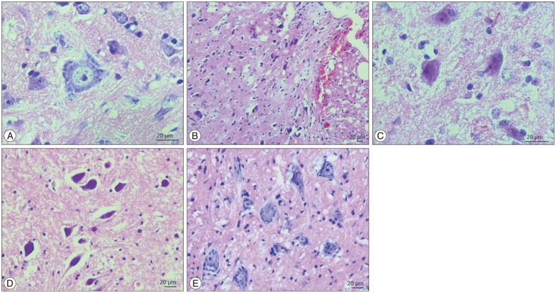
Fig. 1 H&E staining of spinal cord tissues. Control (A), trauma 3rd day (B), trauma 7th day (C), S-15176-treated 3rd day (D), S-15176-treated 7th day (E) groups (bar : 20 µm).
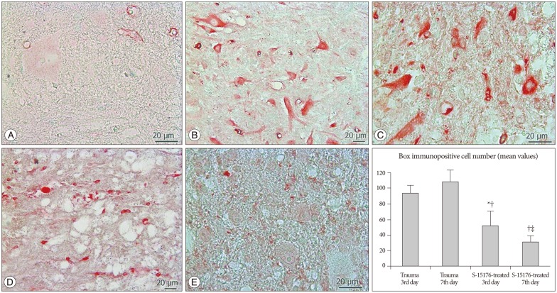
Fig. 2 Bax immunostaining of spinal cord tissue samples. Control (A), trauma 3rd day (B), trauma 7th day (C), S-15176-treated 3rd day (D), S-15176-treated 7th day (E) groups (bar : 20 µm). Evaluation of bax immunopositive cell numbers in the trauma groups; *p<0.01 vs. trauma 3rd, †p<0.001 vs. trauma 7th, ‡p<0.001 vs. trauma 3rd days.
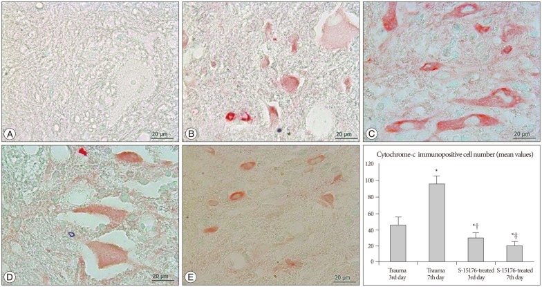
Fig. 3 Cytochrome-c immunostaining of spinal cord tissue samples. Control (A), trauma 3rd day (B), trauma 7th day (C), S-15176-treated 3rd day (D), S-15176-treated 7th day (E) groups (bar : 20 µm). Evaluation of cytochrome-c immunopositive cell numbers in the trauma groups; *p<0.01 vs. trauma 3rd, †p<0.001 vs. trauma 7th, ‡p<0.001 vs. trauma 3rd days.
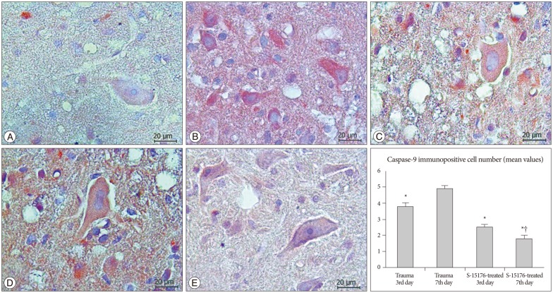
Fig. 4 Caspase-9 immunostaining of spinal cord tissue samples. Control (A), trauma 3rd day (B), trauma 7th day (C), S-15176-treated 3rd day (D), S-15176-treated 7th day (E) groups (bar : 20 µm). Evaluation of caspase-9 immunopositive cell numbers in the trauma groups; *p<0.01 vs. trauma 3rd and †p<0.001 vs. trauma 7th days.
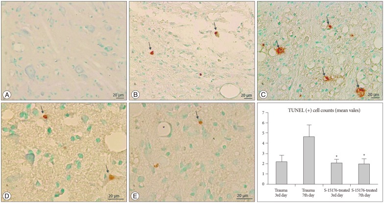
Fig. 5 TUNEL staining of spinal cord tissue samples. Control (A), trauma 3rd day (B), trauma 7th day (C), S-15176-treated 3rd day (D), S-15176-treated 7th day (E) groups (arrows : apoptotic cells) (bar : 20 µm). Graphs showing the mean values of apoptotic cell numbers in the trauma groups; *p<0.01 vs. trauma 3rd day.
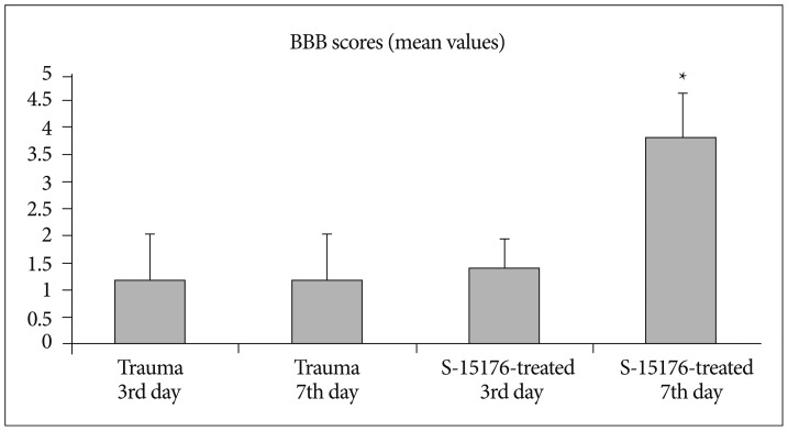
Fig. 6 Changes in the mean BBB scores of the trauma groups (mean±SD). *p<0.05 vs. all groups.
Reference
-
1. Albengres E, Le Louët H, d’Athis P, Tillement JP. S15176 and S16950 interaction with Cyclosporin A antiproliferative effect on cultured human lymphocytes. Fundam Clin Pharmacol. 2001; 15:41–46. PMID: 11468012.
Article2. Allen AR. Surgery of experimental lesion of spinal cord equivalent to crush injury of fracture dislocation of spinal column : a preliminary report. J Am Med Assoc. 1911; 57:877–880.3. Amar AP, Levy ML. Pathogenesis and pharmacological strategies for mitigating secondary damage in acute spinal cord injury. Neurosurgery. 1999; 44:1027–1039. discussion 1039-1040PMID: 10232536.
Article4. Basso DM, Beattie MS, Bresnahan JC. A sensitive and reliable locomotor rating scale for open field testing in rats. J Neurotrauma. 1995; 12:1–21. PMID: 7783230.
Article5. Bernardi P. The permeability transition pore. Control points of a cyclosporin A-sensitive mitochondrial channel involved in cell death. Biochim Biophys Acta. 1996; 1275:5–9. PMID: 8688451.
Article6. Bernardi P, Broekemeier KM, Pfeiffer DR. Recent progress on regulation of the mitochondrial permeability transition pore; a cyclosporin-sensitive pore in the inner mitochondrial membrane. J Bioenerg Biomembr. 1994; 26:509–517. PMID: 7896766.
Article7. Broekemeier KM, Dempsey ME, Pfeiffer DR. Cyclosporin A is a potent inhibitor of the inner membrane permeability transition in liver mitochondria. J Biol Chem. 1989; 264:7826–7830. PMID: 2470734.
Article8. Chávez E, Rodríguez JS, García G, García N, Correa F. Oligomycin strengthens the effect of cyclosporin A on mitochondrial permeability transition by inducing phosphate uptake. Cell Biol Int. 2005; 29:551–558. PMID: 15979905.
Article9. Cheng H, Cao Y, Olson L. Spinal cord repair in adult paraplegic rats : partial restoration of hind limb function. Science. 1996; 273:510–513. PMID: 8662542.
Article10. Crompton M. The mitochondrial permeability transition pore and its role in cell death. Biochem J. 1999; 341(Pt 2):233–249. PMID: 10393078.
Article11. Crompton M, Costi A, Hayat L. Evidence for the presence of a reversible Ca2+-dependent pore activated by oxidative stress in heart mitochondria. Biochem J. 1987; 245:915–918. PMID: 3117053.
Article12. Crowe MJ, Bresnahan JC, Shuman SL, Masters JN, Beattie MS. Apoptosis and delayed degeneration after spinal cord injury in rats and monkeys. Nat Med. 1997; 3:73–76. PMID: 8986744.
Article13. Dumont RJ, Okonkwo DO, Verma S, Hurlbert RJ, Boulos PT, Ellegala DB, et al. Acute spinal cord injury, part I : pathophysiologic mechanisms. Clin Neuropharmacol. 2001; 24:254–264. PMID: 11586110.14. Dumont RJ, Verma S, Okonkwo DO, Hurlbert RJ, Boulos PT, Ellegala DB, et al. Acute spinal cord injury, part II : contemporary pharmacotherapy. Clin Neuropharmacol. 2001; 24:265–279. PMID: 11586111.15. Elimadi A, Jullien V, Tillement JP, Morin D. S-15176 inhibits mitochondrial permeability transition via a mechanism independent of its antioxidant properties. Eur J Pharmacol. 2003; 468:93–101. PMID: 12742516.
Article16. Elimadi A, Sapena R, Settaf A, Le Louet H, Tillement J, Morin D. Attenuation of liver normothermic ischemia--reperfusion injury by preservation of mitochondrial functions with S-15176, a potent trimetazidine derivative. Biochem Pharmacol. 2001; 62:509–516. PMID: 11448461.
Article17. Elimadi A, Settaf A, Morin D, Sapena R, Lamchouri F, Cherrah Y, et al. Trimetazidine counteracts the hepatic injury associated with ischemia-reperfusion by preserving mitochondrial function. J Pharmacol Exp Ther. 1998; 286:23–28. PMID: 9655837.18. Fiskum G. Mitochondrial participation in ischemic and traumatic neural cell death. J Neurotrauma. 2000; 17:843–855. PMID: 11063052.
Article19. Fournier N, Ducet G, Crevat A. Action of cyclosporine on mitochondrial calcium fluxes. J Bioenerg Biomembr. 1987; 19:297–303. PMID: 3114244.
Article20. Friberg H, Ferrand-Drake M, Bengtsson F, Halestrap AP, Wieloch T. Cyclosporin A, but not FK 506, protects mitochondria and neurons against hypoglycemic damage and implicates the mitochondrial permeability transition in cell death. J Neurosci. 1998; 18:5151–5159. PMID: 9651198.
Article21. Geisler FH, Dorsey FC, Coleman WP. Recovery of motor function after spinal-cord injury--a randomized, placebo-controlled trial with GM-1 ganglioside. N Engl J Med. 1991; 324:1829–1838. PMID: 2041549.
Article22. Green DR, Reed JC. Mitochondria and apoptosis. Science. 1998; 281:1309–1312. PMID: 9721092.
Article23. Halestrap AP, Connern CP, Griffiths EJ, Kerr PM. Cyclosporin A binding to mitochondrial cyclophilin inhibits the permeability transition pore and protects hearts from ischaemia/reperfusion injury. Mol Cell Biochem. 1997; 174:167–172. PMID: 9309682.
Article24. Hamburger V. Cell death in the development of the lateral motor column of the chick embryo. J Comp Neurol. 1975; 160:535–546. PMID: 1123466.
Article25. Hamdan M, Urien S, Le Louet H, Tillement JP, Morin D. Inhibition of mitochondrial carnitine palmitoyltransferase-1 by a trimetazidine derivative, S-15176. Pharmacol Res. 2001; 44:99–104. PMID: 11516258.
Article26. Hauet T, Mothes D, Goujon JM, Caritez JC, Carretier M, le Moyec L, et al. Trimetazidine prevents renal injury in the isolated perfused pig kidney exposed to prolonged cold ischemia. Transplantation. 1997; 64:1082–1086. PMID: 9381535.
Article27. Hu Y, Benedict MA, Ding L, Núñez G. Role of cytochrome c and dATP/ATP hydrolysis in Apaf-1-mediated caspase-9 activation and apoptosis. EMBO J. 1999; 18:3586–3595. PMID: 10393175.
Article28. Joshi DC, Bakowska JC. Determination of mitochondrial membrane potential and reactive oxygen species in live rat cortical neurons. J Vis Exp. 2011; 23(51):Pii: 2704.
Article29. Katoh K, Ikata T, Katoh S, Hamada Y, Nakauchi K, Sano T, et al. Induction and its spread of apoptosis in rat spinal cord after mechanical trauma. Neurosci Lett. 1996; 216:9–12. PMID: 8892379.
Article30. Kawashima S, Yamamoto T, Horiuchi Y, Fujiwara K, Gouda S, Yoshimura Y, et al. S-15176 and its methylated derivative suppress the CsA-insensitive mitochondrial permeability transition and subsequent cytochrome c release induced by silver ion, and show weak protonophoric activity. Mol Cell Biochem. 2011; 358:45–51. PMID: 21688046.
Article31. Keane RW, Kraydieh S, Lotocki G, Bethea JR, Krajewski S, Reed JC, et al. Apoptotic and anti-apoptotic mechanisms following spinal cord injury. J Neuropathol Exp Neurol. 2001; 60:422–429. PMID: 11379817.
Article32. Kluck RM, Bossy-Wetzel E, Green DR, Newmeyer DD. The release of cytochrome c from mitochondria : a primary site for Bcl-2 regulation of apoptosis. Science. 1997; 275:1132–1136. PMID: 9027315.
Article33. Kroemer G, Dallaporta B, Resche-Rigon M. The mitochondrial death/life regulator in apoptosis and necrosis. Annu Rev Physiol. 1998; 60:619–642. PMID: 9558479.
Article34. Kroemer G, Petit P, Zamzami N, Vayssière JL, Mignotte B. The biochemistry of programmed cell death. FASEB J. 1995; 9:1277–1287. PMID: 7557017.
Article35. Liu XZ, Xu XM, Hu R, Du C, Zhang SX, McDonald JW, et al. Neuronal and glial apoptosis after traumatic spinal cord injury. J Neurosci. 1997; 17:5395–5406. PMID: 9204923.
Article36. Lu J, Ashwell KW, Waite P. Advances in secondary spinal cord injury : role of apoptosis. Spine (Phila Pa 1976). 2000; 25:1859–1866. PMID: 10888960.37. Ly JD, Grubb DR, Lawen A. The mitochondrial membrane potential (deltapsi(m)) in apoptosis; an update. Apoptosis. 2003; 8:115–128. PMID: 12766472.38. Morin D, Papadopoulos V, Tillement JP. Prevention of cell damage in ischemia : novel molecular targets in mitochondria. Expert Opin Ther Targets. 2002; 6:315–334.
Article39. Morin D, Zini R, Berdeaux A, Tillement JP. Effect of the mitochondrial transition pore inhibitor, S-15176, on rat liver mitochondria : ATP synthase modulation and mitochondrial uncoupling induction. Biochem Pharmacol. 2006; 72:911–918. PMID: 16879802.
Article40. Novgorodov SA, Gudz TI, Brierley GP, Pfeiffer DR. Magnesium ion modulates the sensitivity of the mitochondrial permeability transition pore to cyclosporin A and ADP. Arch Biochem Biophys. 1994; 311:219–228. PMID: 8203884.
Article41. Rupp H, Zarain-Herzberg A, Maisch B. The use of partial fatty acid oxidation inhibitors for metabolic therapy of angina pectoris and heart failure. Herz. 2002; 27:621–636. PMID: 12439634.
Article42. Sayeed I, Parvez S, Wali B, Siemen D, Stein DG. Direct inhibition of the mitochondrial permeability transition pore : a possible mechanism for better neuroprotective effects of allopregnanolone over progesterone. Brain Res. 2009; 1263:165–173. PMID: 19368823.
Article43. Schwab ME, Bartholdi D. Degeneration and regeneration of axons in the lesioned spinal cord. Physiol Rev. 1996; 76:319–370. PMID: 8618960.
Article44. Settaf A, Morin D, Lamchouri F, Elimadi A, Cherrah Y, Tillement JP. Trimetazidine ameliorates the hepatic injury associated with ischaemia-reperfusion in rats. Pharmacol Res. 1999; 39:211–216. PMID: 10094846.
Article45. Settaf A, Zahidy M, Elimadi A, Sapena R, Alsamad IA, Tillement J, et al. S-15176 reduces the hepatic injury in rats subjected to experimental ischemia and reperfusion. Eur J Pharmacol. 2000; 406:281–292. PMID: 11020492.
Article46. Shuman SL, Bresnahan JC, Beattie MS. Apoptosis of microglia and oligodendrocytes after spinal cord contusion in rats. J Neurosci Res. 1997; 50:798–808. PMID: 9418967.
Article47. Sullivan PG, Thompson MB, Scheff SW. Cyclosporin A attenuates acute mitochondrial dysfunction following traumatic brain injury. Exp Neurol. 1999; 160:226–234. PMID: 10630207.
Article48. Tator CH. Biology of neurological recovery and functional restoration after spinal cord injury. Neurosurgery. 1998; 42:696–707. discussion 707-708PMID: 9574633.
Article49. Tator CH, Fehlings MG. Review of the secondary injury theory of acute spinal cord trauma with emphasis on vascular mechanisms. J Neurosurg. 1991; 75:15–26. PMID: 2045903.
Article50. Tatton WG, Chalmers-Redman RM. Mitochondria in neurodegenerative apoptosis : an opportunity for therapy. Ann Neurol. 1998; 44(3 Suppl 1):S134–S141. PMID: 9749585.51. Tunçdemir M, Yıldırım A, Karaoğlan A, Akdemir O, Oztürk M. AR-A014418 as a glycogen synthase kinase-3 inhibitor : anti-apoptotic and therapeutic potential in experimental spinal cord injury. Neurocirugia (Astur). 2013; 24:22–32. PMID: 23116585.
Article52. Tunçdemir M, Ozturk M. The effects of ACE inhibitor and angiotensin receptor blocker on clusterin and apoptosis in the kidney tissue of streptozotocin-diabetic rats. J Mol Histol. 2008; 39:605–616. PMID: 18949565.
Article53. Yılmaz MB, Tönge M, Emmez H, Kaymaz F, Kaymaz M. Neuroprotective effects of quetiapine on neuronal apoptosis following experimental transient focal cerebral ischemia in rats. J Korean Neurosurg Soc. 2013; 54:1–7. PMID: 24044072.
Article54. Yong C, Arnold PM, Zoubine MN, Citron BA, Watanabe I, Berman NE, et al. Apoptosis in cellular compartments of rat spinal cord after severe contusion injury. J Neurotrauma. 1998; 15:459–472. PMID: 9674550.
Article55. Youdim MB, Bar Am O, Yogev-Falach M, Weinreb O, Maruyama W, Naoi M, et al. Rasagiline : neurodegeneration, neuroprotection, and mitochondrial permeability transition. J Neurosci Res. 2005; 79:172–179. PMID: 15573406.
Article56. Young W. Secondary injury mechanisms in acute spinal cord injury. J Emerg Med. 1993; 11(Suppl 1):13–22. PMID: 8445198.
- Full Text Links
-
- Actions
-
Cited
- CITED
-
- Close
- Share
-
- Similar articles
-
- Failure of topical DMSO to improve blood flow or evoked potentials in rat spinal cord injury
- Changes of motor evoked potentials and spinal cord evoked potentials following spinal cord injury in rats
- Functional Recovery in Complete Spinal Cord Injury after Transplantation of Human Umbilical Cord Blood Cells in Rats
- The Effect of Spinal Cord Injury on Pituitary-Testicular Hormone Axis in Rats
- Effects of Tumor Necrosis Factor Alpha Blocker Adalimumab in Experimental Spinal Cord Injury
