Effect of microgrooves and fibronectin conjugation on the osteoblast marker gene expression and differentiation
- Affiliations
-
- 1Department of Biomaterials & Prosthodontics, Kyung Hee University Hospital at Gangdong, Institute of Oral Biology, School of Dentistry, Kyung Hee University, Seoul, Republic of Korea. ysprosth@hanmail.net
- 2Department of Dentistry, Graduate School of Dentistry, Kyung Hee University, Seoul, Republic of Korea.
- 3ED Dental Clinic, Seoul, Republic of Korea.
- 4Department of Oral and Maxillofacial Surgery, Kyung Hee University Hospital at Gangdong, Institute of Oral Biology, School of Dentistry, Kyung Hee University, Seoul, Republic of Korea.
- 5Department of Prosthodontics, National Health Insurance Medical Center Ilsan Hospital, Goyang, Gyeonggi, Republic of Korea.
- 6Department of Maxillofacial Biomedical Engineering and Institute of Oral Biology, School of Dentistry, Kyung Hee University, Seoul, Republic of Korea. schlee@khu.ac.kr
- KMID: 2176625
- DOI: http://doi.org/10.4047/jap.2015.7.6.496
Abstract
- PURPOSE
To determine the effect of fibronectin (FN)-conjugated, microgrooved titanium (Ti) on osteoblast differentiation and gene expression in human bone marrow-derived mesenchymal stem cells (MSCs).
MATERIALS AND METHODS
Photolithography was used to fabricate the microgrooved Ti, and amine functionalization (silanization) was used to immobilize fibronectin on the titanium surfaces. Osteoblast differentiation and osteoblast marker gene expression were analyzed by means of alkaline phosphatase activity assay, extracellular calcium deposition assay, and quantitative real-time PCR.
RESULTS
The conjugation of fibronectin on Ti significantly increased osteoblast differentiation in MSCs compared with non-conjugated Ti substrates. On the extracellular calcium deposition assays of MSCs at 21 days, an approximately two-fold increase in calcium concentration was observed on the etched 60-microm-wide/10-microm-deep microgrooved surface with fibronectin (E60/10FN) compared with the same surface without fibronectin (E60/10), and a more than four-fold increase in calcium concentration was observed on E60/10FN compared with the non-etched control (NE0) and etched control (E0) surfaces. Through a series of analyses to determine the expression of osteoblast marker genes, a significant increase in all the marker genes except type I collagen alpha1 mRNA was seen with E60/10FN more than with any of the other groups, as compared with NE0.
CONCLUSION
The FN-conjugated, microgrooved Ti substrate can provide an effective surface to promote osteoblast differentiation and osteoblast marker gene expression in MSCs.
Keyword
MeSH Terms
Figure
-
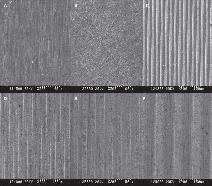
Fig. 1 Images taken of A- NE0 (×500), B- E0(×500), C- NE15/3.5 (×200), D- E15/3.5 (×200), E- E30/10 (×200), and F- E60/10 (×200) using field emission scanning electron microscopy.
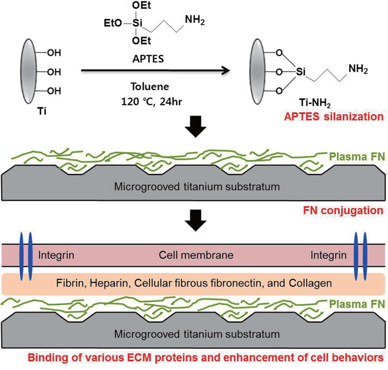
Fig. 2 Schematic illustration of the fabrication of FN-conjugated microgrooved titanium substrata and the expected enhancement of cellular activity.
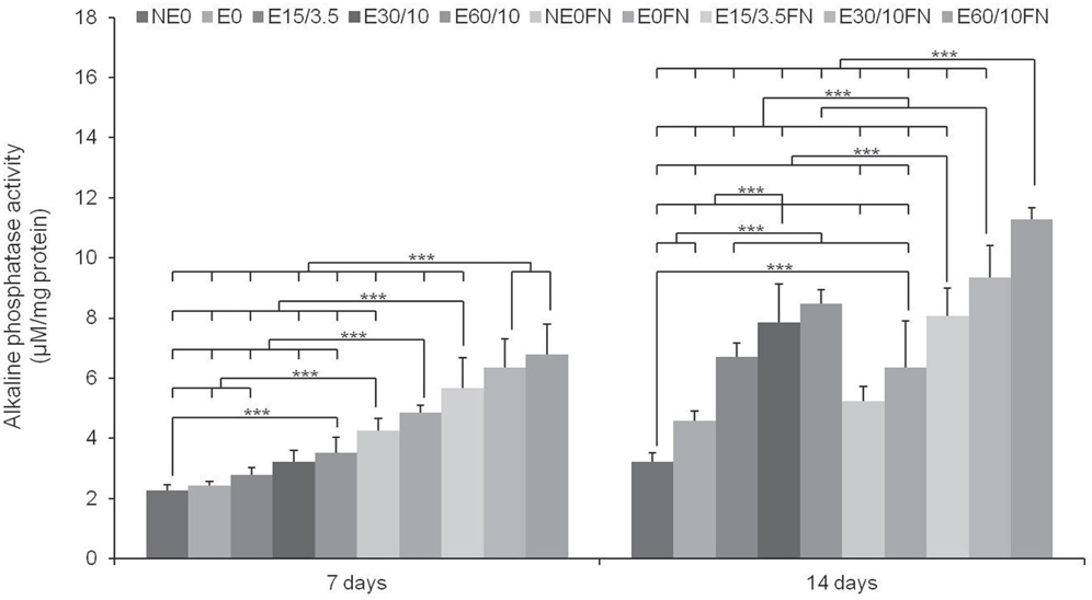
Fig. 3 The multiple comparison result of alkaline phosphatase (ALP) activity in human bone marrow-derived mesenchymal stem cells (MSCs) after 7 and 14 days of osteogenic culture on titanium substrata with specific surface topographies and fibronectin conjugation signaled by the ALP activity assay. One-way ANOVA (n = 5). ***: significant difference (P < .001).
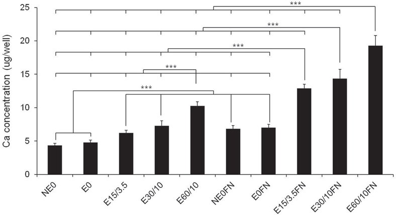
Fig. 4 The Multiple comparison result of osteoblast differentiation in human bone marrow-derived mesenchymal stem cells after 21 days of osteogenic culture on titanium substrata with specific surface topographies and fibronectin conjugation signaled by the extracellular calcium (Ca) deposition assay. One-way ANOVA (n = 5). ***: significant difference (P < .001).

Fig. 5 The relative fold change osteoblast-marker-gene expression in human bone marrow-derived mesenchymal stem cells (MSCs) after 2-day confluence and 14 days of osteogenic culture on titanium substrata with specific surface topographies and fibronectin conjugation by quantitative real-time PCR. Note that the results are presented as a ratio to the mRNA expression levels of the reference GAPDH gene, followed by a standardization of the NE0's Ct (threshold cycle) expression as 1. One-way ANOVA (n = 5). ***: significant difference (P < .001).
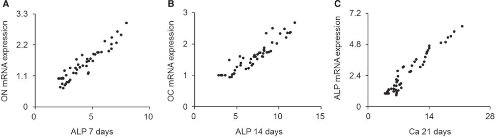
Fig. 6 The scatter-plot of correlation results between the expressed genes determined as greatest influential factors for osteoblastic differentiation and relevant assays. Significant correlations were present for A, B, and C (P < .01) (n = 30).
Cited by 1 articles
-
Regulation of human gingival fibroblast gene expression on microgrooves: A DNA microarray study
Kyungho Lee, Richard Leesungbok, Su-Jin Ahn, Su-Jung Park, Suk Won Lee
J Korean Acad Prosthodont. 2017;55(4):361-371. doi: 10.4047/jkap.2017.55.4.361.
Reference
-
1. Lee SW, Kim SY, Lee MH, Lee KW, Leesungbok R, Oh N. Influence of etched microgrooves of uniform dimension on in vitro responses of human gingival fibroblasts. Clin Oral Implants Res. 2009; 20:458–466.2. Lee MH, Oh N, Lee SW, Leesungbok R, Kim SE, Yun YP, Kang JH. Factors influencing osteoblast maturation on microgrooved titanium substrata. Biomaterials. 2010; 31:3804–3815.3. Park JA, Leesungbok R, Ahn SJ, Lee SW. Effect of etched microgrooves on hydrophilicity of titanium and osteoblast responses: A pilot study. J Adv Prosthodont. 2010; 2:18–24.4. Kim SY, Oh N, Lee MH, Kim SE, Leesungbok R, Lee SW. Surface microgrooves and acid etching on titanium substrata alter various cell behaviors of cultured human gingival fibroblasts. Clin Oral Implants Res. 2009; 20:262–272.5. Guida L, Annunziata M, Rocci A, Contaldo M, Rullo R, Oliva A. Biological response of human bone marrow mesenchymal stem cells to fluoride-modified titanium surfaces. Clin Oral Implants Res. 2010; 21:1234–1241.6. Lee MH, Kang JH, Lee SW. The significance of differential expression of genes and proteins in human primary cells caused by microgrooved biomaterial substrata. Biomaterials. 2012; 33:3216–3234.7. Engvall E, Ruoslahti E. Binding of soluble form of fibroblast surface protein, fibronectin, to collagen. Int J Cancer. 1977; 20:1–5.8. Dean JW 3rd, Culbertson KC, D'Angelo AM. Fibronectin and laminin enhance gingival cell attachment to dental implant surfaces in vitro. Int J Oral Maxillofac Implants. 1995; 10:721–728.9. Gallant ND, Michael KE, Garcia AJ. Cell adhesion strengthening: contributions of adhesive area, integrin binding, and focal adhesion assembly. Mol Biol Cell. 2005; 16:4329–4340.10. Fini M, Savarino L, Nicoli Aldini N, Martin L, Giavaresi G, Rizzi G, Martini D, Ruggeri A, Giunti A, Giardino R. Biomechanical and histomorphometric investigations on two morphologically differing titanium surfaces with and without fluorohydroxyapatite coating: an experimental study in sheep tibiae. Biomaterials. 2003; 24:3183–3192.11. Rezania A, Thomas CH, Healy KE. A probabilistic approach to measure the strength of bone cell adhesion to chemically modified surfaces. Ann Biomed Eng. 1997; 25:190–203.12. Middleton CA, Pendegrass CJ, Gordon D, Jacob J, Blunn GW. Fibronectin silanized titanium alloy: a bioinductive and durable coating to enhance fibroblast attachment in vitro. J Biomed Mater Res A. 2007; 83:1032–1038.13. Pendegrass CJ, Middleton CA, Gordon D, Jacob J, Blunn GW. Measuring the strength of dermal fibroblast attachment to functionalized titanium alloys in vitro. J Biomed Mater Res A. 2010; 92:1028–1037.14. Chou L, Firth JD, Uitto VJ, Brunette DM. Substratum surface topography alters cell shape and regulates fibronectin mRNA level, mRNA stability, secretion and assembly in human fibroblasts. J Cell Sci. 1995; 108(Pt 4):1563–1573.15. Lee SW, Kim SY, Rhyu IC, Chung WY, Leesungbok R, Lee KW. Influence of microgroove dimension on cell behavior of human gingival fibroblasts cultured on titanium substrata. Clin Oral Implants Res. 2009; 20:56–66.16. Jaiswal N, Haynesworth SE, Caplan AI, Bruder SP. Osteogenic differentiation of purified, culture-expanded human mesenchymal stem cells in vitro. J Cell Biochem. 1997; 64:295–312.17. Branemark PI, Albrektsson T. Titanium implants permanently penetrating human skin. Scand J Plast Reconstr Surg. 1982; 16:17–21.18. Dalby MJ, McCloy D, Robertson M, Wilkinson CD, Oreffo RO. Osteoprogenitor response to defined topographies with nanoscale depths. Biomaterials. 2006; 27:1306–1315.19. den Braber ET, de Ruijter JE, Ginsel LA, von Recum AF, Jansen JA. Quantitative analysis of fibroblast morphology on microgrooved surfaces with various groove and ridge dimensions. Biomaterials. 1996; 17:2037–2044.20. Li Z, Hassan MQ, Jafferji M, Aqeilan RI, Garzon R, Croce CM, van Wijnen AJ, Stein JL, Stein GS, Lian JB. Biological functions of miR-29b contribute to positive regulation of osteoblast differentiation. J Biol Chem. 2009; 284:15676–15684.21. Valencia S, Gretzer C, Cooper LF. Surface nanofeature effects on titanium-adherent human mesenchymal stem cells. Int J Oral Maxillofac Implants. 2009; 24:38–46.22. Mendonca G, Mendonca DB, Simoes LG, Araujo AL, Leite ER, Duarte WR, Aragao FJ, Cooper LF. The effects of implant surface nanoscale features on osteoblast-specific gene expression. Biomaterials. 2009; 30:4053–4062.23. Mendonca G, Mendonca DB, Aragao FJ, Cooper LF. The combination of micron and nanotopography by H(2)SO(4)/H(2)O(2) treatment and its effects on osteoblast-specific gene expression of hMSCs. J Biomed Mater Res A. 2010; 94:169–179.24. Chau JF, Leong WF, Li B. Signaling pathways governing osteoblast proliferation, differentiation and function. Histol Histopathol. 2009; 24:1593–1606.25. Nakashima K, Zhou X, Kunkel G, Zhang Z, Deng JM, Behringer RR, de Crombrugghe B. The novel zinc fingercontaining transcription factor osterix is required for osteoblast differentiation and bone formation. Cell. 2002; 108:17–29.26. Balloni S, Calvi EM, Damiani F, Bistoni G, Calvitti M, Locci P, Becchetti E, Marinucci L. Effects of titanium surface roughness on mesenchymal stem cell commitment and differentiation signaling. Int J Oral Maxillofac Implants. 2009; 24:627–635.27. Jayaraman M, Meyer U, Buhner M, Joos U, Wiesmann HP. Influence of titanium surfaces on attachment of osteoblastlike cells in vitro. Biomaterials. 2004; 25:625–631.28. Sato M, Morii E, Komori T, Kawahata H, Sugimoto M, Terai K, Shimizu H, Yasui T, Ogihara H, Yasui N, Ochi T, Kitamura Y, Ito Y, Nomura S. Transcriptional regulation of osteopontin gene in vivo by PEBP2alphaA/CBFA1 and ETS1 in the skeletal tissues. Oncogene. 1998; 17:1517–1525.29. Komori T. Regulation of bone development and extracellular matrix protein genes by RUNX2. Cell Tissue Res. 2010; 339:189–195.30. Ducy P, Starbuck M, Priemel M, Shen J, Pinero G, Geoffroy V, Amling M, Karsenty G. A Cbfa1-dependent genetic pathway controls bone formation beyond embryonic development. Genes Dev. 1999; 13:1025–1036.31. Maruyama Z, Yoshida CA, Furuichi T, Amizuka N, Ito M, Fukuyama R, Miyazaki T, Kitaura H, Nakamura K, Fujita T, Kanatani N, Moriishi T, Yamana K, Liu W, Kawaguchi H, Nakamura K, Komori T. Runx2 determines bone maturity and turnover rate in postnatal bone development and is involved in bone loss in estrogen deficiency. Dev Dyn. 2007; 236:1876–1890.
- Full Text Links
-
- Actions
-
Cited
- CITED
-
- Close
- Share
-
- Similar articles
-
- Macrophage-Stimulating Protein Enhances Osteoblastic Differentiation via the Recepteur d'Origine Nantais Receptor and Extracellular Signal-Regulated Kinase Signaling Pathway
- Heat or radiofrequency plasma glow discharge treatment of a titanium alloy stimulates osteoblast gene expression in the MC3T3 osteoprogenitor cell line
- Zinc modulation of osterix in MC3T3-E1 cells
- The biological effects of fibronectin typeIII 7-10 to MC3T3-E1 osteoblast
- Zinc upregulates bone-specific transcription factor Runx2 expression via BMP-2 signaling and Smad-1 phosphorylation in osteoblasts
