The location of midfacial landmarks according to the method of establishing the midsagittal reference plane in three-dimensional computed tomography analysis of facial asymmetry
- Affiliations
-
- 1School of Dentistry, Dental Science Research Institute, Chonnam National University, Gwangju, Korea.
- 2Department of Oral Anatomy, School of Dentistry, Dental Science Research Institute, Chonnam National University, Gwangju, Korea.
- 3Department of Nursing, Kwangju Women's University, Gwangju, Korea.
- 4Department of Oral and Maxillofacial Radiology, School of Dentistry, Dental Science Research Institute, Chonnam National University, Gwangju, Korea. yoonfr@chonnam.ac.kr
- KMID: 2132935
- DOI: http://doi.org/10.5624/isd.2015.45.4.227
Abstract
- PURPOSE
The purpose of this study was to evaluate the influence of methods of establishing the midsagittal reference plane (MRP) on the locations of midfacial landmarks in the three-dimensional computed tomography (CT) analysis of facial asymmetry.
MATERIALS AND METHODS
A total of 24 patients (12 male and 12 female; mean age, 22.5 years; age range, 18.2-29.7 years) with facial asymmetry were included in this study. The MRP was established using two different methods on each patient's CT image. The x-coordinates of four midfacial landmarks (the menton, nasion, upper incisor, and lower incisor) were obtained by measuring the distance and direction of the landmarks from the MRP, and the two methods were compared statistically. The direction of deviation and the severity of asymmetry found using each method were also compared.
RESULTS
The x-coordinates of the four anatomic landmarks all showed a statistically significant difference between the two methods of establishing the MRP. For the nasion and lower incisor, six patients (25.0%) showed a change in the direction of deviation. The severity of asymmetry also changed in 16 patients (66.7%).
CONCLUSION
The results of this study suggest that the locations of midfacial landmarks change significantly according to the method used to establish the MRP.
Figure
-
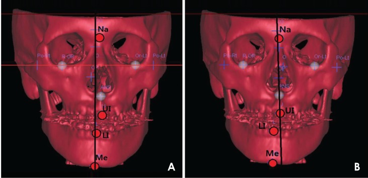
Fig. 1 The establishment of the midsagittal reference plane by two methods. A. In Method 1, the horizontal reference plane is established using the PoR, PoL, and OrL landmarks, the midsagittal reference plane (MRP) by using the Cg and P, and the coronal reference plane by using the Op. The planes are perpendicular to one another. B. In Method 2, the Op, Cg, and ANS landmarks identified in Method 1 are used to establish the MRP. Midfacial landmarks are indicated as circles, and the MRP is shown as a black vertical line. PoR: right porion, PoL: left porion, OrL: left orbitale, Cg: crista galli, P: prechiasmatic groove, Op: opisthion, ANS: anterior nasal spine.
Cited by 2 articles
-
Novel three-dimensional position analysis of the mandibular foramen in patients with skeletal class III mandibular prognathism
Sang-Hoon Kang, Yeon-Ho Kim, Yu-Jin Won, Moon-Key Kim
Imaging Sci Dent. 2016;46(2):77-85. doi: 10.5624/isd.2016.46.2.77.Pierre Robin sequence with severe scoliosis in an adult: A case report of clinical and radiological features
Jae-Jun Kim, Dong-Soon Choi, Insan Jang, Bong-Kuen Cha, In-Woo Park
Imaging Sci Dent. 2019;49(4):323-329. doi: 10.5624/isd.2019.49.4.323.
Reference
-
1. Hwang HS, Hwang CH, Lee KH, Kang BC. Maxillofacial 3-dimensional image analysis for the diagnosis of facial asymmetry. Am J Orthod Dentofacial Orthop. 2006; 130:779–785. PMID: 17169741.
Article2. Grummons DC, Kappeyne van de Coppello MA. A frontal asymmetry analysis. J Clin Orthod. 1987; 21:448–465. PMID: 3476493.3. Ferguson JW. Cephalometric interpretation and assessment of facial asymmetry secondary to congenital torticollis. The significance of cranial base reference lines. Int J Oral Maxillofac Surg. 1993; 22:7–10. PMID: 8459125.
Article4. Haraguchi S, Takada K, Yasuda Y. Facial asymmetry in subjects with skeletal Class III deformity. Angle Orthod. 2002; 72:28–35. PMID: 11843270.5. Ahn JS, Hwang HS. Relationship between perception of facial asymmetry and posteroanterior cephalometric measurements. Korean J Orthod. 2001; 31:489–498.6. You KH, Lee KJ, Lee SH, Baik HS. Three-dimensional computed tomography analysis of mandibular morphology in patients with facial asymmetry and mandibular prognathism. Am J Orthod Dentofacial Orthop. 2010; 138:540.e1–540.e8. PMID: 21055584.
Article7. Kwon TG, Park HS, Ryoo HM, Lee SH. A comparison of craniofacial morphology in patients with and without facial asymmetry - a three-dimensional analysis with computed tomography. Int J Oral Maxillofac Surg. 2006; 35:43–48. PMID: 15925488.8. Hwang HS, Min YS, Lee SC, Sun MK, Lim HS. Change of lip-line cant after 1-jaw orthognathic surgery in patients with mandibular asymmetry. Am J Orthod Dentofacial Orthop. 2009; 136:564–569. PMID: 19815160.
Article9. Baek SH, Cho IS, Chang YI, Kim MJ. Skeletodental factors affecting chin point deviation in female patients with class III malocclusion and facial asymmetry: a three-dimensional analysis using computed tomography. Oral Surg Oral Med Oral Pathol Oral Radiol Endod. 2007; 104:628–639. PMID: 17656131.
Article10. Jung YJ, Kim MJ, Baek SH. Hard and soft tissue changes after correction of mandibular prognathism and facial asymmetry by mandibular setback surgery: three-dimensional analysis using computerized tomography. Oral Surg Oral Med Oral Pathol Oral Radiol Endod. 2009; 107:763–771.e8. PMID: 19272814.
Article11. Matteson SR, Bechtold W, Phillips C, Staab EV. A method for three-dimensional image reformation for quantitative cephalometric analysis. J Oral Maxillofac Surg. 1989; 47:1053–1061. PMID: 2795298.
Article12. Cavalcanti MG, Vannier MW. Quantitative analysis of spiral computed tomography for craniofacial clinical applications. Dentomaxillofac Radiol. 1998; 27:344–350. PMID: 10895633.
Article13. Olszewski R, Zech F, Cosnard G, Nicolas V, Macq B, Reychler H. Three-dimensional computed tomography cephalometric craniofacial analysis: experimental validation in vitro. Int J Oral Maxillofac Surg. 2007; 36:828–833. PMID: 17825530.
Article14. Tuncer BB, Atac MS, Yüksel S. A case report comparing 3-D evaluation in the diagnosis and treatment planning of hemimandibular hyperplasia with conventional radiography. J Craniomaxillofac Surg. 2009; 37:312–319. PMID: 19289289.
Article15. Kragskov J, Bosch C, Gyldensted C, Sindet-Pedersen S. Comparison of the reliability of craniofacial anatomic landmarks based on cephalometric radiographs and three-dimensional CT scans. Cleft Palate Craniofac J. 1997; 34:111–116. PMID: 9138504.
Article16. Maeda M, Katsumata A, Ariji Y, Muramatsu A, Yoshida K, Goto S, et al. 3D-CT evaluation of facial asymmetry in patients with maxillofacial deformities. Oral Surg Oral Med Oral Pathol Oral Radiol Endod. 2006; 102:382–390. PMID: 16920547.
Article17. Kim TY, Baik JS, Park JY, Chae HS, Huh KH, Choi SC. Determination of midsagittal plane for evaluation of facial asymmetry using three-dimensional computed tomography. Imaging Sci Dent. 2011; 41:79–84. PMID: 21977479.
Article18. Yoon KW, Yoon SJ, Kang BC, Kim YH, Kook MS, Lee JS, et al. Deviation of landmarks in accordance with methods of establishing reference planes in three-dimensional facial CT evaluation. Imaging Sci Dent. 2014; 44:207–212. PMID: 25279341.
Article19. Bookstein FL, Grayson B, Cutting CB, Kim HC, McCarthy JG. Landmarks in three dimensions: reconstruction from cephalograms versus direct observation. Am J Orthod Dentofacial Orthop. 1991; 100:133–140. PMID: 1867164.
Article20. Ferrario VF, Sforza C, Poggio CE, Tartaglia G. Distance from symmetry: a three-dimensional evaluation of facial asymmetry. J Oral Maxillofac Surg. 1994; 52:1126–1132. PMID: 7965306.
Article21. Pelo S, Deli R, Correra P, Boniello R, Gasparini G, Moro A. Evaluation of 2 different reference planes used for the study of asymmetric facial malformations. J Craniofac Surg. 2009; 20:41–45. PMID: 19164986.
Article22. Severt TR, Proffit WR. The prevalence of facial asymmetry in the dentofacial deformities population at the University of North Carolina. Int J Adult Orthodon Orthognath Surg. 1997; 12:171–176. PMID: 9511487.23. Edler R, Wertheim D, Greenhill D. Comparison of radiographic and photographic measurement of mandibular asymmetry. Am J Orthod Dentofacial Orthop. 2003; 123:167–174. PMID: 12594423.
Article24. Michiels LY, Tourne LP. Nasion true vertical: a proposed method for testing the clinical validity of cephalometric measurements applied to a new cephalometric reference line. Int J Adult Orthodon Orthognath Surg. 1990; 5:43–52. PMID: 2373912.
- Full Text Links
-
- Actions
-
Cited
- CITED
-
- Close
- Share
-
- Similar articles
-
- Comparison of midsagittal reference plane in PA cephalogram and 3D CT
- Deviation of landmarks in accordance with methods of establishing reference planes in three-dimensional facial CT evaluation
- Determination of midsagittal plane for evaluation of facial asymmetry using three-dimensional computed tomography
- Validity of midsagittal reference planes constructed in 3D CT images
- Differences in positions of cone-beam computed tomography landmarks in patients with skeletal Class III facial asymmetry according to midsagittal planes
