Deviation of landmarks in accordance with methods of establishing reference planes in three-dimensional facial CT evaluation
- Affiliations
-
- 1School of Dentistry, Dental Science Research Institute, Chonnam National University, Gwangju, Korea.
- 2Department of Oral and Maxillofacial Radiology, School of Dentistry, Dental Science Research Institute, Chonnam National University, Gwangju, Korea. yoonfr@chonnam.ac.kr
- 3Department of Oral and Maxillofacial Radiology, Hallym University Sacred Heart Hospital, Anyang, Korea.
- 4Department of Oral and Maxillofacial Surgery, School of Dentistry, Dental Science Research Institute, Chonnam National University, Gwangju, Korea.
- 5Department of Orthodontics, School of Dental Medicine, Case Western Reserve University, Cleveland, OH, USA.
- KMID: 1974484
- DOI: http://doi.org/10.5624/isd.2014.44.3.207
Abstract
- PURPOSE
This study aimed to investigate the deviation of landmarks from horizontal or midsagittal reference planes according to the methods of establishing reference planes.
MATERIALS AND METHODS
Computed tomography (CT) scans of 18 patients who received orthodontic and orthognathic surgical treatment were reviewed. Each CT scan was reconstructed by three methods for establishing three orthogonal reference planes (namely, the horizontal, midsagittal, and coronal reference planes). The horizontal (bilateral porions and bilateral orbitales) and midsagittal (crista galli, nasion, prechiasmatic point, opisthion, and anterior nasal spine) landmarks were identified on each CT scan. Vertical deviation of the horizontal landmarks and horizontal deviation of the midsagittal landmarks were measured.
RESULTS
The porion and orbitale, which were not involved in establishing the horizontal reference plane, were found to deviate vertically from the horizontal reference plane in the three methods. The midsagittal landmarks, which were not used for the midsagittal reference plane, deviated horizontally from the midsagittal reference plane in the three methods.
CONCLUSION
In a three-dimensional facial analysis, the vertical and horizontal deviations of the landmarks from the horizontal and midsagittal reference planes could vary depending on the methods of establishing reference planes.
Figure
-
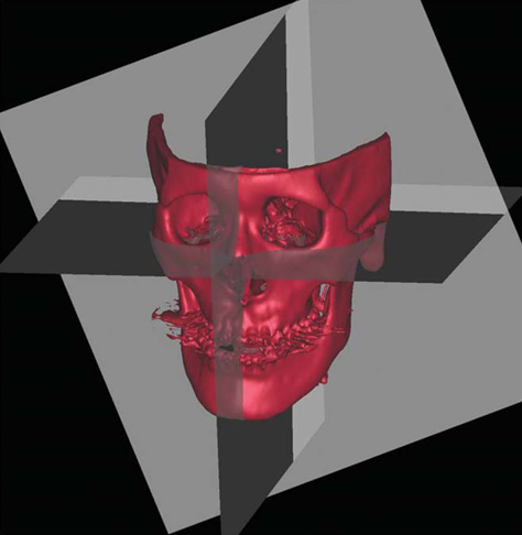
Fig. 1 Three orthogonal planes are established for each subject.
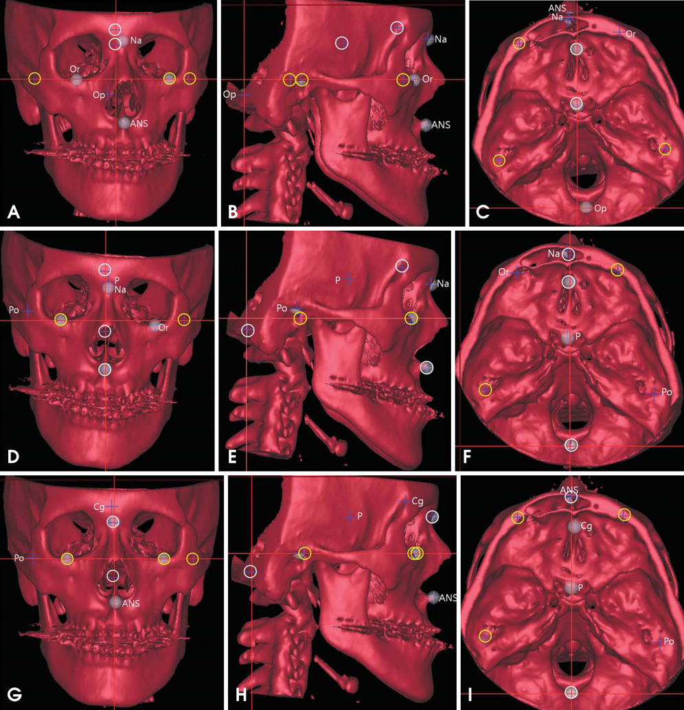
Fig. 2 One subject is analyzed by three methods (A-C: M1; D-F: M2; and G-I: M3). The landmarks used for the HRP are marked as yellow circles; those for the MRP as white circles. The landmarks that are not involved in establishing the HRP and MRP are named. A-C: The Na, Op, and ANS deviated horizontally from the MRP. D-F: The P and Na deviated horizontally from the MRP, and the Po and Or vertically from the HRP. G-I: The Cg and ANS deviated horizontally from the MRP.
Cited by 2 articles
-
A comparative study of the deviation of the menton on posteroanterior cephalograms and three-dimensional computed tomography
Hee Jin Lee, Sungeun Lee, Eun Joo Lee, In Ja Song, Byung-Cheol Kang, Jae-Seo Lee, Hoi-Jeong Lim, Suk-Ja Yoon
Imaging Sci Dent. 2016;46(1):33-38. doi: 10.5624/isd.2016.46.1.33.The location of midfacial landmarks according to the method of establishing the midsagittal reference plane in three-dimensional computed tomography analysis of facial asymmetry
Min Sun Kim, Eun Joo Lee, In Ja Song, Jae-Seo Lee, Byung-Cheol Kang, Suk-Ja Yoon
Imaging Sci Dent. 2015;45(4):227-232. doi: 10.5624/isd.2015.45.4.227.
Reference
-
1. Katsumata A, Fujishita M, Maeda M, Ariji Y, Ariji E, Langlais RP. 3D-CT evaluation of facial asymmetry. Oral Surg Oral Med Oral Pathol Oral Radiol Endod. 2005; 99:212–220.
Article2. Maeda M, Katsumata A, Ariji Y, Muramatsu A, Yoshida K, Goto S, et al. 3D-CT evaluation of facial asymmetry in patients with maxillofacial deformities. Oral Surg Oral Med Oral Pathol Oral Radiol Endod. 2006; 102:382–390.
Article3. Kwon TG, Park HS, Ryoo HM, Lee SH. A comparison of craniofacial morphology in patients with and without facial asymmetry-a three-dimensional analysis with computed tomography. Int J Oral Maxillofac Surg. 2006; 35:43–48.
Article4. Park SH, Yu HS, Kim KD, Lee KJ, Baik HS. A proposal for a new analysis of craniofacial morphology by 3-dimensional computed tomography. Am J Orthod Dentofacial Orthop. 2006; 129:600.e23–600.e34.
Article5. Yoon SJ, Lim HJ, Kang BC, Hwang HS. Three dimensional CT analysis of facial asymmetry. Korean J Oral Maxillofac Radiol. 2007; 37:45–52.6. Baek SH, Cho IS, Chang YI, Kim MJ. Skeletodental factors affecting chin point deviation in female patients with class III malocclusion and facial asymmetry: a three-dimensional analysis using computed tomography. Oral Surg Oral Med Oral Pathol Oral Radiol Endod. 2007; 104:628–639.
Article7. Yoon SJ, Wang RF, Hwang HS, Kang BC, Lee JS, Palomo JM. Application of spherical coordinate system to facial asymmetry analysis in mandibular prognathism patients. Imaging Sci Dent. 2011; 41:95–100.
Article8. Hwang HS, Hwang CH, Lee KH, Kang BC. Maxillofacial 3-dimensional image analysis for the diagnosis of facial asymmetry. Am J Orthod Dentofacial Orthop. 2006; 130:779–785.
Article9. Kim EJ, Palomo JM, Kim SS, Lim HJ, Lee KM, Hwang HS. Maxillofacial characteristics affecting chin deviation between mandibular retrusion and prognathism patients. Angle Orthod. 2011; 81:988–993.
Article10. Baek C, Paeng JY, Lee JS, Hong J. Morphologic evaluation and classification of facial asymmetry using 3-dimensional computed tomography. J Oral Maxillofac Surg. 2012; 70:1161–1169.
Article11. Grumons DC, Kappeyne van de Coppello MA. A frontal asymmetry analysis. J Clin Orthod. 1987; 21:448–465.12. Haraguchi S, Takada K, Yasuda Y. Facial asymmetry in subjects with skeletal Class III deformity. Angle Orthod. 2002; 72:28–35.13. Kim TY, Baik JS, Park JY, Chae HS, Huh KH, Choi SC. Determination of midsagittal plane for evaluation of facial asymmetry using three-dimensional computed tomography. Imaging Sci Dent. 2011; 41:79–84.
Article14. Pelo S, Deli R, Correra P, Boniello R, Gasparini G, Moro A. Evaluation of 2 different reference planes used for the study of asymmetric facial malformations. J Craniofac Surg. 2009; 20:41–45.
Article15. Farman AG, Scarfe WC. Development of imaging selection criteria and procedures should precede cephalometric assessment with cone-beam computed tomography. Am J Orthod Dentofacial Orthop. 2006; 130:257–265.
Article16. Muramatsu A, Kimura M, Nawa H, Yoshida K, Maeda M, Katsumata A, et al. Reproducibility of maxillofacial anatomic landmarks on 3-dimensional computed tomographic images determined with the 95% confidence ellipse method. Angle Orthod. 2008; 78:396–402.
Article17. Ramírez-Sotelo LR, Almeida S, Ambrosano GM, Bóscolo F. Validity and reproducibility of cephalometric measurements performed in full and hemifacial reconstructions derived from cone beam computed tomography. Angle Orthod. 2012; 82:827–832.
Article18. Trpkova B, Prasad NG, Lam EW, Raboud D, Glover KE, Major PW. Assessment of facial asymmetries from posteroanterior cephalograms: validity of reference lines. Am J Orthod Dentofacial Orthop. 2003; 123:512–520.
Article19. Ferguson JW. Cephalometric interpretation and assessment of facial asymmetry secondary to congenital torticollis. The significance of cranial base reference lines. Int J Oral Maxillofac Surg. 1993; 22:7–10.
Article
- Full Text Links
-
- Actions
-
Cited
- CITED
-
- Close
- Share
-
- Similar articles
-
- Comparison of midsagittal reference plane in PA cephalogram and 3D CT
- The location of midfacial landmarks according to the method of establishing the midsagittal reference plane in three-dimensional computed tomography analysis of facial asymmetry
- Quantification of three-dimensional facial asymmetry for diagnosis and postoperative evaluation of orthognathic surgery
- Determination of midsagittal plane for evaluation of facial asymmetry using three-dimensional computed tomography
- Validity of midsagittal reference planes constructed in 3D CT images
