J Korean Orthop Assoc.
2008 Feb;43(1):139-142. 10.4055/jkoa.2008.43.1.139.
Gout Tophi in the Bipartite Patella: A Case Report
- Affiliations
-
- 1Department of Orthopedic Surgery, Inha University College of Medicine, Incheon, Korea. TJLEE@inha.ac.kr
- 2Department of Pathology, Inha University College of Medicine, Incheon, Korea.
- KMID: 2106431
- DOI: http://doi.org/10.4055/jkoa.2008.43.1.139
Abstract
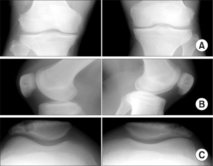
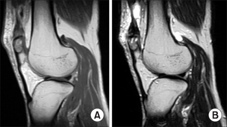
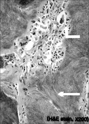
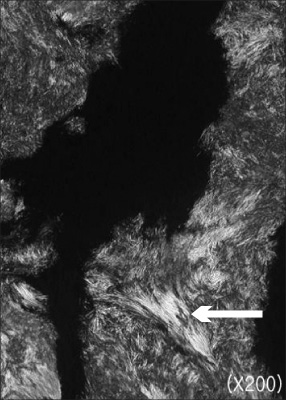
- Osteolysis of the patella occurs in benign or malignant bone tumors, metastatic disease, osteolytic infections, degenerative, or metabolic bone disease. Several cases of patellar destruction secondary to gout have been reported. However, there has only been one case of bipartite patellar bone destruction secondary to gout reported in the literature. We encountered a patient with an osteolytic lesion of the bipartite patella suggesting a bone tumor or metabolic bone disease. A biopsy and histology examination suggested a diagnosis of gout tophi. This case demonstrated bilateral bipartite patella with gout involvement on the plain roentgenograms.
Keyword
Figure
Reference
-
1. Cohn BT, Ibarra JA, Jackson DW. Erosion of the patella end in gout. A case report. Am J Sports Med. 1988. 16:421–423.2. Dahlin DC, Unni KK. Bone tumors: General aspects and data on 8542 cases. 1986. 4th ed. Springfield: Charles C. Thomas.3. Ishikawa H, Sakurai A, Hirata S, et al. Painful bipartite patella in young atheletes. The diagnostic value of skyline views taken in squatting position and the results of surgical excision. Clin Orthop Relat Res. 1994. 305:223–228.4. Kanbe K, Nagase M, Kobuna Y, Kimura M. Tophaceous gout of patella partita. J Rheumatol. 1993. 20:1456–1457.5. Kransdorf MJ, Moser RP Jr, Vinh TN, Aoki J, Callaghan JJ. Primary tumors of the patella. A review of 42 cases. Skeletal Radiol. 1989. 18:365–371.6. Reber P, Crevoisier X, Noesberger B. Unusual localisation of tophaceous gout. A report of four cases and review of the literature. Arch Orthop Trauma Surg. 1996. 115:297–299.7. Sai T, Hayami Y, Yukioka M, et al. A case of painful bipartite patella associated with gout [Japanese]. Orthop Surg Traumatol. 1988. 31:1691–1694.8. Tashiro S, Sugita T, Nakamura S, Kurata Y. Gout tophus in the bipartite patella. Orthopedics. 2002. 25:1295–1296.
Article9. Toda K, Sakagami M, Kusaka O, Itikawa M, Fujimoto E. A case of painful bipartite patella associated with gout [Japanese]. Rinsho Seikei Geka. 1991. 26:857–860.10. Walot I, Staple TW. Case report 539. Tophaceous gout of the patella. Skeletal Radiol. 1989. 18:233–236.
- Full Text Links
- Actions
-
Cited
- CITED
-
- Close
- Share
- Similar articles
-
- Traumatic Separation of Bipartite Patella Underlying Gout
- Tophaceous Gout Involving the Bipartitle Patella: A Case Report
- Giant Tophi Involving Both Suprapatellar Pouches and Upper Poles of the Patellae: Treatment with Febuxostat and the 6 Years Follow-Up
- A Case of Gout Presented with Tophi as an Initial Manifestation
- Symptomatic Tophaceous Gout in the Bilateral Patellae


