Giant Tophi Involving Both Suprapatellar Pouches and Upper Poles of the Patellae: Treatment with Febuxostat and the 6 Years Follow-Up
- Affiliations
-
- 1Department of Orthopedic Surgery and Traumatology, Cheju Halla General Hospital, Jeju, Korea. hallaos7777@naver.com
- KMID: 2438970
- DOI: http://doi.org/10.4055/jkoa.2019.54.1.78
Abstract
- Tophi is one of the clinical manifestations of gout. On the other hand, it does not draw the patient's attention when it is asymptomatic, which leads to delayed management. The current case is a typical example of delayed diagnosis and management. The authors' preferred management of tophi was medical not surgical, even though the hitherto therapeutic issue has been conservative versus surgical. The authors chose conservative treatment in the osteolytic lesion resulting from huge tophi in the patella, and the report the results of 6 years follow-up.
Keyword
Figure
-
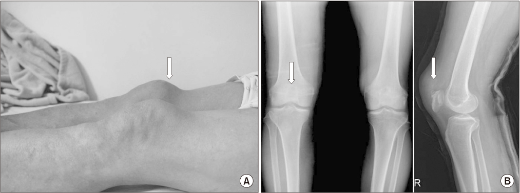
Figure 1 (A) Gross findings of both knees shows the protruded upper pole of the right patella by huge tohi (arrow). (B) Simpe anteroposterior radiograph of both knees shows irregularly defined ring-shaped radiopaque shadows in the upper pole of both patellae. Lateral radipgraph shows the swollen soft tissue shadows with calcific spots in the upper poles of the right patella and eroded anterior cortices of the patella (arrows).
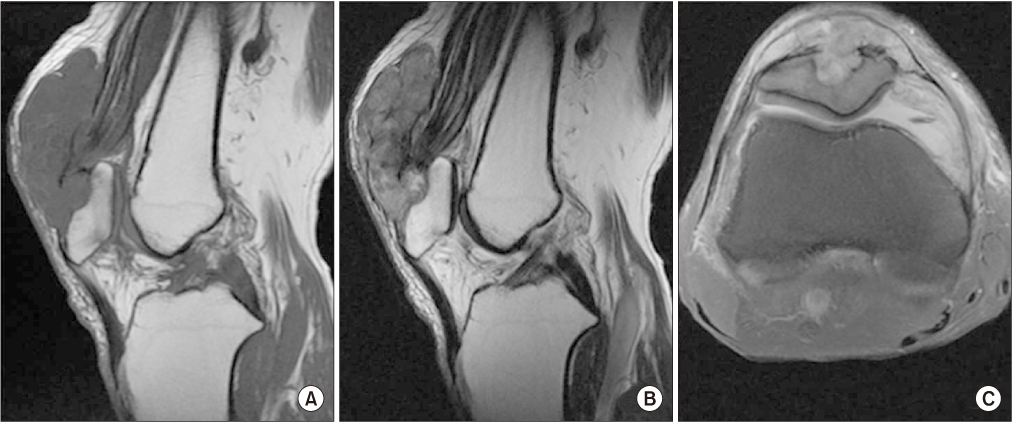
Figure 2 Sagittal and tangential views of a magnetic resonance imaging of the right knee showing a hypo-intense (T1) (A), hyper-intense (T2) (B), large mass in the supra-patellar pouches and upper poles of the patellae, with osteolytic lesion (C) in the superior patella region.

Figure 3 Needle biopsy showing white chalk-like material.

Figure 4 Polarizing microscopy image (×400) showing a birefringent needle shaped urate crystal (arrows).

Figure 5 Tophi crystals are surrounded by inflammatory cells including macrophages, lymphocytes, foreign body giant cells (H&E, ×400).

Figure 6 Radiographs at one week later (A), 2 years later (B), 4 years later (C), 6 years later (D). After 4 years, the tophi size decreased and the osteolytic lesion was filled with new bone (arrows).
Reference
-
1. Kim MK, Moon KH, Lee TJ, Kim L, Lee JS. Gout tophi in the bipartite patella. J Korean Orthop Assoc. 2008; 43:139–142.2. Kim JO, An KY, Park BJ, Min WB. Surgical treatment of gouty tophi in bilateral popliteal cysts. J Korean Orthop Assoc. 2016; 51:178–182.
Article3. Gentili A. The advanced imaging of gouty tophi. Curr Rheumatol Rep. 2006; 8:231–235.
Article4. Ehara S, Khurana JS, Kattapuram SV, Rosenberg AE, el-Khoury GY, Rosenthal DI. Osteolytic lesions of the patella. AJR Am J Roentgenol. 1989; 153:103–106.
Article5. Gerster JC, Landry M, Dufresne L, Meuwly JY. Imaging of tophaceous gout: computed tomography provides specific images compared with magnetic resonance imaging and ultrasonography. Ann Rheum Dis. 2002; 61:52–54.
Article6. Reber P, Crevoisier X, Noesberger B. Unusual localisation of tophaceous gout. A report of four cases and review of the literature. Arch Orthop Trauma Surg. 1996; 115:297–299.7. Price MD, Padera RF, Harris MB, Vrahas MS. Case reports: pathologic fracture of the patella from a gouty tophus. Clin Orthop Relat Res. 2006; 445:250–253.
Article8. Becker MA, Schumacher HR Jr, Wortmann RL, et al. Febuxostat compared with allopurinol in patients with hyperuricemia and gout. N Engl J Med. 2005; 353:2450–2461.
Article9. Kasper IR, Juriga MD, Giurini JM, Shmerling RH. Treatment of tophaceous gout: When medication is not enough. Semin Arthritis Rheum. 2016; 45:669–674.
Article10. Huang X, Du H, Gu J, et al. An allopurinol-controlled, multicenter, randomized, double-blind, parallel between-group, comparative study of febuxostat in Chinese patients with gout and hyperuricemia. Int J Rheum Dis. 2014; 17:679–686.
Article
- Full Text Links
-
- Actions
-
Cited
- CITED
-
- Close
- Share
-
- Similar articles
-
- Febuxostat for the Treatment of Chronic Tophaceous Gout in a Patient on Continuous Ambulatory Peritoneal Dialysis
- A retrospective observational study of the appropriate starting dose of febuxostat in patients with gout
- Arch Form Pathologic Suprapatellar Plica: A Case Report
- A Case of Gout Presented with Tophi as an Initial Manifestation
- Symptomatic Tophaceous Gout in the Bilateral Patellae
