Korean J Urol.
2014 Sep;55(9):581-586. 10.4111/kju.2014.55.9.581.
Pilot Study of Low-Dose Nonenhanced Computed Tomography With Iterative Reconstruction for Diagnosis of Urinary Stones
- Affiliations
-
- 1Department of Urology, Chung-Ang University College of Medicine, Seoul, Korea. kim14141@hanafos.com
- 2Department of Urology, Severance Hospital, Urological Science Institute, Yonsei University College of Medicine, Seoul, Korea.
- KMID: 2069775
- DOI: http://doi.org/10.4111/kju.2014.55.9.581
Abstract
- PURPOSE
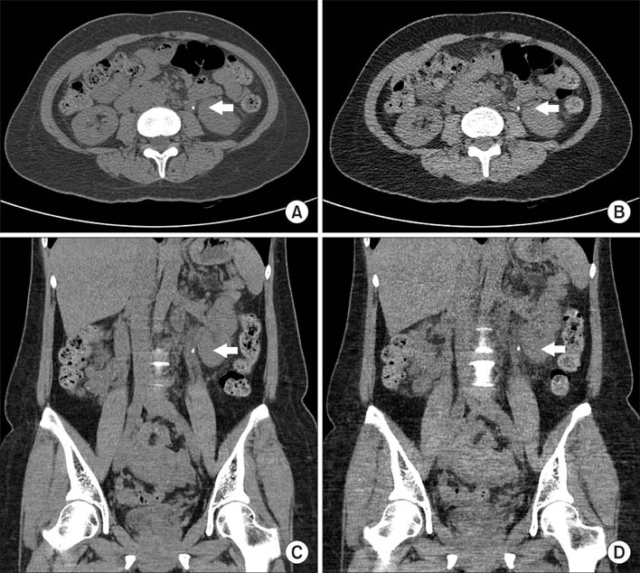
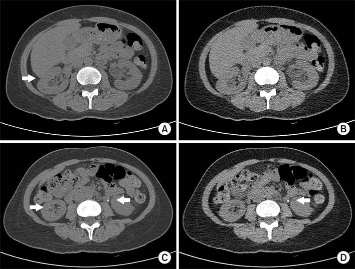
To evaluate the efficacy of low-dose computed tomography (LDCT) for detecting urinary stones with the use of an iterative reconstruction technique for reducing radiation dose and image noise.
MATERIALS AND METHODS
A total of 101 stones from 69 patients who underwent both conventional nonenhanced computed tomography (CCT) and LDCT were analyzed. Interpretations were made of the two scans according to stone characteristics (size, volume, location, Hounsfield unit [HU], and skin-to-stone distance [SSD]) and radiation dose by dose-length product (DLP), effective dose (ED), and image noise. Diagnostic performance for detecting urinary stones was assessed by statistical evaluation.
RESULTS
No statistical differences were found in stone characteristics between the two scans. The average DLP and ED were 384.60+/-132.15 mGy and 5.77+/-1.98 mSv in CCT and 90.08+/-31.80 mGy and 1.34+/-0.48 mSv in LDCT, respectively. The dose reduction rate of LDCT was nearly 77% for both DLP and ED (p<0.01). The mean objective noise (standard deviation) from three different areas was 23.0+/-2.5 in CCT and 29.2+/-3.1 in LDCT with a significant difference (p<0.05); the slight increase was 21.2%. For stones located throughout the kidney and ureter, the sensitivity and specificity of LDCT remained 96.0% and 100%, with positive and negative predictive values of 100% and 96.2%, respectively.
CONCLUSIONS
LDCT showed significant radiation reduction while maintaining high image quality. It is an attractive option in the diagnosis of urinary stones.
MeSH Terms
Figure
Cited by 1 articles
-
Low-Dose Unenhanced Computed Tomography with Iterative Reconstruction for Diagnosis of Ureter Stones
Byung Hoon Chi, In Ho Chang, Dong Hoon Lee, Sung Bin Park, Kyung Do Kim, Young Tae Moon, Taekyu Hur
Yonsei Med J. 2018;59(3):389-396. doi: 10.3349/ymj.2018.59.3.389.
Reference
-
1. Chua ME, Gatchalian GT, Corsino MV, Reyes BB. Diagnostic utility of attenuation measurement (Hounsfield units) in computed tomography stonogram in predicting the radio-opacity of urinary calculi in plain abdominal radiographs. Int Urol Nephrol. 2012; 44:1349–1355.2. Yilmaz S, Sindel T, Arslan G, Ozkaynak C, Karaali K, Kabaalioglu A, et al. Renal colic: comparison of spiral CT, US and IVU in the detection of ureteral calculi. Eur Radiol. 1998; 8:212–217.3. Dalrymple NC, Verga M, Anderson KR, Bove P, Covey AM, Rosenfield AT, et al. The value of unenhanced helical computerized tomography in the management of acute flank pain. J Urol. 1998; 159:735–740.4. Lim GS, Jang SH, Son JH, Lee JW, Hwang JS, Lim CH, et al. Comparison of non-contrast-enhanced computed tomography and intravenous pyelogram for detection of patients with urinary calculi. Korean J Urol. 2014; 55:120–123.5. Abramson S, Walders N, Applegate KE, Gilkeson RC, Robbin MR. Impact in the emergency department of unenhanced CT on diagnostic confidence and therapeutic efficacy in patients with suspected renal colic: a prospective survey. 2000 ARRS President's Award. American Roentgen Ray Society. AJR Am J Roentgenol. 2000; 175:1689–1695.6. John BS, Patel U, Anson K. What radiation exposure can a patient expect during a single stone episode? J Endourol. 2008; 22:419–422.7. Brenner DJ, Hall EJ. Computed tomography: an increasing source of radiation exposure. N Engl J Med. 2007; 357:2277–2284.8. Lopez M, Hoppe B. History, epidemiology and regional diversities of urolithiasis. Pediatr Nephrol. 2010; 25:49–59.9. Brenner D, Elliston C, Hall E, Berdon W. Estimated risks of radiation-induced fatal cancer from pediatric CT. AJR Am J Roentgenol. 2001; 176:289–296.10. Committee to Assess Health Risks from Exposure to Low Levels of Ionizing Radiation. Board on Radiation Effects Research (BRER). Division on Earth and Life Studies (DELS). National Research Council. Health risks from exposure to low levels of ionizing radiation: BEIR VII phase 2. Washington, D.C.: The National Academies Press;2006.11. Gorycki T, Lasek I, Kamiński K, Studniarek M. Evaluation of radiation doses delivered in different chest CT protocols. Pol J Radiol. 2014; 79:1–5.12. Kim K, Kim YH, Kim SY, Kim S, Lee YJ, Kim KP, et al. Low-dose abdominal CT for evaluating suspected appendicitis. N Engl J Med. 2012; 366:1596–1605.13. Hansmann J, Schoenberg GM, Brix G, Henzler T, Meyer M, Attenberger UI, et al. CT of urolithiasis: comparison of image quality and diagnostic confidence using filtered back projection and iterative reconstruction techniques. Acad Radiol. 2013; 20:1162–1167.14. Heneghan JP, McGuire KA, Leder RA, DeLong DM, Yoshizumi T, Nelson RC. Helical CT for nephrolithiasis and ureterolithiasis: comparison of conventional and reduced radiation-dose techniques. Radiology. 2003; 229:575–580.15. Chargari C, Cosset JM. The issue of low doses in radiation therapy and impact on radiation-induced secondary malignancies. Bull Cancer. 2013; 100:1333–1342.16. Rabes HM, Klugbauer S. Radiation-induced thyroid carcinomas in children: high prevalence of RET rearrangement. Verh Dtsch Ges Pathol. 1997; 81:139–144.17. Bartoletti R, Cai T, Mondaini N, Melone F, Travaglini F, Carini M, et al. Epidemiology and risk factors in urolithiasis. Urol Int. 2007; 79:Suppl 1. 3–7.18. Curhan GC. Epidemiology of stone disease. Urol Clin North Am. 2007; 34:287–293.19. Koenig TR, Wolff D, Mettler FA, Wagner LK. Skin injuries from fluoroscopically guided procedures: part 1, characteristics of radiation injury. AJR Am J Roentgenol. 2001; 177:3–11.20. Cardis E, Vrijheid M, Blettner M, Gilbert E, Hakama M, Hill C, et al. The 15-Country Collaborative Study of Cancer Risk among Radiation Workers in the Nuclear Industry: estimates of radiation-related cancer risks. Radiat Res. 2007; 167:396–416.21. Vardhanabhuti V, Ilyas S, Gutteridge C, Freeman SJ, Roobottom CA. Comparison of image quality between filtered back-projection and the adaptive statistical and novel model-based iterative reconstruction techniques in abdominal CT for renal calculi. Insights Imaging. 2013; 4:661–669.22. Hara AK, Paden RG, Silva AC, Kujak JL, Lawder HJ, Pavlicek W. Iterative reconstruction technique for reducing body radiation dose at CT: feasibility study. AJR Am J Roentgenol. 2009; 193:764–771.23. Kambadakone AR, Chaudhary NA, Desai GS, Nguyen DD, Kulkarni NM, Sahani DV. Low-dose MDCT and CT enterography of patients with Crohn disease: feasibility of adaptive statistical iterative reconstruction. AJR Am J Roentgenol. 2011; 196:W743–W752.24. Kulkarni NM, Uppot RN, Eisner BH, Sahani DV. Radiation dose reduction at multidetector CT with adaptive statistical iterative reconstruction for evaluation of urolithiasis: how low can we go? Radiology. 2012; 265:158–166.25. Prakash P, Kalra MK, Kambadakone AK, Pien H, Hsieh J, Blake MA, et al. Reducing abdominal CT radiation dose with adaptive statistical iterative reconstruction technique. Invest Radiol. 2010; 45:202–210.26. May MS, Wust W, Brand M, Stahl C, Allmendinger T, Schmidt B, et al. Dose reduction in abdominal computed tomography: intraindividual comparison of image quality of full-dose standard and half-dose iterative reconstructions with dual-source computed tomography. Invest Radiol. 2011; 46:465–470.
- Full Text Links
- Actions
-
Cited
- CITED
-
- Close
- Share
- Similar articles
-
- Detectability of Extrahepatic Duct Stones: A Comparison between Nonenhanced and Enhanced CT
- Low-Dose Unenhanced Computed Tomography with Iterative Reconstruction for Diagnosis of Ureter Stones
- Dosimetric Effects of Low Dose 4D CT Using a Commercial Iterative Reconstruction on Dose Calculation in Radiation Treatment Planning: A Phantom Study
- Effects of Iterative Reconstruction Algorithm, Automatic Exposure Control on Image Quality, and Radiation Dose: Phantom Experiments with Coronary CT Angiography Protocols
- Contrast-Enhanced CT with Knowledge-Based Iterative Model Reconstruction for the Evaluation of Parotid Gland Tumors: A Feasibility Study


