Prog Med Phys.
2017 Mar;28(1):27-32. 10.14316/pmp.2017.28.1.27.
Dosimetric Effects of Low Dose 4D CT Using a Commercial Iterative Reconstruction on Dose Calculation in Radiation Treatment Planning: A Phantom Study
- Affiliations
-
- 1Department of Radiation Oncology, Soonchunhyang University Hospital, Seoul, Korea. changaram@schmc.ac.kr
- KMID: 2382802
- DOI: http://doi.org/10.14316/pmp.2017.28.1.27
Abstract
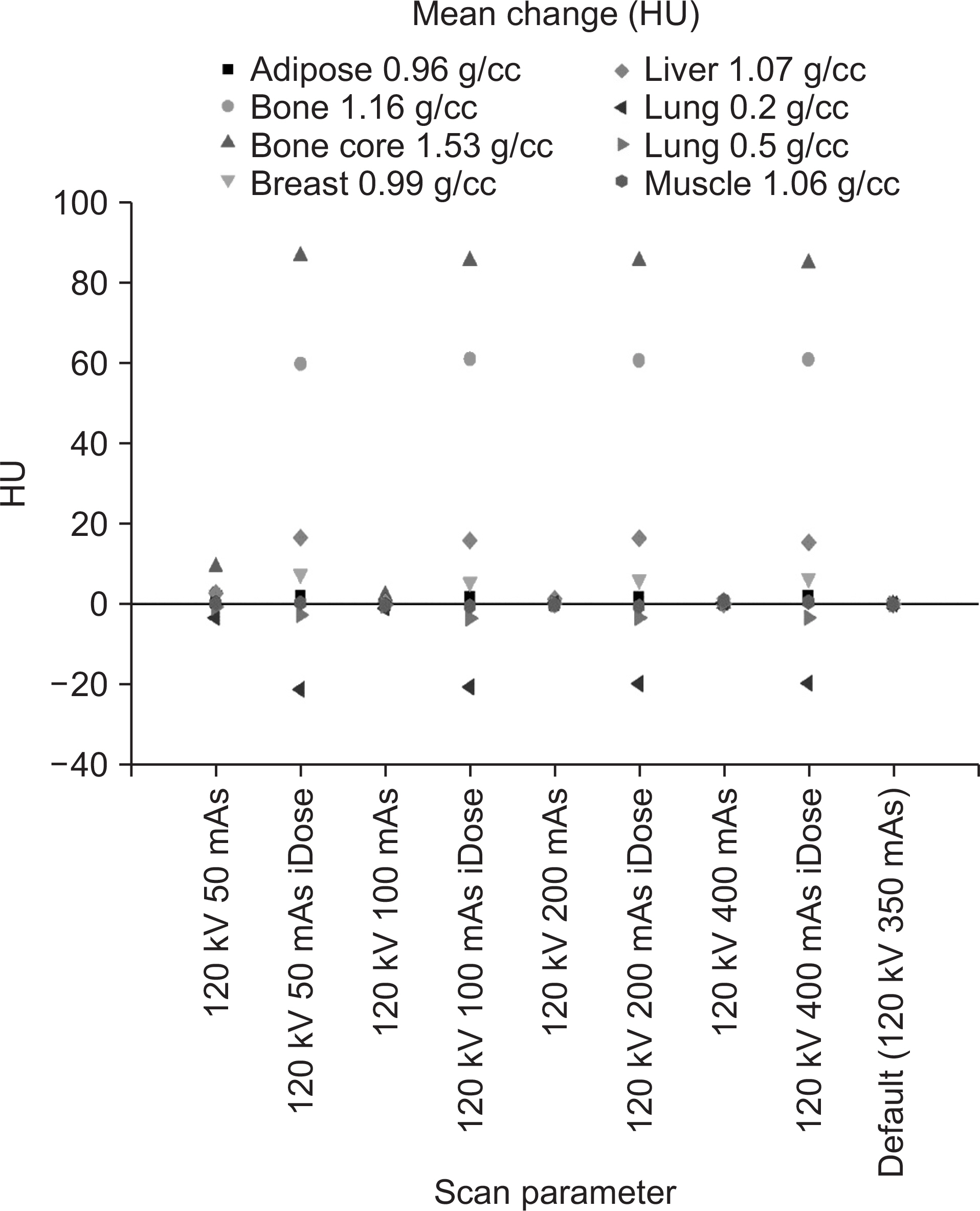
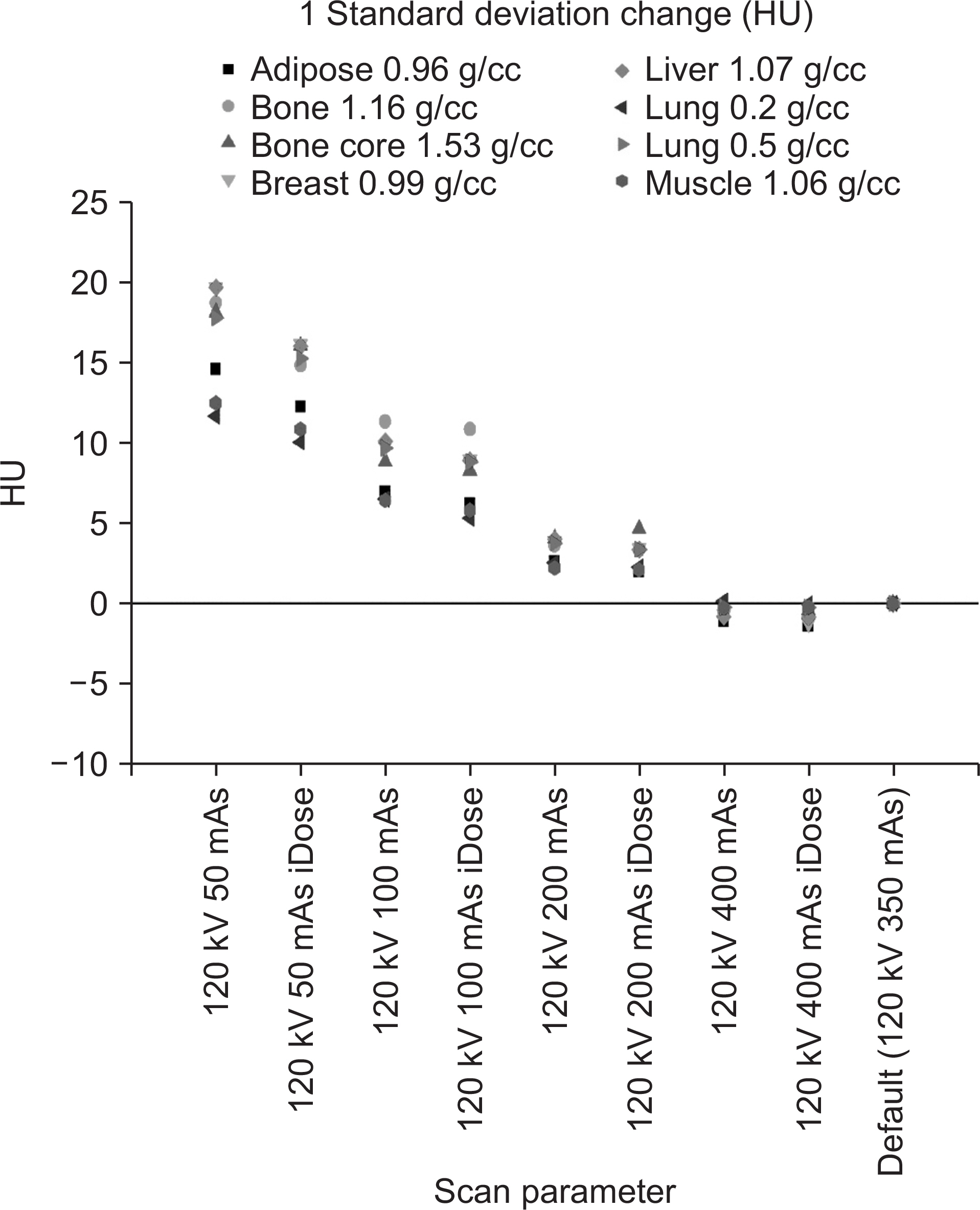
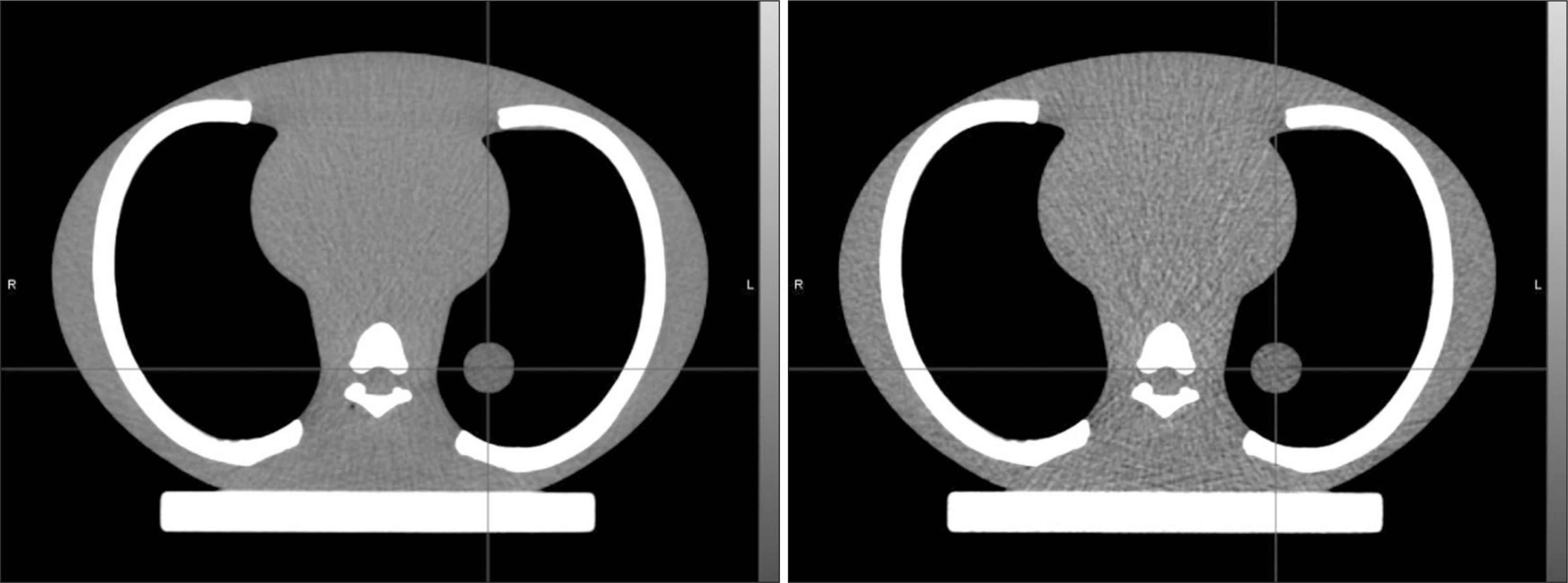
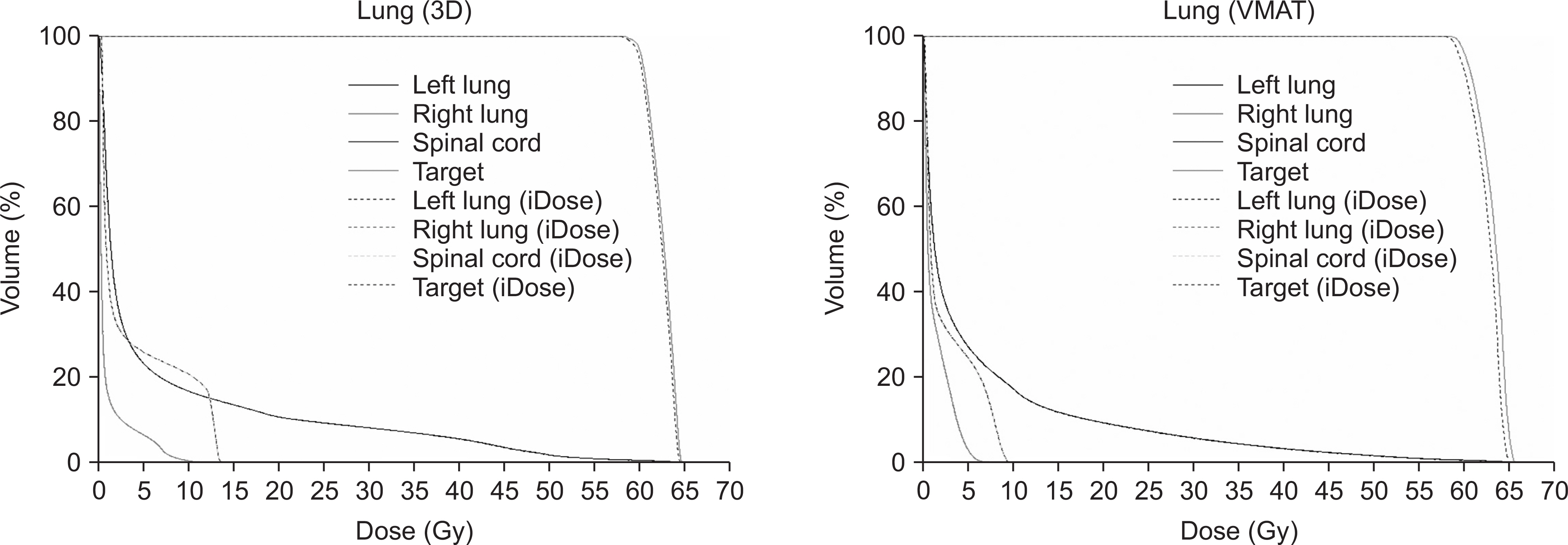
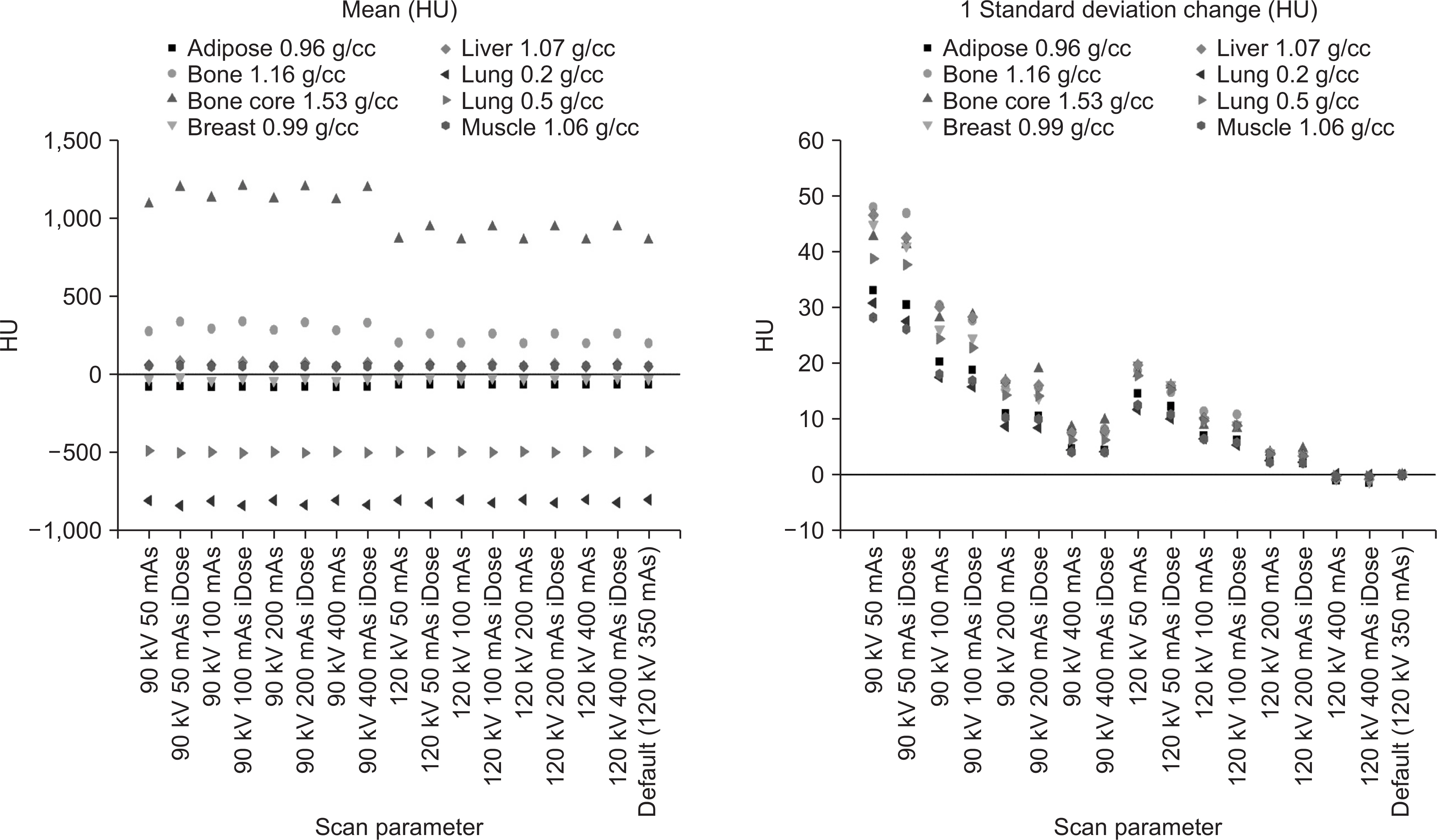
- We investigated the effect of a commercial iterative reconstruction technique (iDose, Philips) on the image quality and the dose calculation for the treatment plan. Using the electron density phantom, the 3D CT images with five different protocols (50, 100, 200, 350 and 400 mAs) were obtained. Additionally, the acquired data was reconstructed using the iDose with level 5. A lung phantom was used to acquire the 4D CT with the default protocol as a reference and the low dose (one third of the default protocol) 4D CT using the iDose for the spine and lung plans. When applying the iDose at the same mAs, the mean HU value was changed up to 85 HU. Although the 1 SD was increased with reducing the CT dose, it was decreased up to 4 HU due to the use of iDose. When using the low dose 4D CT with iDose, the dose change relative to the reference was less than 0.5% for the target and OARs in the spine plan. It was also less than 1.1% in the lung plan. Therefore, our results suggests that this dose reduction technique is applicable to the 4D CT image acquisition for the radiation treatment planning.
Figure
Reference
-
1. Balter JM, Ten Haken RK, Lawrence TS, Lam KL, Robertson JM. Uncertainties in CT-based radiation therapy treatment planning associated with patient breathing. Int J Radiat Oncol Biol Phys. 1996; 36:167–174.
Article2. Keall PJ, Starkschall G, Shukla H, Forster KM, Ortiz V, Stevens CW, et al. Acquiring 4D thoracic CT scans using a multislice helical method. Phys Med Biol. 2004; 49:2053–2067.
Article3. Ohara K, Okumura T, Akisada M, Inada T, Mori T, Yokota H, et al. Irradiation synchronized with respiration gate. Int J Radiat Oncol Biol Phys. 1989; 17:853–857.
Article4. Hubbard P, Callahan J, Cramb J, Budd R, Kron T. Audit of radiation dose delivered in time-resolved four-dimensional computed tomography in a radiotherapy department. J Med Imaging Radiat Oncol. 2015; 59:346–352.
Article5. Low DA, Nystrom M, Kalinin E, Parikh P, Dempsey JF, Bradley JD, et al. A method for the reconstruction of four-dimensional synchronized CT scans acquired during free breathing. Med Phys. 2003; 30:1254–1263.
Article6. Matsuzaki Y, Fujii K, Kumagai M, Tsuruoka I, Mori S. Effective and organ doses using helical 4DCT for thoracic and abdominal therapies. J Radiat Res. 2013; 54:962–970.
Article7. Mori S, Ko S, Ishii T, Nishizawa K. Effective doses in four-dimensional computed tomography for lung radiotherapy planning. Med Dosim. 2009; 34:87–90.
Article8. Hara AK, Paden RG, Silva AC, Kujak JL, Lawder HJ, Pavlicek W. Iterative reconstruction technique for reducing body radiation dose at CT: feasibility study. AJR Am J Roentgenol. 2009; 193:764–771.
Article9. Kim Y, Kim YK, Lee BE, Lee SJ, Ryu YJ, Lee JH, et al. Ultralow-dose CT of the thorax using iterative reconstruction: evaluation of image quality and radiation dose reduction. American Journal of Roentgenology. 2015; 204:1197–1202.
Article10. Raman SP, Johnson PT, Deshmukh S, Mahesh M, Grant KL, Fishman EK. CT dose reduction applications: available tools on the latest generation of CT scanners. Journal of the American College of Radiology. 2013; 10:37–41.
Article11. Arapakis I, Efstathopoulos E, Tsitsia V, Kordolaimi S, Economopoulos N, Argentos S, et al. Using “iDose4” iterative reconstruction algorithm in adults' chest–abdomen–pelvis CT examinations: effect on image quality in relation to patient radiation exposure. The British journal of radiology. 2014; 87:20130613.
Article12. Mehta D, Thompson R, Morton T, Dhanantwari A, Shefer E. Iterative model reconstruction: simultaneously lowered computed tomography radiation dose and improved image quality. Med Phys Int J. 2013; 2:147–155.13. Olsson M-L, Norrgren K. An investigation of the iterative reconstruction method iDose4 on a Philips CT Brilliance 64 using a Catphan 600 phantom. SPIE Medical Imaging. 2012. 831348.
Article14. Li H, Lee D, Low D, Gay H, Michalski J, Mutic S. SU-E-J-167: Iterative Reconstruction Techniques for Radiation Therapy CT Simulations: A Phantom Study. Medical Physics. 2013; 40:189–189.
Article
- Full Text Links
- Actions
-
Cited
- CITED
-
- Close
- Share
- Similar articles
-
- Effects of Iterative Reconstruction Algorithm, Automatic Exposure Control on Image Quality, and Radiation Dose: Phantom Experiments with Coronary CT Angiography Protocols
- Analysis of Dose Distribution on Critical Organs for Radiosurgery with CyberKnife Real-Time Tumor Tracking System
- Dosimetric Evaluation of Acuros XB for Treatment Plan on Multiphasic Contrast Enhanced CT
- Quantitative Image Quality and Histogram-Based Evaluations of an Iterative Reconstruction Algorithm at Low-to-Ultralow Radiation Dose Levels: A Phantom Study in Chest CT
- Evaluations of a Commercial CLEANBOLUS-WHITE for Clinical Application

