Prog Med Phys.
2024 Mar;35(1):10-15. 10.14316/pmp.2024.35.1.10.
Evaluations of a Commercial CLEANBOLUS-WHITE for Clinical Application
- Affiliations
-
- 1Department of Radiation Oncology, Seoul National University Hospital, Seoul, Korea
- 2Institute of Radiation Medicine, Seoul National University Medical Research Center, Seoul, Korea
- KMID: 2554467
- DOI: http://doi.org/10.14316/pmp.2024.35.1.10
Abstract
- Purpose
This study aimed to comprehensively investigate the diverse characteristics of a novel commercial bolus, CLEANBOLUS-WHITE (CBW), to ascertain its suitability for clinical application.
Methods
The evaluation of CBW encompassed both physical and biological assessments. Physical parameters such as mass density and shore hardness were measured alongside analyses of element composition. Biological evaluations included assessments for skin irritation and cytotoxicity. Dosimetric properties were examined by calculating surface dose and beam quality using a treatment planning system (TPS). Additionally, doses were measured at maximum and reference depths, and the results were compared with those obtained using a solid water phantom. The effect of air gap on dose measurement was also investigated by comparing measured doses on the RANDO phantom, under the bolus, with doses calculated from the TPS.
Results
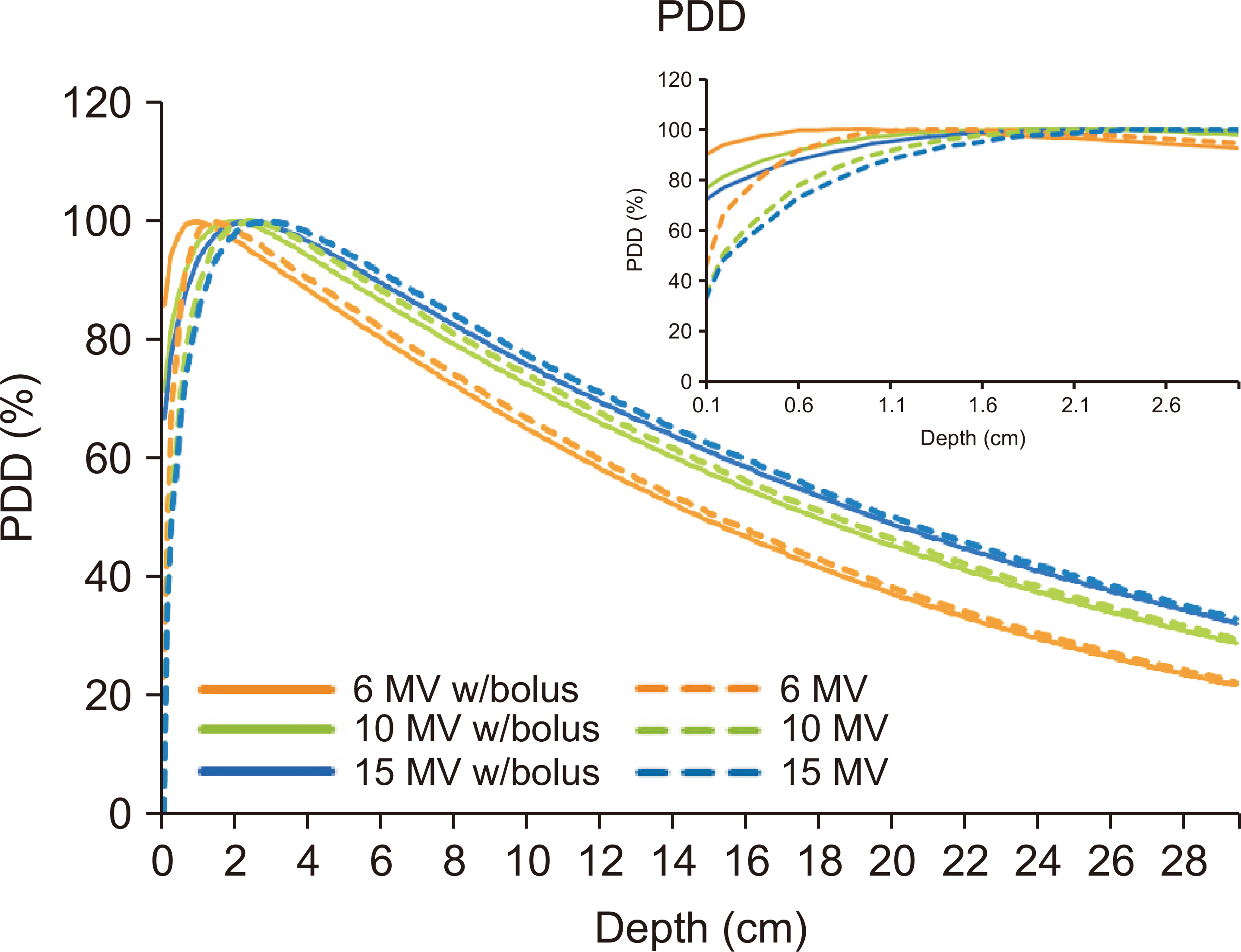
Biological evaluation confirmed that CBW is non-cytotoxic, nonirritant, and nonsensitizing. The bolus exhibited a mass density of 1.02 g/cm 3 and 14 shore 00. Dosimetric evaluations revealed that using the 0.5 cm CBW resulted in less than a 1% difference compared to using the solid water phantom. Furthermore, beam quality calculations in the TPS indicated increased surface dose with the bolus. The air gap effect on dose measurement was deemed negligible, with a difference of approximately 1% between calculated and measured doses, aligning with measurement uncertainty.
Conclusions
CBW demonstrates outstanding properties for clinical utilization. The dosimetric evaluation underscores a strong agreement between calculated and measured doses, validating its reliability in both planning and clinical settings.
Keyword
Figure
Reference
-
References
1. McKenna MG, Chen XG, Altschuler MD, Bloch P. 1995; Calculation of the dose in the build-up region for high energy photon beam. Treatment planning when beam spoilers are employed. Radiother Oncol. 34:63–68. DOI: 10.1016/0167-8140(95)01504-A. PMID: 7792400.
Article2. Ishmael Parsai E, Shvydka D, Pearson D, Gopalakrishnan M, Feldmeier JJ. 2008; Surface and build-up region dose analysis for clinical radiotherapy photon beams. Appl Radiat Isot. 66:1438–1442. DOI: 10.1016/j.apradiso.2008.02.089. PMID: 18434173.
Article3. Butson MJ, Cheung T, Yu P, Metcalfe P. 2000; Effects on skin dose from unwanted air gaps under bolus in photon beam radiotherapy. Radiat Meas. 32:201–204. DOI: 10.1016/S1350-4487(99)00276-0.
Article4. Khan Y, Villarreal-Barajas JE, Udowicz M, Sinha R, Muhammad W, Abbasi AN, et al. 2013; Clinical and dosimetric implications of air gaps between bolus and skin surface during radiation therapy. J Cancer Ther. 4:1251–1255. DOI: 10.4236/jct.2013.47147.
Article5. Lobo D, Banerjee S, Srinivas C, Ravichandran R, Putha SK, Prakash Saxena PU, et al. 2020; Influence of air gap under bolus in the dosimetry of a clinical 6 MV photon beam. J Med Phys. 45:175–181. DOI: 10.4103/jmp.JMP_53_20. PMID: 33487930. PMCID: PMC7810143.
Article6. Wang KM, Rickards AJ, Bingham T, Tward JD, Price RG. 2022; Technical note: evaluation of a silicone-based custom bolus for radiation therapy of a superficial pelvic tumor. J Appl Clin Med Phys. 23:e13538. DOI: 10.1002/acm2.13538. PMID: 35084098. PMCID: PMC8992939.
Article7. Wang X, Wang X, Xiang Z, Zeng Y, Liu F, Shao B, et al. 2021; The clinical application of 3D-printed boluses in superficial tumor radiotherapy. Front Oncol. 11:698773. DOI: 10.3389/fonc.2021.698773. PMID: 34490095. PMCID: PMC8416990.
Article8. Burleson S, Baker J, Hsia AT, Xu Z. 2015; Use of 3D printers to create a patient-specific 3D bolus for external beam therapy. J Appl Clin Med Phys. 16:5247. DOI: 10.1120/jacmp.v16i3.5247. PMID: 26103485. PMCID: PMC5690114.
Article9. Park JM, Son J, An HJ, Kim JH, Wu HG, Kim JI. 2019; Bio-compatible patient-specific elastic bolus for clinical implementation. Phys Med Biol. 64:105006. DOI: 10.1088/1361-6560/ab1c93. PMID: 31022714.
Article10. Almond PR, Biggs PJ, Coursey BM, Hanson WF, Huq MS, Nath R, et al. 1999; AAPM's TG-51 protocol for clinical reference dosimetry of high-energy photon and electron beams. Med Phys. 26:1847–1870. DOI: 10.1118/1.598691. PMID: 10505874.
Article11. McEwen M, DeWerd L, Ibbott G, Followill D, Rogers DW, Seltzer S, et al. 2014; Addendum to the AAPM's TG-51 protocol for clinical reference dosimetry of high-energy photon beams. Med Phys. 41:041501. DOI: 10.1118/1.4866223. PMID: 24694120. PMCID: PMC5148035.
Article12. Son J, Jung S, Park JM, Choi CH, Kim JI. 2021; Assessing commercial CLEANBOLUS based on silicone for clinical use. Prog Med Phys. 32:159–164. DOI: 10.14316/pmp.2021.32.4.159.
Article
- Full Text Links
- Actions
-
Cited
- CITED
-
- Close
- Share
- Similar articles
-
- Assessing Commercial CLEANBOLUS Based on Silicone for Clinical Use
- Corrigendum: Assessing Commercial CLEANBOLUS Based on Silicone for Clinical Use
- Comparison of Skin Prick Test Results between Crude Allergen Extracts from Foods and Commercial Allergen Extracts in Atopic Dermatitis by Double-Blind Placebo- Controlled Food Challenge for Milk, Egg, and Soybean
- Influence of Different Supplements on the Commercial Cultivation of Milky White Mushroom
- A Case of White Superficial Onychomycosis


