Three-dimensional analysis of the distal movement of maxillary 1st molars in patients fitted with mini-implant-aided trans-palatal arches
- Affiliations
-
- 1Department of Orthodontics, Faculty of Dentistry, Hamadan University of Medical Sciences, Hamadan, Iran. nasrinne@yahoo.com
- 2Hamadan Dental Research Centre, Hamadan University of Medical Sciences, Hamadan, Iran.
- 3Department of Biostatistics, Faculty of Public Health, Hamadan University of Medical Sciences, Hamadan, Iran.
- KMID: 2068518
- DOI: http://doi.org/10.4041/kjod.2015.45.5.236
Abstract
OBJECTIVE
The aim of this study was to investigate three-dimensional molar displacement after distalization via miniscrews and a horizontal modification of the trans-palatal-arch (TPA).
METHODS
The subjects in this clinical trial were 26 Class II patients. After the preparation of a complete set of diagnostic records, miniscrews were inserted between the maxillary 2nd premolar and 1st molar on the palatal side. Elastic modules connected to the TPA exerting an average force of 150-200 g/side parallel to the occlusal plane were applied. Cone-beam computed tomography was utilized to evaluate the position of the miniscrews relative to the adjacent teeth and maxillary sinus, and the direction of force relative to molar furcation. The distances from the central point of the incisive papilla to the mesiopalatal cusps of the 1st maxillary molars and the distances between the mesiopalatal cusps of the left and right molars were measured to evaluate displacement of the maxillary molars on the horizontal plane. Interocclusal space was used to evaluate vertical changes.
RESULTS
Mean maxillary 1st molar distalization was 2.3 +/- 1.1 mm, at a rate of 0.4 +/- 0.2 mm/month, and rotation was not significant. Intermolar width increased by 2.9 +/- 1.8 mm. Molars were intruded relative to the neighboring teeth, from 0.1 to 0.8 mm.
CONCLUSIONS
Distalization of molars was possible without extrusion, using the appliance investigated. The intrusive component of force reduced the rate of distal movement.
MeSH Terms
Figure
-
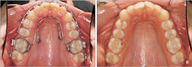
Figure 1 Mini-implant-aided trans palatal arch molar distalizing appliances include a horseshoe shaped palatal bar inserted in the palatal sheath, and two miniscrews between the 1st molar and 2nd premolar. They exert a traction force from the anterior helix to the miniscrews. A, Before the start of treatment; B, after the completion of distalization.
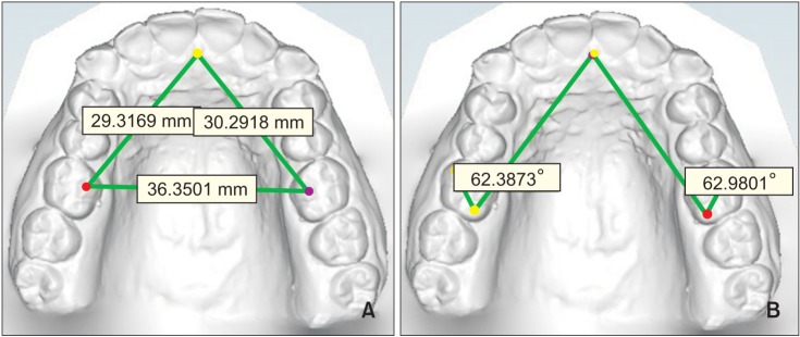
Figure 2 The measurements on digital models. A, Distance between mesiopalatal cusps of both side first molars and incisive papilla; B, angle between the lines connecting incisive papilla to distopalatal cusp of the first molar and mesiopalatal cusp to distopalatal cusp of the first molar.
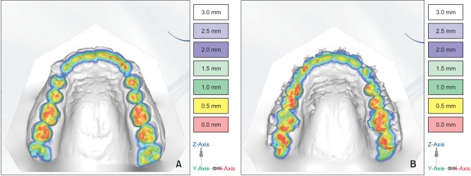
Figure 3 Evaluation methods of vertical teeth movement by three-dimensional digital model. A, Before treatment; B, after treatment. The red areas are contact areas, which are more marked on the right first maxillary molars before treatment than after treatment. Red marks are stronger on premolars and second molars after treatment, which is an additional sign of first molar intrusion.
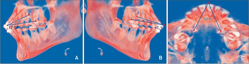
Figure 4 Demonstrations of the lines of traction force by cone-beam computed tomography (CBCT) images. A, Right sagittal view; B, left sagittal biew; C, coronal view. In right and left sagittal views of the CBCT scan, the angle between the lines of traction from the helix to the minscrews relative to the occlusal plane clearly indicate the apical direction of distal driving force. The coronal view shows the divergent distal driving force as the expansion component of applied force.
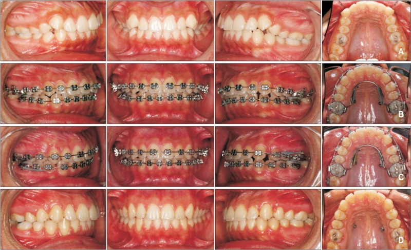
Figure 5 The sequence of changes in dentition. A, Before treatment; B, at the start of appliance placement and leveling; C, after the completion of distalization, and D, after debonding.
Cited by 1 articles
-
Three-dimensional evaluation of tooth movement in Class II malocclusions treated without extraction by orthodontic mini-implant anchorage
Dler Ali, Hnd Mohammed, Seung-Hwan Koo, Kyung-Hwa Kang, Sang-Cheol Kim
Korean J Orthod. 2016;46(5):280-289. doi: 10.4041/kjod.2016.46.5.280.
Reference
-
1. Nalcaci R, Bicakci AA, Ozan F. Noncompliance screw supported maxillary molar distalization in a parallel manner. Korean J Orthod. 2010; 40:250–259.
Article2. Choi YJ, Lee JS, Cha JY, Park YC. Total distalization of the maxillary arch in a patient with skeletal Class II malocclusion. Am J Orthod Dentofacial Orthop. 2011; 139:823–833. PMID: 21640890.
Article3. Choi NC, Park YC, Lee HA, Lee KJ. Treatment of Class II protrusion with severe crowding using indirect miniscrew anchorage. Angle Orthod. 2007; 77:1109–1118. PMID: 18004931.
Article4. Grec RH, Janson G, Branco NC, Moura-Grec PG, Patel MP, Castanha Henriques JF. Intraoral distalizer effects with conventional and skeletal anchorage: a meta-analysis. Am J Orthod Dentofacial Orthop. 2013; 143:602–615. PMID: 23631962.
Article5. Sugawara J, Kanzaki R, Takahashi I, Nagasaka H, Nanda R. Distal movement of maxillary molars in nongrowing patients with the skeletal anchorage system. Am J Orthod Dentofacial Orthop. 2006; 129:723–733. PMID: 16769490.
Article6. Kinzinger GS, Eren M, Diedrich PR. Treatment effects of intraoral appliances with conventional anchorage designs for non-compliance maxillary molar distalization: a literature review. Eur J Orthod. 2008; 30:558–571. PMID: 18820306.
Article7. Sodagar A, Ahmad Akhoundi MS, Rafighii A, Arab S. Fabrication and evaluation of a noncompliant molar distalizing appliance: bonded molar distalizer. J Dent (Tehran). 2011; 8:107–116. PMID: 22457837.8. McSherry PF, Bradley H. Class II correction-reducing patient compliance: a review of the available techniques. J Orthod. 2000; 27:219–225. PMID: 11099554.
Article9. Mavropoulos A, Karamouzos A, Kiliaridis S, Papadopoulos MA. Efficiency of noncompliance simultaneous first and second upper molar distalization: a three-dimensional tooth movement analysis. Angle Orthod. 2005; 75:532–539. PMID: 16097221.10. Gelgör IE, Büyükyilmaz T, Karaman AI, Dolanmaz D, Kalayci A. Intraosseous screw-supported upper molar distalization. Angle Orthod. 2004; 74:838–850. PMID: 15673149.11. Cornelis MA, De Clerck HJ. Maxillary molar distalization with miniplates assessed on digital models: a prospective clinical trial. Am J Orthod Dentofacial Orthop. 2007; 132:373–377. PMID: 17826606.
Article12. Gelgor IE, Karaman AI, Buyukyilmaz T. Comparison of 2 distalization systems supported by intraosseous screws. Am J Orthod Dentofacial Orthop. 2007; 131:161.e1–161.e8. PMID: 17276855.
Article13. Kuroda S, Yamada K, Deguchi T, Kyung HM, Takano-Yamamoto T. Class II malocclusion treated with miniscrew anchorage: comparison with traditional orthodontic mechanics outcomes. Am J Orthod Dentofacial Orthop. 2009; 135:302–309. PMID: 19268827.
Article14. Park HS, Lee SK, Kwon OW. Group distal movement of teeth using microscrew implant anchorage. Angle Orthod. 2005; 75:602–609. PMID: 16097229.15. Blaya MG, Blaya DS, Guimaraes MB, Hirakata LM, Marquezan M. Patient's perception on mini-screws used for molar distalization. Rev Odonto Ciênc. 2010; 25:266–270.16. Park HS, Kwon TG, Sung JH. Nonextraction treatment with microscrew implants. Angle Orthod. 2004; 74:539–549. PMID: 15387034.17. Goyal A, Shivalinga B, Jyothikiran H, Patel V. Mini-implant supported molar distalization. J Dent Implant. 2013; 2:136–140.
Article18. Villela HM, Santos Sampaio AL, Bezerra F. Use of orthodontic miniscrews in asymmetrical corrections. Dental Press J Orthod. 2008; 13:107–117.19. Kang YG, Kim JY, Nam JH. Control of maxillary dentition with 2 midpalatal orthodontic miniscrews. Am J Orthod Dentofacial Orthop. 2011; 140:879–885. PMID: 22133954.
Article20. Yamada K, Kuroda S, Deguchi T, Takano-Yamamoto T, Yamashiro T. Distal movement of maxillary molars using miniscrew anchorage in the buccal interradicular region. Angle Orthod. 2009; 79:78–84. PMID: 19123698.
Article21. Graber LW, Vanarsdall RL, Vig K. Orthodontics, current principles and techniques biomechanical considerations with temporary anchorage devices. 5th ed. Philadelphia, PA: Mosby;2012. p. 381–420.22. Oberti G, Villegas C, Ealo M, Palacio JC, Baccetti T. Maxillary molar distalization with the dual-force distalizer supported by mini-implants: a clinical study. Am J Orthod Dentofacial Orthop. 2009; 135:282.e1–282.e5. discussion 282-3PMID: 19268824.
Article23. Almeida MA, Phillips C, Kula K, Tulloch C. Stability of the palatal rugae as landmarks for analysis of dental casts. Angle Orthod. 1995; 65:43–48. PMID: 7726462.24. Kircelli BH, Pektaş ZO, Kircelli C. Maxillary molar distalization with a bone-anchored pendulum appliance. Angle Orthod. 2006; 76:650–659. PMID: 16808573.25. Escobar SA, Tellez PA, Moncada CA, Villegas CA, Latorre CM, Oberti G. Distalization of maxillary molars with the bone-supported pendulum: a clinical study. Am J Orthod Dentofacial Orthop. 2007; 131:545–549. PMID: 17418723.
Article26. Lai EH, Yao CC, Chang JZ, Chen I, Chen YJ. Three-dimensional dental model analysis of treatment outcomes for protrusive maxillary dentition: comparison of headgear, miniscrew, and miniplate skeletal anchorage. Am J Orthod Dentofacial Orthop. 2008; 134:636–645. PMID: 18984395.
Article27. Polat-Ozsoy O, Kircelli BH, Arman-Ozçirpici A, Pektaş ZO, Uçkan S. Pendulum appliances with 2 anchorage designs: conventional anchorage vs bone anchorage. Am J Orthod Dentofacial Orthop. 2008; 133:339.e9–339.e17. PMID: 18331928.
Article
- Full Text Links
-
- Actions
-
Cited
- CITED
-
- Close
- Share
-
- Similar articles
-
- Three-dimensional evaluation of tooth movement in Class II malocclusions treated without extraction by orthodontic mini-implant anchorage
- A study of incidence of palatal side abfractions in maxillary posterior teeth
- The pattern of movement and stress distribution during retraction of maxillary incisors using a 3-D finite element method
- Maxillary space closure using a digital manufactured Mesialslider in a single appointment workflow
- Three dimensional finite element analysis of continuous and segmented arches with use of orthodontic miniscrews
