A study of incidence of palatal side abfractions in maxillary posterior teeth
- Affiliations
-
- 1Department of Prosthodontics, College of Dentistry, Chosun University, Gwangju, Republic of Korea. lkj1998@chosun.ac.kr
- KMID: 2470625
- DOI: http://doi.org/10.14368/jdras.2019.35.4.206
Abstract
- PURPOSE
Most of studies dealing with abfractions are limited to the buccal surfaces of the teeth. In this study, we analyzed the cause for abfraction by investigating the incidence of palatal side abfractions in maxillary posterior teeth.
MATERIALS AND METHODS
We investigated a total of 3193 maxillary posterior teeth by an intraoral examination, model observation, and observation of virtual model fabricated using model scanning. We recorded the results and classified them depending on the type of teeth, age, gender, and side of arches. We also performed Chi-square test to evaluate the statistical significance among the groups (α = 0.05).
RESULTS
The incidence of palatal side abfraction of the maxillary molars (10.8%) was higher than the premolars (6.8%), and among them, the incidence of the 1st molars (39.1%) were the highest. The incidence of palatal side abfraction increased with age and was statistically significant (P < 0.05). There was no statistical significance in the difference by gender (P > 0.05); in the case of arches, left arch showed higher incidence and it was statistically significant (P < 0.05).
CONCLUSION
Palatal side abfraction in maxillary posterior teeth was frequently observed in the maxillary 1st molars, and the incidence increased with age. This result suggests that the main reason for abfraction is due to occlusal force.
Figure
-
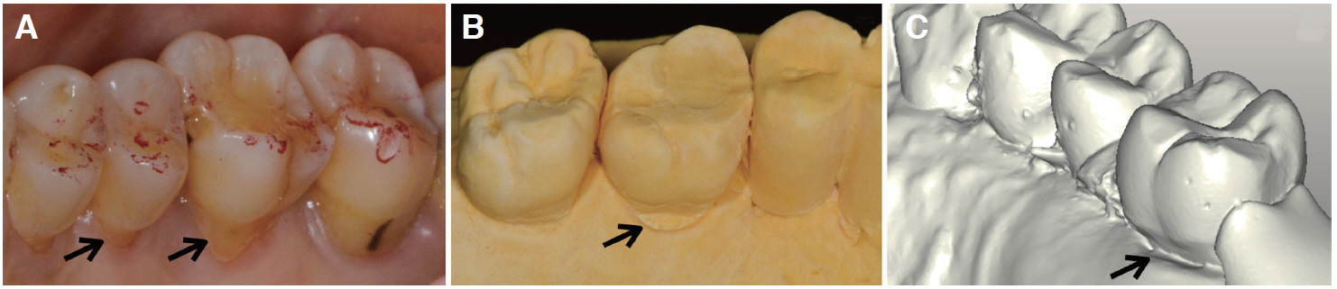
Fig. 1 Palatal side abfractions (arrow). (A) Oral examination, (B) Cast, (C) CAD (Computer Aided Design).
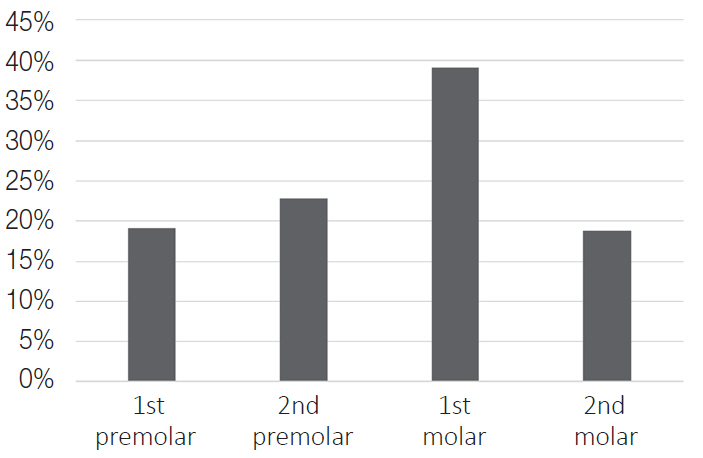
Fig. 2 Distribution of palatal side abfractions in maxillary molar and premolar. Out of 3193 teeth, 276 teeth exhibited palatal side abfractions. When manifestation of palatal side abfraction was classified depending on the type of teeth, 1st premolar accounted for 19.2% (53 teeth), 2nd premolar 22.8% (63 teeth), 1st molar 39.1% (108 teeth), and 2nd molar accounted for 18.8% (52 teeth).
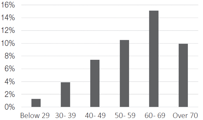
Fig. 3 Distribution of palatal side abfractions according to the age. The ratio of palatal side abfraction in maxillary posterior teeth surveyed for each age was 1.3% under 29 years of age, 3.9% for 30 - 39, 7.4% for 40 - 49, 10.5% for 50 - 59, 15.1% for 60 - 69, and 9.9% for 70 years old and above.
Reference
-
References
1. Mjor IA. 2001; Pulp-dentin biology in restorative dentistry. Part 5: Clinical management and tissue changes associated with wear and trauma. Quintessence Int. 32:771–88. PMID: 11820046.2. Wood I, Jawad Z, Paisley C, Brunton P. 2008; Non-carious cervical tooth surface loss: a literature review. J Dent. 36:759–66. DOI: 10.1016/j.jdent.2008.06.004. PMID: 18656296.3. Miller WD. 1907; Experiments and observations on the wasting of tooth tissue variously designated as erosion, abrasion, chemical abrasion, denudation, etc. Dent Cosm. 49:1–23.4. Lee WC, Eakle WS. 1984; Possible role of tensile stress in the etiology of cervical erosive lesions of teeth. J Prosthet Dent. 52:374–80. DOI: 10.1016/0022-3913(84)90448-7. PMID: 6592336.5. Grippo JO. 1991; Abfractions: a new classification of hard tissue lesions of teeth. J Esthet Dent. 3:14–9. DOI: 10.1111/j.1708-8240.1991.tb00799.x. PMID: 1873064.6. Goel VK, Khera SC, Ralston JL, Chang KH. 1991; Stresses at the dentinoenamel junction of human teeth-a finite element investigation. J Prosthet Dent. 66:451–9. DOI: 10.1016/0022-3913(91)90504-P. PMID: 1791555.7. Chen KK, Miyake K, Terashita M. 1999; Cervical strains induced by occlusal loading. J Dent Res. 78:474.8. Nohl FS, McCabe JF, Walls AWG. 1999; The effect of load angle on strains induced in maxillary premolars in vitro. J Dent Res. 78:1059.9. Dejak B, Mlotkowski A, Romanowicz M. 2005; Finite element analysis of mechanism of cervical lesion formation in simulated molars during mastication and parafunction. J Prosthet Dent. 94:520–9. DOI: 10.1016/j.prosdent.2005.10.001. PMID: 16316798.10. Lee HE, Lin CL, Wang CH, Cheng CH, Chang CH. 2002; Stresses at the cervical lesion of maxillary premolar-a finite element investigation. J Dent. 30:283–90. DOI: 10.1016/S0300-5712(02)00020-9. PMID: 12554108.11. Rees JS. 2006; The biomechanics of abfraction. Proc Inst Mech Eng H. 220:69–80. DOI: 10.1243/095441105X69141. PMID: 16459447.12. Pintado MR, Delong R, Ko CC, Sakaguchi RL, Douglas WH. 2000; Correlation of noncarious cervical lesion size and occlusal wear in a single adult over a 14-year time span. J Prosthet Dent. 84:436–43. DOI: 10.1067/mpr.2000.109477. PMID: 11044852.13. Sawlani K, Lawson NC, Burgess JO, Lemons JE, Kinderknecht KE, Givan DA, Ramp L. 2016; Factors influencing the progression of noncarious cervical lesions: a 5-year prospective clinical evaluation. J Prosthet Dent. 115:571–7. DOI: 10.1016/j.prosdent.2015.10.021. PMID: 26774320.14. Brandini DA, Trevisan CL, Panzarini SR, Pedrini D. 2012; Clinical evaluation of the association between noncarious cervical lesions and occlusal forces. J Prosthet Dent. 108:298–303. DOI: 10.1016/S0022-3913(12)60180-2. PMID: 23107237.15. Antonelli JR, Hottel TL, Brandt R, Scarbecz M, Patel T. 2013; The role of occlusal loading in the pathogenesis of non-carious cervical lesions. Am J Dent. 26:86–92.16. Wood ID, Kassir AS, Brunton PA. 2009; Effect of lateral excursive movements on the progression of abfraction lesions. Oper Dent. 34:273–9. DOI: 10.2341/08-100. PMID: 19544815.17. Estafan A, Furnari PC, Goldstein G, Hittelman EL. 2005; In vivo correlation of noncarious cervical lesions and occlusal wear. J Prosthet Dent. 93:221–6. DOI: 10.1016/j.prosdent.2004.12.012. PMID: 15775922.18. Nascimento MM, Dilbone DA, Pereira PN, Duarte WR, Geraldeli S, Delgado AJ. 2016; Abfraction lesions: etiology, diagnosis, and treatment options. Clin Cosmet Investig Dent. 8:79–87. DOI: 10.2147/CCIDE.S63465. PMID: 27217799. PMCID: PMC4861607.19. Shulman EH, Robinson HB. 1948; Salivary citrate content and erosion of the teeth. J Dent Res. 27:541–4. DOI: 10.1177/00220345480270041501. PMID: 18875186.20. Bergstrom J, Eliasson S. 1988; Cervical abrasion in relation to toothbrushing and periodontal health. Scand J Dent Res. 96:405–11. DOI: 10.1111/j.1600-0722.1988.tb01575.x. PMID: 3201112.21. Levitch LC, Bader JD, Shugars DA, Heymann HO. 1994; Non-carious cervical lesions. J Dent. 22:195–207. DOI: 10.1016/0300-5712(94)90107-4. PMID: 7962894.22. Smith WA, Marchan S, Rafeek RN. 2008; The prevalence and severity of non-carious cervical lesions in a group of patients attending a university hospital in Trinidad. J Oral Rehabil. 35:128–34. PMID: 18197846.23. Nascimento MM, Gordan VV, Qvist V, Bader JD, Rindal DB, Williams OD, Gewartowski D, Fellows JL, Litaker MS, Gilbert GH; Dental Practice-Based Research Network Collaborative Group. 2011; Restoration of noncarious tooth defects by dentists in The Dental Practice-Based Research Network. J Am Dent Assoc. 142:1368–75. DOI: 10.14219/jada.archive.2011.0138. PMID: 22130438.24. Ichim IP, Schmidlin PR, Li Q, Kieser JA, Swain MV. 2007; Restoration of non-carious cervical lesions Part II. Restorative material selection to minimise fracture. 23:1562–9. DOI: 10.1016/j.dental.2007.02.002. PMID: 17391747.25. Rees JS, Hammadeh M, Jagger DC. 2003; Abfraction lesion formation in maxillary incisors, canines and premolars: a finite element study. Eur J Oral Sci. 111:149–54. DOI: 10.1034/j.1600-0722.2003.00018.x. PMID: 12648267.26. Radentz WH, Barnes GP, Cutright DE. 1976; A survey of factors possibly associated with cervical abrasion of tooth surfaces. J Periodontol. 47:148–54. DOI: 10.1902/jop.1976.47.3.148. PMID: 1062558.27. Grippo JO, Simring M, Coleman TA. 2012; Abfraction, abrasion, biocorrosion, and the enigma of noncarious cervical lesions: a 20-year perspective. J Esthet Restor Dent. 24:10–23. DOI: 10.1111/j.1708-8240.2011.00487.x. PMID: 22296690.28. Ferrario VF, Sforza C, Serrao G, Dellavia C, Tartaglia GM. 2004; Single tooth bite forces in healthy young adults. J Oral Rehabil. 31:18–22. DOI: 10.1046/j.0305-182X.2003.01179.x. PMID: 15125591.29. Harvey WL, Hatch RA, Osborne JW. 1991; Computerized occlusal analysis: an evaluation of the sensors. J Prosthet Dent. 65:89–92. DOI: 10.1016/0022-3913(91)90056-3. PMID: 2033554.30. Yaffe A, Ehrlich J. 1987; The functional range of tooth contact in lateral gliding movements. J Prosthet Dent. 57:730–3. DOI: 10.1016/0022-3913(87)90373-8. PMID: 3473233.31. Staninec M, Nalla RK, Hilton JF, Ritchie RO, Watanabe LG, Nonomura G, Marshall GW, Marshall SJ. 2005; Dentin erosion simulation by cantilever beam fatigue and pH change. J Dent Res. 84:371–5. DOI: 10.1177/154405910508400415. PMID: 15790746.32. Whitehead SA, Wilson NH, Watts DC. 1999; Development of noncarious cervical notch lesions in vitro. J Esthet Dent. 11:332–7. DOI: 10.1111/j.1708-8240.1999.tb00416.x. PMID: 10825868.33. Sessle BJ. 1981. Mastication, swallowing, and related activities. Oral Biol St Louis CV Mosby Co. p. 51–89.
- Full Text Links
-
- Actions
-
Cited
- CITED
-
- Close
- Share
-
- Similar articles
-
- A study on the occlusal wear patterns in maxillary posterior teeth with palatal side abfractions
- Maxillary complete denture fabrication cases with posterior palatal seal considering palatal form and tissue displacement
- Photoelastic evaluation of maxillary posterior crossbite appliance
- A Comparative study on the palatal mucosa thickness measurements using periodontal probe and pltrasonic device
- Evaluation of Impacted Maxillary Canine Position Using Panoramic Radiographs and Cone-beam Computed Tomography
