Three-dimensional evaluation of tooth movement in Class II malocclusions treated without extraction by orthodontic mini-implant anchorage
- Affiliations
-
- 1Department of Orthodontics, School of Dentistry, Wonkwang University, Iksan, Korea. sangkim@wku.ac.kr
- 2Department of Prosthodontics, School of Dentistry, Wonkwang University, Iksan, Korea.
- KMID: 2352290
- DOI: http://doi.org/10.4041/kjod.2016.46.5.280
Abstract
OBJECTIVE
The aim of this study was to analyze tooth movement and arch width changes in maxillary dentition following nonextraction treatment with orthodontic mini-implant (OMI) anchorage in Class II division 1 malocclusions.
METHODS
Seventeen adult patients diagnosed with Angle's Class II division 1 malocclusion were treated by nonextraction with OMIs as anchorage for distalization of whole maxillary dentition. Three-dimensional virtual maxillary models were superimposed with the best-fit method at the pretreatment and post-treatment stages. Linear, angular, and arch width variables were measured using Rapidform 2006 software, and analyzed by the paired t-test.
RESULTS
All maxillary teeth showed statistically significant movement posteriorly (p < 0.05). There were no significant changes in the vertical position of the maxillary teeth, except that the second molars were extruded (0.86 mm, p < 0.01). The maxillary first and second molars were rotated distal-in (4.5°, p < 0.001; 3.0°, p < 0.05, respectively). The intersecond molar width increased slightly (0.1 mm, p > 0.05) and the intercanine, interfirst premolar, intersecond premolar, and interfirst molar widths increased significantly (2.2 mm, p < 0.01; 2.2 mm, p < 0.05; 1.9 mm, p < 0.01; 2.0 mm, p < 0.01; respectively).
CONCLUSIONS
Nonextraction treatment with OMI anchorage for Class II division 1 malocclusions could retract the whole maxillary dentition to achieve a Class I canine and molar relationship without a change in the vertical position of the teeth; however, the second molars were significantly extruded. Simultaneously, the maxillary arch was shown to be expanded with distal-in rotation of the molars.
Keyword
Figure
-

Figure 1 Superimposition of three-dimensional pretreatment and post-treatment virtual maxillary models.
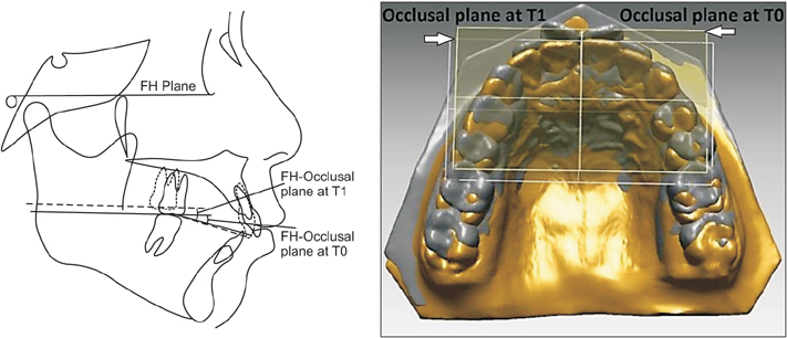
Figure 2 Confirmation of the accuracy of superimposition of the three-dimensional (3D) virtual maxillary models. The angle between the Frankfort horizontal (FH) plane and the maxillary occlusal plane in the lateral cephalogram was measured pretreatment (T0) and post-treatment (T1) to verify a change in the occlusal plane of the 3D virtual maxillary models between T0 and T1. If the angular difference between the 3D virtual models and lateral cephalograms was greater than 5o, superimposition of the 3D models was repeated to correct the error.
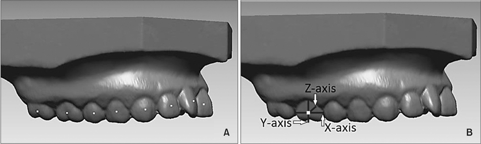
Figure 3 A, Facial axis (FA) points. B, Definition of the coordinate system established at the FA point; the X-axis is the horizontal one, the Y-axis is the vertical one, and the Z-axis is a sagittal axis perpendicular to the X-axis and Y-axis.
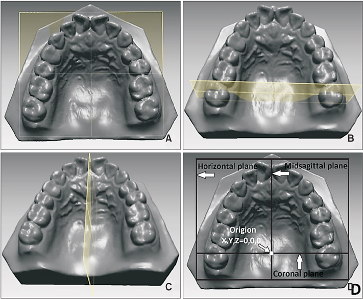
Figure 4 Definition of reference planes. A, The horizontal plane was set to the uppermost region in the midpalatal area, and was parallel to the occlusal plane. B, The coronal plane was set to be perpendicular to the horizontal plane connecting the facial axis points of the upper right and left second molars at the pretreatment (T0) stage. C, The midsagittal plane was set to be perpendicular to the horizontal and coronal planes passing through a midpoint between the maxillary right and left central incisor edges. D, The point of origin was set at the intersection of the horizontal, coronal, and mid-sagittal planes.
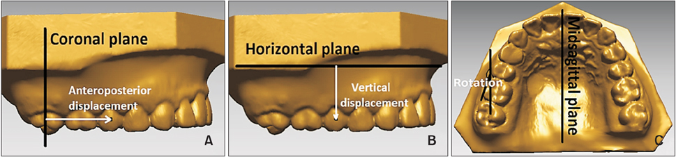
Figure 5 Definitions of linear and angular variables. A, Anteroposterior displacement (mm): distance between the facial axis (FA) point and the coronal plane. For the difference between pretreatment (T0) and post-treatment (T1), i.e., T0–T1, positive means posterior movement and negative means anterior movement. B, Vertical displacement (mm): distance between the FA point and the horizontal plane. For T0–T1, positive means intrusion and negative means extrusion. C, Molar rotation (°): the angle made with the x-axis of an individual molar tooth and the midsagittal plane on the occlusal plane. For T0–T1, positive means mesial-in rotation and negative means distal-in rotation.
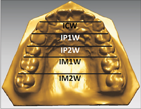
Figure 6 Definitions of arch width variables. Intercanine width (ICW), distance between the cusp tip of right and left maxillary canines; interfirst premolar width (IP1W), distance between the cusp tip of right and left maxillary first premolars; intersecond premolar width (IP2W), distance between the cusp tip of right and left maxillary second premolars; interfirst molar width (IM1W), distance between the mesiobuccal cusp tip of right and left maxillary first molars; intersecond molar width (IM2W), distance between the mesiobuccal cusp tip of right and left maxillary second molars.
Cited by 2 articles
-
Clinical application of an intraoral scanner for serial evaluation of orthodontic tooth movement: A preliminary study
Dalsun Yun, Dong-Soon Choi, Insan Jang, Bong-Kuen Cha
Korean J Orthod. 2018;48(4):262-267. doi: 10.4041/kjod.2018.48.4.262.Three-dimensional analysis of tooth movement in Class II malocclusion treatment using arch wire with continuous tip-back bends and intermaxillary elastics
Ji-Yea Lee, Sung-Kwon Choi, Tae-Hoon Kwon, Kyung-Hwa Kang, Sang-Cheol Kim
Korean J Orthod. 2019;49(6):349-359. doi: 10.4041/kjod.2019.49.6.349.
Reference
-
1. Park HS, Lee SK, Kwon OW. Group distal movement of teeth using microscrew implant anchorage. Angle Orthod. 2005; 75:602–609.2. Egolf RJ, BeGole EA, Upshaw HS. Factors associated with orthodontic patient compliance with intraoral elastic and headgear wear. Am J Orthod Dentofacial Orthop. 1990; 97:336–348.
Article3. Keles A, Sayinsu K. A new approach in maxillary molar distalization: intraoral bodily molar distalizer. Am J Orthod Dentofacial Orthop. 2000; 117:39–48.
Article4. Hilgers JJ. The pendulum appliance for Class II noncompliance therapy. J Clin Orthod. 1992; 26:706–714.5. Gianelly AA, Bednar J, Dietz VS. Japanese NiTi coils used to move molars distally. Am J Orthod Dentofacial Orthop. 1991; 99:564–566.
Article6. Gianelly AA, Vaitas AS, Thomas WM. The use of magnets to move molars distally. Am J Orthod Dentofacial Orthop. 1989; 96:161–167.
Article7. Locatelli R, Bednar J, Dietz VS, Gianelly AA. Molar distalization with superelastic NiTi wire. J Clin Orthod. 1992; 26:277–279.8. Carano A, Testa M. The distal jet for upper molar distalization. J Clin Orthod. 1996; 30:374–380.9. Keles A. Maxillary unilateral molar distalization with sliding mechanics: a preliminary investigation. Eur J Orthod. 2001; 23:507–515.
Article10. Gelgör IE, Büyükyilmaz T, Karaman AI, Dolanmaz D, Kalayci A. Intraosseous screw-supported upper molar distalization. Angle Orthod. 2004; 74:838–850.11. Grec RH, Janson G, Branco NC, Moura-Grec PG, Patel MP, Castanha Henriques JF. Intraoral distalizer effects with conventional and skeletal anchorage: a meta-analysis. Am J Orthod Dentofacial Orthop. 2013; 143:602–615.
Article12. Mah SJ, Kim JE, Ahn EJ, Nam JH, Kim JY, Kang YG. Analysis of midpalatal miniscrew-assisted maxillary molar distalization patterns with simultaneous use of fixed appliances: A preliminary study. Korean J Orthod. 2016; 46:55–61.
Article13. Miresmaeili A, Sajedi A, Moghimbeigi A, Farhadian N. Three-dimensional analysis of the distal movement of maxillary 1st molars in patients fitted with miniimplant-aided trans-palatal arches. Korean J Orthod. 2015; 45:236–244.
Article14. Caprioglio A, Cafagna A, Fontana M, Cozzani M. Comparative evaluation of molar distalization therapy using pendulum and distal screw appliances. Korean J Orthod. 2015; 45:171–179.
Article15. Park HS, Kwon TG, Sung JH. Nonextraction treatment with microscrew implants. Angle Orthod. 2004; 74:539–549.16. Trpkova B, Major P, Prasad N, Nebbe B. Cephalometric landmarks identification and reproducibility: a meta analysis. Am J Orthod Dentofacial Orthop. 1997; 112:165–170.
Article17. Hatcher DC, Aboudara CL. Diagnosis goes digital. Am J Orthod Dentofacial Orthop. 2004; 125:512–515.
Article18. Moles R. The SureSmile system in orthodontic practice. J Clin Orthod. 2009; 43:161–174.19. Joffe L. OrthoCAD: digital models for a digital era. J Orthod. 2004; 31:344–347.20. Cha BK, Lee JY, Jost-Brinkmann PG, Yoshida N. Analysis of tooth movement in extraction cases using three-dimensional reverse engineering technology. Eur J Orthod. 2007; 29:325–331.
Article21. Thiruvenkatachari B, Al-Abdallah M, Akram NC, Sandler J, O'Brien K. Measuring 3-dimensional tooth movement with a 3-dimensional surface laser scanner. Am J Orthod Dentofacial Orthop. 2009; 135:480–485.
Article22. Cho MY, Choi JH, Lee SP, Baek SH. Three-dimensional analysis of the tooth movement and arch dimension changes in Class I malocclusions treated with first premolar extractions: a guideline for virtual treatment planning. Am J Orthod Dentofacial Orthop. 2010; 138:747–757.
Article23. Park HM, Kim BH, Yang IH, Baek SH. Preliminary three-dimensional analysis of tooth movement and arch dimension change of the maxillary dentition in Class II division 1 malocclusion treated with first premolar extraction: conventional anchorage vs. mini-implant anchorage. Korean J Orthod. 2012; 42:280–290.
Article24. Hoggan BR, Sadowsky C. The use of palatal rugae for the assessment of anteroposterior tooth movements. Am J Orthod Dentofacial Orthop. 2001; 119:482–488.
Article25. Choi DS, Jeong YM, Jang I, Jost-Brinkmann PG, Cha BK. Accuracy and reliability of palatal superimposition of three-dimensional digital models. Angle Orthod. 2010; 80:497–503.
Article26. Chen G, Chen S, Zhang XY, Jiang RP, Liu Y, Shi FH, et al. Stable region for maxillary dental cast superimposition in adults, studied with the aid of stable miniscrews. Orthod Craniofac Res. 2011; 14:70–79.
Article27. Andrews LF. The six keys to normal occlusion. Am J Orthod. 1972; 62:296–309.
Article28. Oh YH, Park HS, Kwon TG. Treatment effects of microimplant-aided sliding mechanics on distal retraction of posterior teeth. Am J Orthod Dentofacial Orthop. 2011; 139:470–481.
Article29. Lai EH, Yao CC, Chang JZ, Chen I, Chen YJ. Three-dimensional dental model analysis of treatment outcomes for protrusive maxillary dentition: comparison of headgear, miniscrew, and miniplate skeletal anchorage. Am J Orthod Dentofacial Orthop. 2008; 134:636–645.
Article30. Yamada K, Kuroda S, Deguchi T, Takano-Yamamoto T, Yamashiro T. Distal movement of maxillary molars using miniscrew anchorage in the buccal interradicular region. Angle Orthod. 2009; 79:78–84.
Article31. Sung EH, Kim SJ, Chun YS, Park YC, Yu HS, Lee KJ. Distalization pattern of whole maxillary dentition according to force application points. Korean J Orthod. 2015; 45:20–28.
Article32. Jeon JM, Yu HS, Baik HS, Lee JS. En-masse distalization with miniscrew anchorage in Class II nonextract ion treatment. J Clin Orthod. 2006; 40:472–476.33. Giuntini V, Baccetti T, Defraia E, Cozza P, Franchi L. Mesial rotation of upper first molars in Class II division 1 malocclusion in the mixed dentition: a controlled blind study. Prog Orthod. 2011; 12:107–113.
Article
- Full Text Links
-
- Actions
-
Cited
- CITED
-
- Close
- Share
-
- Similar articles
-
- Preliminary three-dimensional analysis of tooth movement and arch dimension change of the maxillary dentition in Class II division 1 malocclusion treated with first premolar extraction: conventional anchorage vs. mini-implant anchorage
- Correction of a maxillary canine-first premolar transposition using mini-implant anchorage
- A new protocol of the sliding mechanics with micro-implant anchorage(M.I.A.)
- Mode of tooth movement according to the timing of orthodontic force application after extraction
- Three dimensional analysis of tooth movement using different sizes of NiTi wire on NiTi scissors-bite corrector
