Korean J Orthod.
2011 Oct;41(5):371-378. 10.4041/kjod.2011.41.5.371.
Correction of a maxillary canine-first premolar transposition using mini-implant anchorage
- Affiliations
-
- 1Department of Orthodontics, Faculty of Dentistry, Yeditepe University, Turkey. ooztoprak@hotmail.com
- KMID: 1975394
- DOI: http://doi.org/10.4041/kjod.2011.41.5.371
Abstract
- Transposition is defined as a dental anomaly manifested by a positional interchange of 2 adjacent teeth within the same quadrant of the dental arch. Maxillary canine-first premolar [Mx4-3] transposition is the most frequent tooth transposition reported in the literature. In this case report, an orthodontic correction of a transposition of the maxillary left canine and first premolar with the help of palatally located mini-implant anchorage is described. Esthetic and occlusal evaluations suggested alignment of the transposed teeth to their correct anatomic positions in the dental arch. The clinical result at the end of the treatment was satisfactory. Alignment was obtained, and intercuspation was adequate. Nevertheless, the maxillary canine showed facial recession, probably because it was initially positioned buccally. Supporting tissue was examined after treatment and no alveolar bone damage was observed.
MeSH Terms
Figure
-
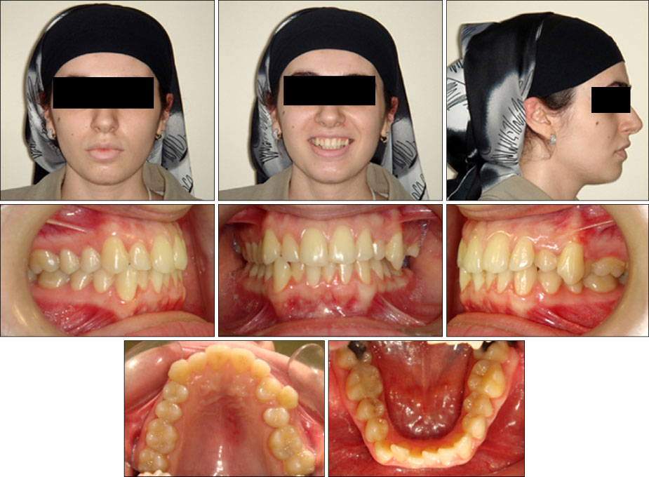
Fig. 1 Pre-treatment extraoral and intraoral photographs.

Fig. 2 Pre-treatment study models.
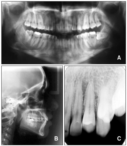
Fig. 3 Radiographs of patient before treatment. Pre-treatment panoramic radiograph (A), lateral cephalometric radiograph (B), and periapical radiograph (C).
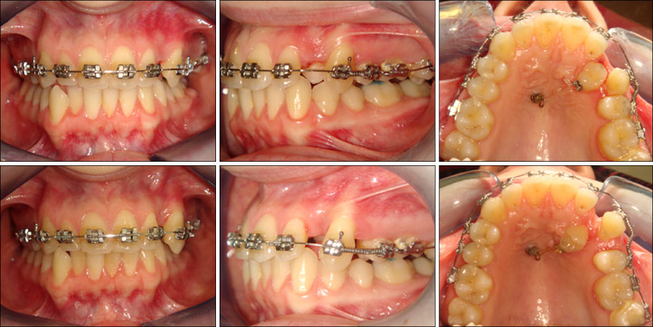
Fig. 4 Mini-implant placement.
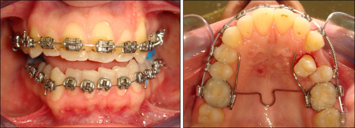
Fig. 5 Activation of transpalatal arch.
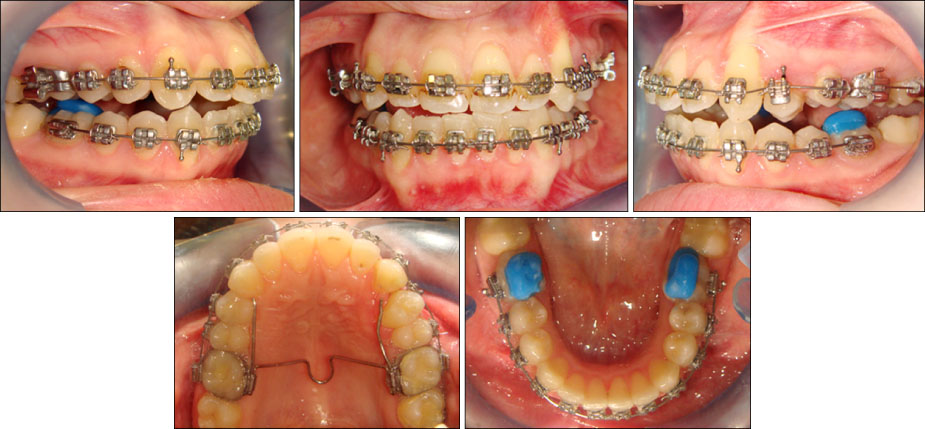
Fig. 6 Progress intraoral photographs.
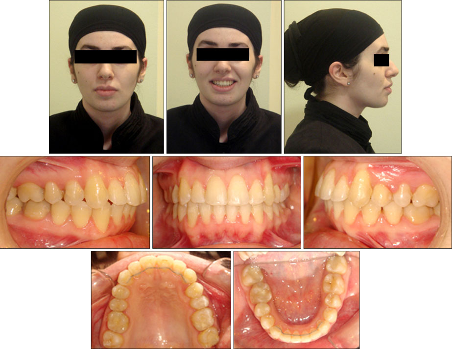
Fig. 7 Post-treatment extraoral and intraoral photographs.
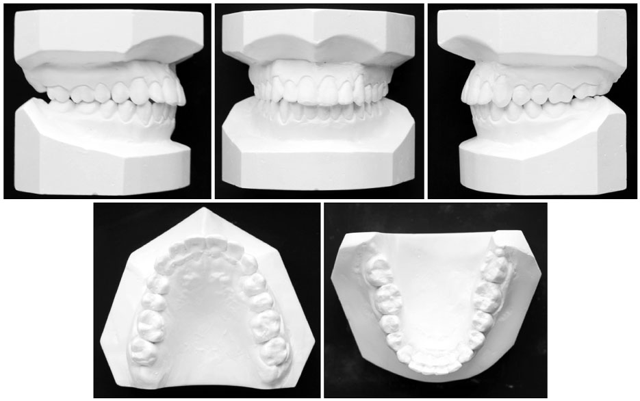
Fig. 8 Post-treatment study models.

Fig. 9 Radiographs of patient after treatment. Post-treatment panoramic radiograph (A), lateral cephalometric radiograph (B), and periapical radiograph (C).

Fig. 10 24-month post-retention intraoral photographs and periapical radiograph.
Reference
-
1. Peck L, Peck S, Attia Y. Maxillary canine-first premolar transposition, associated dental anomalies and genetic basis. Angle Orthod. 1993. 63:99–109.2. Shapira Y, Kuftinec MM. Tooth transpositions--a review of the literature and treatment considerations. Angle Orthod. 1989. 59:271–276.3. Shapira Y, Kuftinec MM. Early detection and prevention of mandibular tooth transposition. J Dent Child (Chic). 2003. 70:204–207.4. Peck S, Peck L, Kataja M. Mandibular lateral incisor-canine transposition, concomitant dental anomalies, and genetic control. Angle Orthod. 1998. 68:455–466.5. Peck S, Peck L. Classification of maxillary tooth transpositions. Am J Orthod Dentofacial Orthop. 1995. 107:505–517.
Article6. Chattopadhyay A, Srinivas K. Transposition of teeth and genetic etiology. Angle Orthod. 1996. 66:147–152.7. Shapira Y, Kuftinec MM. Maxillary tooth transpositions: characteristic features and accompanying dental anomalies. Am J Orthod Dentofacial Orthop. 2001. 119:127–134.
Article8. Ely NJ, Sherriff M, Cobourne MT. Dental transposition as a disorder of genetic origin. Eur J Orthod. 2006. 28:145–151.
Article9. Yilmaz HH, Türkkahraman H, Sayin MO. Prevalance of tooth transpositions and associated dental anomalies in a Turkish population. Dentomaxillofac Radiol. 2005. 34:32–35.10. Shapira Y. Bilateral transposition of mandibular canines and lateral incisors: orthodontic management of a case. Br J Orthod. 1978. 5:207–209.
Article11. Joshi MR, Bhatt NA. Canine transposition. Oral Surg Oral Med Oral Pathol. 1971. 31:49–54.
Article12. Shapira Y. Transposition of canines. J Am Dent Assoc. 1980. 100:710–712.
Article13. Shapira Y, Kuftinec MM, Stom D. Maxillary canine-lateral incisor transposition--orthodontic management. Am J Orthod Dentofacial Orthop. 1989. 95:439–444.
Article14. Jackson M. Transposition of upper canine and lateral incisor. Br Dent J. 1951. 90:158.15. Wasserstein A, Tzur B, Brezniak N. Incomplete canine transposition and maxillary central incisor impaction--a case report. Am J Orthod Dentofacial Orthop. 1997. 111:635–639.
Article16. Jackson M. Upper canine in position of upper central incisor. Br Dent J. 1951. 90:243.17. Joshi MR, Gaitonde SS. Canine transposition of extensive degree: a case report. Br Dent J. 1966. 121:221–222.18. Hallet GE. A maxillary canine erupting in the first molar region. Br Dent J. 1942. 72:191–192.19. Kokich VG, Nappen DL, Shapiro PA. Gingival contour and cilinical crown length: their effect on the esthetic appearance of maxillary anterior teeth. Am J Orthod. 1984. 86:89–94.
Article20. Kuroda S, Kuroda Y. Nonextraction treatment of upper canine-premolar transposition in an adult patient. Angle Orthod. 2005. 75:472–477.
- Full Text Links
-
- Actions
-
Cited
- CITED
-
- Close
- Share
-
- Similar articles
-
- The pattern of movement and stress distribution during retraction of maxillary incisors using a 3-D finite element method
- Three-dimensional evaluation of tooth movement in Class II malocclusions treated without extraction by orthodontic mini-implant anchorage
- Preliminary three-dimensional analysis of tooth movement and arch dimension change of the maxillary dentition in Class II division 1 malocclusion treated with first premolar extraction: conventional anchorage vs. mini-implant anchorage
- A clinical study on anchorage control of molar anchoring spring(MAS) during retraction of the maxillary canine
- Surgical orthodontic treatment of skeletal Class III malocclusion using mini-implant: correction of horizontal and vertical dental compensation
